Copyright 0 1995 by the Genetics Society of America
Spatial and
Temporal Patterns
of
Zin-12
Expression
During
C.
elegans Hermaphrodite Development
Hilary
A.
Wilkinson””and Iva Greenwald*’+
*Department of Biochemist? and Molecular Biophysics and tHoward Hughes Medical Institute, Columbia University, College of Physicians and Surgeons, New York, New York 1 0 0 3 2
Manuscript received April 11, 1995
Accepted for publication June 22, 1995
ABSTRACT
The lin-12 gene encodes a receptor that mediates certain cellcell interactions during Caenmhabditis
elegans development. We have examined the expression of a lin-12::lacZ reporter gene in individual
cells during the development of C. elegans hermaphrodites. lin-12::lacZis expressed in a discrete spatial and temporal pattern during development and the lin-12::lacZ reporter gene will provide a useful
marker for other studies, particularly of somatic gonadal and vulval development. In general, the cells that express lin-I2::lacZ correspond to cells whose fates are known to be altered in lin-12 mutants implying that restriction of lin-12 expression may be an important regulatory mechanism; the exceptions to this statement may reveal the cellular defects that underlie aspects of the lin-12 phenotype that have
not been previously explained. For decisions that are not naturally variable, lin-12::lacZ expression does not appear to change before or upon commitment to a cell fate implying that in these cases posttranscriptional regulation of lin-12 activity may control cell fate specification.
D
URING animal development, cell-cell interactions play important roles in causing initially equivalent cells to adopt different fates. Receptors such as lin-12,a member of the “lin-l2/Notch family”, mediate such interactions (reviewed in GREENWALD and RUBIN 1 9 9 2 ) . Genetic and anatomic studies of lin-12 mutants that have either elevated or reduced levels of lin-12 activity revealed that lin-12 controls certain binary decisions during Caenorhabditis elegans development ( GREENWALD
et al. 1983). The phenotype of lin-12 null [lin-12(0)]
mutants revealed that several different cell fate deci- sions are mediated by Zin-12 five cell fate transforma- tions are highly penetrant in lin-12(0) mutant hermaph- rodites (GREENWALD et al. 1 9 8 3 ; NEWMAN et al. 1 9 9 5 ) . Additional cell fate decisions are mediated either by
lin-12 or a related gene gQ-1, and are altered with low penetrance in lin-12(0) mutants (LAMBIE and KIMBLE
1991). Mutations that activate lin-12 [lin-lZ(d)] can cause cell fate transformations that are the opposite of those caused by lin-12(0) mutations (GREENWALD et al. 1983; GREENWALD and SEYDOUX 1 9 9 0 ; STRUHL et al. 1993; NEWMAN et al. 1 9 9 5 ) .
The cells affected by mutations in lin-12 can generally be grouped by shared developmental potential. In most cases, equivalent developmental potential of cells that invariably have different fates is revealed by laser abla- tion experiments: cells whose fates are altered after ab- lation of their neighbors are inferred to require cell-
Corresponding author: Iva Greenwald, Department of Biochemistry, Columbia University, Hammer Health Sciences Center, Room 720A, 701 West 168th St., New York, NY 10032.
’
Present address: Department of Cellular Biochemistty and Physiol-ogy, Merck Research Laboratories, Rahway, NJ 07065.
Genetics 141: 513-526 (October, 1995)
cell interactions for their correct specification (for dis- cussion, see SULSTON and WHITE 1 9 8 0 ; SULSTON 1 9 8 8 ) .
In a few cases, equivalent developmental potential is suggested by natural variability in the cell lineage and can be confirmed by laser ablation experiments (SULS
TON and HORVITZ 1977; KIMBLE and HIRSH 1979; SULS
TON et al. 1980, 1983; KIMBLE 1 9 8 1 ) .
Because lin-12 encodes a receptor, its activity may be controlled at one or more levels. (1) lin-12 might be ubiquitously expressed, but L I N - 1 2 activity controlled posttranscriptionally. Several different possible posttran- scriptional regulatory mechanisms have been observed for other receptors. The availability of ligand or compo- nents of the signal transduction machinery may regulate the activity of a receptor: for example, the Torso recep tor tyrosine kinase is uniformly distributed in the Dro- sophila early embryo, but the activity of its ligand is spatially localized, leading to Torso activation only at the termini of the embryo (CASANOVA and STRUHL 1 9 9 3 ) . The availability of receptor protein itself may regulate its activity: for example,
glpl
maternal mRNA is found in all of the first eight early blastomeres of C.514 H. A. Wilkinson and I. Greenwald
In a previous study, we examined the expression of
a lin-12::lacZ r e p o r t e r g e n e d u r i n g the specification of two cells, Zl.ppp and Z4.aaa, of the hermaphrodite
gonad (WILKINSON et al. 1994). Unlike other cells af- fected by mutations in lin-12 during hermaphrodite de-
velopment, Z1.ppp a n d Z4.aaa have naturally variable fates, with each cell having the potential to become an
anchor cell (AC) o r a ventral uterine precursor cell
(VU)
(KIMBLE and HIRSH 1979). However, in a given hermaphrodite, interactions between Zl.ppp and Z4.aaa cause only one to become the AC while the other be-comes a VU (KIMBLE 1981; SEYDOUX and GREENWALD
1989). In lin-12(0) animals, both cells become ACs; i n lin-l2(d) animals, both cells become W s . The Zin-
12::lacZ r e p o r t e r gene is initially expressed in both Z1.ppp and Z4.aaa, but before commitment, lin-12 ex- pression is seen only in the presumptive W (WILKIN-
In this study, we have explored the relationship be-
tween lin-12 expression and its activity by examining the expression of a Zin-12::ZacZ r e p o r t e r g e n e i n cells t h r o u g h o u t wild-type hermaphrodite development. We
have f o u n d t h a t lin-12::lacZis expressed in the groups
of equivalent cells that are affected by mutations in Zin- 12. Moreover, we have found that lin-l2::lacZ expres- sion appears to be u n i f o r m a n d c o n s t a n t i n cells with invariant fates, in contrast to what we observed for the naturally variable pair, Z1.ppp and Z4.aaa (WILKINSON
et al. 1994).
SON st al. 1994).
MATERIALS AND METHODS
General methods and strains: Methods for handling and culturing C. elegans have been described by BRENNER (1974). The wild-type parent for all strains used was C. elegans var. Bristol strain N2 (BRENNER 1974). The LGZ mutations used were as follows: smg-l(r861) and une-54(r29?) (HODGKIN et al. 1989); and unc-l3(r51) (BKENNEK 1974). The LGIIl mutations used were as follows: dpy-l7(e164) and unc.-?2(e189) (BRENNER 1974). The LGX mutation used was unc-84(e1410ts). Strain GS956, of genotype smg-1 unc-54;arZsll, was used for this study (see below).
arlsll: The generation ofGS956, the canonical lin-12::lacZ reporter gene strain containing arlsll, is also described in WlI.KINSON et az. (1994). ads11 is an integrated array com- posed of the plasmids pRF4 [ r d 6 (su1006)] (MEILO rt aZ. 1991) and pBGSLE (a lin-12::ZucZ chimeric gene) (WII.KIN-
SON et aZ. 1994). pBGSLE contains all sequences required to rescue a lin-12(0) mutant (FITZGERALD et al. 1993). A modified l a d g e n e (FIRE et nl. 1990) was inserted into the BamHI site at position 6 in frame to the lin-l2ATG. The ZacZgene encodes a P-galactosidase protein that contains a nuclear localization signal and includes its own stop codon but no polyadenylation signal. The mRNA encoded by the lin-12::ZucZgene is there- fore predicted to be Unstable due to the presence of a large
3' untranslated region containing most of the lin-12 coding sequence. For this reason, ,%galactosidase activity is only de- tectable in the presence of a smg-l mutation, which stabilizes mRNAs with long 3' untranslated regions (PUIAK and ANDER-
We have taken a number of precautions to help ensure that expression of d s l l accurately reflects expression of the
SON 1993).
endogenous lin-12 gene (data not shown). First, we estah lished five independent transgenic lines carrying extra- chromosomal arrays of this reporter gene and created seven independent attached lines deriving from two different extra- chromosomal arrays. All of the integrated and extrachromo- somal arrays displayed similar expression patterns, although some lines displayed some variability in the intensity or pene- trance of staining. Second, we analyzed an integrated array to circumvent variability due to mosaicism (FIRE 1986; HERMAY 1995). Third, the marker plasmid used, pRF4, has been used as a coinjection marker with the lin-12(+) rescuing clone and these arrays are able to rescue all of the defects associated with loss of Zin-12 activity (FITZGERUD et al. 1993). T h ~ s , i t
appears that this marker does not interfere with lin-12 expres- sion in the cells in which it is required. Fourth, the expression pattern of the lin-12::lacZis altered in different Zin-12mutants consistent with the cell fate transformations in these mutants (Wll.KlNsON et al. 1994; this work). Fifth, the ZacZ gene used encodes a modified P-galactosidase protein that appears to be unstable and does not appreciably perdure (WAY and C~rl41.Fll'.
We also note that the use of smg-l is not likely to have any effect on the pattern of transcription, because .rmg-1 appears to act exclusively by stabilizing unstable mRNAs (PUIAK and ANDERSON 1993). The stabilization of Zin-12::luc.Z mRNA in smg-1 mutants does not necessarily lead to perdurance of the reporter gene mRNA, since we have observed an apparently rapid change in expression pattern within a cell and nonstain- ing daughters of staining mothers (WII.KINSON rt al. 1994; this
ad51 1 was mapped between dpy-17 and unc-?2 III at 20" in the following manner: unr-13; arlsll hermaphrodites were mated to dpy-17 unc.-32/+
+
males. Cross progeny of the geno- type unc-I?/+; arI,sll/dpy-l7 unc-32 were picked and their progeny scored for the number of Dpy non-Unc-32 recombi- nant animals that had also picked up arlsll; 6/16 Dpy non- Unc-32 animals segregated Roller progeny. These data indi- cate that the array is "1.5 map units (m.u.) to the left of the lin-12 locus.P-galactosidase activity assay and identification of cells: smg-l unc-54;arIsll animals were grown at 25". Individual ani- mals of a particular age were identified under Nomarski dif- ferential interference microscopy and then tested for P-galac- tosidase activity using an acetone fixation protocol described by FIRE (1993). Pictures of the staining pattern were taken at
X1000 with a flash using TMAX400 film (Kodak).
Individual staining nuclei were identified by three criteria: the age of the animal as determined by Nomarski examination before fixation; the size and shape of the nuclei that stained; and the position of the staining nuclei in the animal relative to other nuclei, as defined by S L ~ I . S I O N (1976), SUI.STON and HORVITZ (1977), and SUI.STON et al. (1983). Counterstaining with 4,ddiarnidino-2 phenylindole (DAPI) allowed visualim tion of all nuclei in the animal by fluorescence microscopy (ELLIS and HOKVITZ 1986).
We were particularly interested in the staining pattern ob- served in the ventral hypodermal cells that give rise to thc vulva and examined this pattern in more detail. W C s : The uric-84 gene is required for the correct nuclear migration of Pl-Pl2, the cells that generate P1.p-P12.p in the ventral hypodermis (SUISTON and HOKVITL 1977; FIXSEN 1985). I n
uric-84 mutants the nuclei of the P cells do not migrate to the ventral cord and instead die during the L1 larval stage. A n - mals of the genotype smg-1 unc-~4;urlJll;unc-84 were exam- ined in the L2 larval stage for the staining pattern observed in the ventral cord. In most animals, all P nuclei failed to migrate, and no P-galactosidase staining was observed. In some animals, one or two P nuclei migrated. If P3-P8 m i -
1989; FIKF. Pt Uz. 1990; WI I KI N SO N et al. 1994).
lin-12 Expression Patterns
grated, P-galactosidase staining was observed in the ventral cord. If PI, P2, or P9-Pl2 migrated, no staining was observed.
W C descendants: A monoclonal antibody that recognizes des- mosomal junctions of cells in the ventral hypodermis (MH27)
(FRANCIS and WATERSTON 1985; PFUESS and H I R ~ H 1986) was used to examine the staining pattern observed in the “N”
and “T” cell descendants of P5.p and P7.p. This antibody stains the outline of the grandaughters of P5.p, P6.p and P7.p soon after they are born (KENYON 1986). We used the MH27 antibody in conjunction with a rabbit anti-p-galactosidase anti- body (Cappel Laboratories) for immunofluorescent analysis (using a protocol described by FINNEY and RUVKUN 1990) in the late L3 stage. This examination confirmed that P-galactos- idase protein was not detected in the nuclei of the central four cells that are the T descendants of P6.p and the protein was detected in the nuclei of the N and T descendants of P5.p and P7.p.
RESULTS
In C. ekguns, the best available method for the study of gene expression in larvae is the analysis of transgenic strains carrying gene fusions with reporter genes such as
l a d , which can be assayed for P-galactosidase activity (FIRE et ul. 1990).
As
described previously, we have con- structed a Sn-12::lacZreporter gene that contains all ge- nomic sequences necessary for rescue of a lin-12(0) mu- tant (WILKINSON et al. 1994). Here we report a detailed description of the expression pattern of arlsll, an inte- grated array containing the lin-12::lacZ reporter gene, during larval development. We have taken a number of precautions to help ensure that expression of the re- porter gene accurately reflects expression of the endoge- nous gene, as described in MATERIALS AND METHODS.lin-12: : lacZ expression in somatic gonadal development
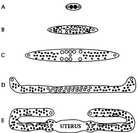
Background: HIRSH et al. (1976) and KIMBLE and HIRSH (1979) described the anatomy and development of the hermaphrodite gonad in C. ekguns (Figure 1).
At hatching, the hermaphrodite gonad consists of two somatic progenitors, Z1 and 24, and two germline pro- genitors, 22 and 23. Gonadal development may be di- vided in two phases, based on the two periods of somatic proliferation. The first phase of gonadal development begins with a period of somatic proliferation during the L1 stage, when Z1 and 24 each produce six descen- dants. During the L2 stage, no further cell divisions occur in the somatic gonad, although there is germline proliferation. In the late L2 stage, the gonad primor- dium forms: the 10 proximal somatic gonadal cells re- arrange themselves a n d displace the germ nuclei into the gonadal arms, and one of these cells, the presump- tive AC, moves to occupy a central position.
The second phase of gonadal development begins in the L3 stage and continues into the L4 stage. During this time the somatic gonadal cells in the primordium (with the exception of the anchor cell and the distal tip cells) all divide. The progeny of these divisions dif-
A
B
515
C
FIGURE 1 .-Overview of gonadal development. Schematic diagram of the first (A-C) and second (D and E) phase of gonadal development (adapted from HODGIUN 1988). 0, so- matically derived nuclei; 0 , germline nuclei. (A) Dorsal view of the gonad during the early L1 stage. There are four cells that give rise to the gonad. (B) Dorsal view of the gonad during the L1 molt. The two somatic progenitors generate
12 progeny. The germ nuclei begin to divide. (C) Dorsal view of the gonad during the L2 molt. The germ nuclei proliferate extensively during the L2 stage and the somatic nuclei re- arrange their position to form the gonad primordium. (D)
Lateral view of the gonad during the L3 stage. The two gonad arms extend and the somatic nuclei proliferate to generate the cells that will form the structures of the gonad. The germ nuclei continue to proliferate. ( E ) Lateral view of the late L4 gonad. The two arms of the gonad reflex and continue to grow out. The somatic nuclei generate the spermatheca (SP), uterus, and the sheath cells that surround the gonad arms. The germ nuclei continue to proliferate and those in the spermatheca differentiate into sperm. After the L4 molt, the germ nuclei in the proximal gonad arms will begin to differen- tiate into oocytes.
ferentiate to give rise to the uterus, anterior and poste- rior sheaths and the anterior and posterior sperma- thecae. During this time the gonad arms continue to elongate and form a reflexed tube. T h e proximal germ nuclei begin to differentiate into sperm. T h e distal germ nuclei remain mitotic. After the L4 molt, oocytes begin to form and are fertilized as they pass through the spermatheca to the uterus.
1272-12 activity is required for at least two cell fate deci- sions in somatic gonad development. In the early L2 stage, Zl.ppa, Zl.ppp, Z4.aap, and Z4.aaa have equiva- lent developmental potential: each of them has the abil- ity to become an AC o r a W (KIMBLE 1981; SEYDOUX and GRFLENWALD 1989; SEYDOUX et al. 1990). Zl.ppa and Z4.aap always become W s in wild type (KIMBLE and
516 H. A. Wilkinson and I. Greenwald
1983). Moreover, in wild-type hermaphrodites, Zl.ppa and Z4.aap have the potential to become ACs early in the L2 stage; in a lin-12(0) mutant, this potential is ex- tended until late in the L2 stage (SEYDOUX et al. 1990). The fates of Zl.ppp and Z4.aaa are naturally variable. Each cell has an equal chance of becoming the AC or a
W. In any given hermaphrodite, only one of these cells will become the AC, while the other becomes a W (KIM- BLE and HIRSH 1979). Interactions between Zl.ppp and Z4.aaa specify their fates (KIMBLE 1981; SEYDOUX and GREENWALD 1989). These interactions are mediated by lin-1.2 in lin-12(0) mutants, both Zl.ppp and Z4.aaa be- come ACs, while in lin-l2(d) mutants, which have ele- vated levels of tin-12 activity, both Zl.ppp and Z4.aaa become W s (GREENWALD et al. 1983). The three W s undergo slightly different lineages (KIMBLE and HIRSH
1979) (see Figure 3). A. NEWMAN, J. G. WHITE and P. W. STERNBERG (1995) have shown that the daughters of the
W s are initially equivalent, but that a signal from the AC appears to induce these cells to generate a certain number of 7r cells, which undergo a characteristic lin- eage. They have also shown that this interaction depends on lin-12activity: in a lin-12(0) mutant, the W daughters do not divide and no 7r cells are generated, whereas in a lin-l2(d) mutant, excess 7r cells are generated.
lin-12: : lacZ is expressed in all three Ws: lin-12 : : lacZ expression in the early phase of the Z 1 and 24 lineages is summarized in Figure 2. Expression begins in Zl.pp and Z4.aa, and is seen in Zl.ppp, Z4.aaa, Zl.ppa and Z4.aap. AI1 four cells continue to express the reporter gene until the middle of the L2 stage. Some additional staining is seen elsewhere before primordium forma- tion at low frequency (see Figure 2 legend). After the primordium has formed, lin-12::lacZ expression is de- tected only in the three W s : Zl.ppa and Z4.aap, and either Zl.ppp or Z4.aaa. During the early L3 stage, lin- 12::lacZ staining disappears in the W s ; staining reap- pears in their daughters just after division (see below). We have described elsewhere how the lin-12::lacZ ex- pression pattern in Zl.ppp or Z4.aaa changes before primordium formation and its relationship to the deci- sion of these cells between the AC and W fates (WIL-
KINSON et al. 1994).
lin-12:: lacZ is expressed in the W daughters: Many different cells express the lin-12::lacZ reporter gene during the second phase of somatic gonadal develop- ment. There are six cells that express the reporter gene in the somatic gonad in the mid-L3 stage (Figure 3). This staining corresponds to the daughters of the WS,
that is W l ( l / r ) , W 2 ( a / p ) and W 3 ( a / p ) [the actual lineage of the cells depends on whether Zl.ppp or Z4.aaa has become an AC: if Zl.ppp adopts the AC fate, then Z4.aap adopts the W 1 fate, Zl.ppa adopts the
W 2 fate, and Z4.aaa adopts the W 3 fate; or if Z4.aaa adopts the AC fate, then Zl.ppa adopts the W1 fate, Z4.aap adopts the W 2 fate, and Zl.ppp adopts the W 3 fate (KIMBLE and HIRSH 1979)l. As noted above, there
appears to be a time during the early L3 stage when the W s no longer express the lin-12::lacZreporter gene, so that the staining that is observed in their daughters appears to represent “new” expression.
The lin-12::lacZ reporter is also expressed in all twelve of the granddaughters of the W s (Figure 3). We have been unable to determine whether the reporter construct is expressed in the great-granddaughters be- cause it is expressed in a cluster of up to 28 cells in the somatic gonad, making it difficult to distinguish individual nuclei.
Significance of the staining pattern observed in the W s and their daughters: During the first phase of go- nadal development, Ein-12::lacZ is expressed in Zl.ppa, Zl.ppp, Z4.aap, and Z4.aaa, the cells whose fates are altered in lin-12 mutants. For Zl.ppp and Z4.aaa, lin-
12::lacZ expression is maintained in the presumptive
W and not in the presumptive AC (WILKINSON et al. 1994). For Zl.ppa and Z4.aap, which both become W s ,
lin-12:: lacZ staining appears to be constant throughout the L2 stage. Thus, for cells that become W s , lin-
12::lacZis expressed throughout the L2 stage. A differ- ence between the two pairs of cells, Zl.ppp/Z4.aaa and Zl.ppa/Z4.aap, emerges from an examination of the staining pattern in a lin-12(0) mutant. In a lin-12(0) mutant staining is observed throughout the L2 stage in Zl.ppa and Z4.aap, but staining is observed in Zl.ppp and Z4.aaa only prior to primordium formation (WIL-
KINSON et al. 1994). This observation is consistent with the interpretation that lin-12 activity is not required
per
se for lin-12 expression to be maintained in Zl.ppa and Z4.aap.In a lin-12(+) background there is no temporal change in the staining pattern observed in the W
daughters or W granddaughters that express the lin- 12::lacZ reporter gene. Ablation studies have indicated that an interaction between the AC and the W daugh- ters is required for the correct number of 7r cells to be generated (NEWMAN et al. 1995). It appears unlikely that lin-12 activity is regulated by restricting its expres- sion only to the 7r cells themselves or to the W daugh- ters that generate them. 0-galctosidase activity is also detected in all six of the W daughters in both lin-
12(0) and lin-lZ(d) mutants; this is consistent with the interpretation that lin-12 activity is not required
per
seto generate the pattern observed (data not shown). lin-12::lacZ is expressed in sheath cells Nos. 1
-
3: There is a total of 20 sheath cells, five pairs in each arm, that arise from four different lineages (KIMBLElin-12 Expression Patterns 317
(AI Z l
z-1
I
I
7
P,7
P,; p P ,
I
P,;
7
P,+,
0
0
0
0
0
m
m
0 0 0
0
o
m
m m m
0
0
I I I
m
0
.
m
FIGURE 2.-1in-12::/acZexpression in the first phase of somatic gonadal development. (A) Schematic diagram of the Z1 and 24 lineages during the first phase of somatic gonad development (adapted from KIMBIX and HIRSH 1979). 0 , cells that express the reporter gene;
0,
staining is only sometimes observed in Z l m and Z4.pp (the distal tip cells) or in 2l.paa or Z4.app. None of these staining patterns is found in a high percentage of animals and the reason for this variability is unknown. (R and C )Nomarski photomicrographs of the somatic gonad of an L2 larva that has been fixed and stained for &galactosidase activity as described in MATERIALS AND METHODS. Anterior is to the left. Lateral view of an L2 animal. R and
c
show different planes offocus. Due to the Roller phenotype, all lin-lZ::lmZ expressing cells in the somatic gonad are visible from this angle. All four cells that have the potential to adopt the W fate, Zl.ppa, Zl.ppp, Z4.aaa and Z4.aap, express the lin-12::lncZ reporter gene at this stage. Note that the VPCs also express the lin-12 transgene at this time and these cells appear as dark spots or patches in the photographs.
Zl.paa and the other is a descendant of Zl.ap; and there are two sheath cells No. 1 in the left arm of the gonad: one is a descendant of Z4.app and the other is a descendant of Z4.pa). Soon after they are born, the morphology of these cells changes. They flatten out
and elongate and either move or are carried out along
the arms of the gonad and eventually wrap around the gonad.
During the L3 stage, P-galactosidase activity is de- tected in two sheath cells in each gonad arm: sheath cells No. 1 (Zl.paaa, Zl.apa, Z4.pap, and Z4.appp) (Fig- ure 4). During the early L4 stage, eight more sheath cells express the transgene: sheath cells No. 2 (Zl.paa- paaa, Zl.appaaa, Z4.paappp, and Z4.appappp) and
sheath cells No. 3 (Zl.paapaap, Zl.appaap, Z4.paappa,
and Z4.appappa). The staining in the sheath cells is very reproducible in the early L4 stage, however, it is less reproducible late in the L4 stage and in the young adult stage. Staining is almost always observed in sheath cells No. 1 in the L3 and L4 stages as well as in the
young adult; however, only a subset of animals consis- tently express the reporter gene in sheath cells No. 2 once the nuclei have begun to migrate far out along the arm. We have never been able to detect staining in sheath cells No. 3 once the nuclei have begun to mi- grate far out along the arm. One important observation to note is that the staining is observed in 12 sheath cells during the late L3/early L4 stage soon after they are born (that is, in all the lineages in which they arise). However, as the cells move out the gonad arm, staining is only detectable in one member of' pair No. 1 and one member of pair No. 2 in each arm. It is unknown which member of the pair stains or whether it is always the same member of the pair that continues to express the lin-12::lacZ gene.
lin-12::lacZ is expressed in a subset of spermathecal cells: There is a subset of cells in the DU (dorsal uter- ine) lineage that contribute to the structure of the sper-
matheca (KIMRLE and HIRSH 1979) (Figure 5). These
518 H . A. Wilkinson and I. Greenwald
( A )
vu2
M
0
avu3
0
I
P
vu1
FIGURE 3.-Zin-12::lncZexpression in the W descendants. (A) Schematic diagram of the partial lineage of the W descendants during the L3 stage (adapted from KIMRLE and HIRSF-I 1979). 0 , cells that express the reporter gene. .rr indicates the cells that adopt the .rr cell fate (see RESUITS). (B) Nomarski photomicrograph of the somatic gonad of an L3 larva that has been fixed
and stained for @-galactosidase activity as described i n MATERIAIS AND METHODS. Anterior is to the left. Dorsal view of the mid-
L3 larval stage. All six W daughters are visible in this plane. Dark smudges indicate other cells that are staining at this time
such as the VPC descendants. (C) Nomarski photomicrograph of the somatic gonad of an L3 larva that has been fixed and
stained for @-galactosidase activity as described in MATERIAIS AND METI-IODS. Lateral view of the late L3 hrval stage. Only the
w
descendants o n the left side of the animal are visible in this plane of focus. Dark smudges indicate other cells that are stainingat this time such as the vulval precursor cell descendants and other somatic gonadal cells.
anterior spermatheca and two contribute to the poste- rior spermatheca. Each progenitor gives rise to four cells that form the part of the spermatheca that is adja- cent to the uterus. One cell from each progenitor con- tributes to the core of the spermathecal-uterine junc- tion, a highly specialized structure formed by the fusion of two cells.
The expression of the reporter gene in the spermathe- cal cells is most easily detected during the
L4
larval stage (Figure 5). There are up to eight spermathecal cells [Zl.papaa(a/p) (d/v), Z4.apaaa(a/p) (d/v), Zl.pappp (a/p) (d/v), and Z4.apapp(a/p) (d/v)] in each arm that express the reporter gene; the position and num- ber is consistent with a group of cells in lineage “B”( K I M R L E and HIRSH 1979) that are present at the sper- matheca] uterine junction. The progenitors of these cells [Zl.papaa(a/p), Z4.apaaa(a/p), Zl.pappp(a/p), and Z4.apapp(a/p)] also appear to express the re- porter gene. There is some variablity in the number of staining cells detected and this description may in- clude only a subset of the total number of spermathe- cal cells that express the reporter construct.
Significance of the stainiig pattern observed in the sheath cells and the spermatheca: During the second phase of gonadal development, lin-12::lacZ expression
in the spermatheca and sheath cells was not predicted from the lin-12 mutant phenotypes. However, lin-12(0) and lin-Z2(d) mutants both have fertility defects: lin- 12(0) mutants are generally sterile, and several lin,-12(d) mutants have reduced brood sizes (GREENWALD et al. 1983). The cellular basis for the sterility is not known, but the lin,-12::lacZ expression pattern suggests that spermatheca and/or sheath defects may underlie the fertility defects. Recently, J. MCCARTER and
T.
SCHEDL(personal communication) have demonstrated that the sheath and spermatheca1 cells are involved in at least three processes in the developing germline: prolifera- tion of germ nuclei, germline sex determination, and oocyte maintenance of meiotic arrest. The germline anatomy of lin-12 mutants has not been well character- ized and may display abnormalities in one or more of these processes. lin-Z2(0) and lin-l2(d) mutants appear to have abnormal spermathecal-uterine junctions
lin-12 Expression Patterns 519
( A )
anterior sheath
Zl.paa
I
Zl.ap
I
0
0
posterior sheath
ZAapp
I
Z4.pa
I
0
0
m
o
m
m
+-I
I
A
0
0
1
0
AA
II
0
+ I +
0
0
0
0
A I 4 A A A A
m m o o - m m o o o o m m
CDooom
I I I I I
CD000000~
l l l l l l l l
1 2 3 4 5 1 2 3 4 5 5 4 3 2
m
o
m
4 l
I
0
- 0
0.0
1 1 1 1 1
CDOOOCD
1 5 4 3 2
0
cp
1
FIGURE 4.-lin-l2::lnc% expression in sheath cells. (A) Schematic diagram of the lineages of the sheath cells (adapted from
KIMRLE and HrRsM 1979). 0 , cells that express the reporter gene. 63, expression that is observed only in one of the two pairs of cells indicated (ie., one of the No. 1 sheath cells and one of the No. 2 sheath cells in each sheath). It is unknown in which No. 1 or No. 2 sheath cell expression of the reporter gene is maintained. Note that the No. 3 sheath cells express the reporter gene during the early L4 stage but do not express the reporter gene in the late L4 stage. (B) Bright field photomicrograph of an L4 larva that has been fixed and stained for &galactosidase activity as described in MATERIALS AND METHODS. Anterior is to the left. Lateral view. Note that only one of the two No. 1 sheath cells has visible staining. Due to the Roller phenotype, both Nos. 2 and both Nos. 3 sheath cells are visible.
.&+I(-) mutant dies in the L1 stage (LAMBIE and KIMBLE
1991), functional redundancy for later developmental events would not have been seen.
Given the observation that Zin-12::ZucZ staining per- sists in only one of the pair of sheath cells, it is intri- guing to speculate that these cells are not equivalent as
was previously thought (KIMDLE and HIFLSH 1979). It
may be that there are subtle differences in the functions of individual sheath cells that are not obvious based on morphogenetic or ablation criteria. We have observed the same sheath cell staining pattern in a Zin-I2(d) mu- tant; however, in a lin-12(0) mutant, staining persists in both members of pairs Nos. 1 and 2 (data not shown).
These observations are similar to those described for Z1 .ppp and Z4.aaa in the sense that the staining pattern observed in these two cells is altered in lin-12 mutants. This may indicate that the sheath cells have a naturally variable cell fate decision that is mediated by Zin-12 and that the level of Zin-12 activity affects the pattern of Zin-
12 expression.
lim12::lacZ expression in vulval development
520 H. A. M'ilkinson and I. Greenwald
( A )
anterior spermatheca posterior spermatheca
2l.papaa-left
Z4.apaaa-right
Zl.pappp-left Z4.apapp-right
I
I
I
I
0
0
0
0
e e
e
e
e
FIGLIRE 5 . - / i n - l 2 : : k r z expres- sion in the spermathecal cells. (A)
Schematic diagram of the lineages
of the cells thought to stain in the
spermatheca (adapted from KI\IRI.E: and HlRsll 1979). 0 , cells that ex-
press thc reporter gcne. (B) Nomar-
ski photomicrograph of young adult
that has been fixed and stained for
D-galactosidase activity as described
in MATERIAIS AND METHODS. Ante- rior is to the left. Lateral view. Due
to the Roller phenotype, cells on
both the left and right sides of the
gonad are visible. Arrows indicate spermathecal cells and arrowheads indicate sheath cells.
dudes execution of vulval cell fates and morphogenesis. In wild-type hermaphrodites, the vulval cells are de- scended from three of six VPCs, consecutively num- bered P3.pP8.p (SUISTON and HORWTZ 1977). Each
VPC
has the potential to adopt one of three fates, termed "l"," "2"" and "3O" (SUISTON and WHITE 1980; STERNBERC and HORVITL 1986). Normally, P6.p adopts the 1" fate, P5.p and P7.p adopt the 2" fate, and P3.p, P4.p and P8.p adopt the 3" fate (SULSTON and HORVITZ 1977; SULSTON and WHITE 1980; STERNBERC and HOR- VITZ 1986). The 3" fate is to produce daughters that fuse with the hypodermal syncytium, hyp7. The 1" and 2" fates are to generate the vulval cells that form the opening through which the eggs are laid. The 1" and 2" fates can be distinguished from each other by the plane of the terminal division of the lineage and by the adhesive properties of the penultimate and ultimate cells (STERN-BERG and HORVITZ 1986) (see Figures 6 and 8). There are at least three signaling systems involved in
VPC
specification (HORVITZ and STERNRERG 1991), but the decision to adopt the 2" fate is controlled by lin,-12 activity: in lin-12(0) hermaphrodites, none of the VPCsexpress the 2" fate, while when lin-12 is activated, all
VPCs
express the 2" fate (GREENM'ALD et nl. 1983; STRUHI. Pt al. 1993). 1i.n.-12 is thought to function as thereceptor for a lateral signal among the VPCs (STERN-
BERG 1988; STERNRERC and HORVITZ 1989).
In wild-type hermaphrodites the structure of the vulva is formed by a series of morphogenetic move- ments and cell fusions that take place during the LSL4
stage (SULSTON and HORVITZ 1977;J. WHITE, personal communication). The phase comprising the execution
of vulval cell fates and morphogenesis of the vulval cells during the L3 and L4 stages has not been as well charac- terized as the earlier phase of VPC specification. By
phenotypic analysis and/or genetic epistasis, several genes have been placed downstream of events involved in VPC specification, and hence appear to be involved in execution and morphogenesis (FERGUSON et al. 1987; FREYD 1991; SEYDOUX et al. 1993; T. HERMAN and H.
R.
HORVITZ, personal communciation;D.
LEVITAN and I.GREEMVALD, unpublished observations). These studies
did not provide direct evidence that lin-12 is involved in these processes. Temperature shift studies of lin-12 hypomorphs have demonstrated a requirement for lin-
12 activity for proper egg-laying during the L4 stage when these events occur, however, the identity of the cells that require lin-12activity at this time has not been determined (SUNDARAM and GREENWALD 1993).
lin-12::lacZ is expressed in all six
VPCs
and their daughters: During the L2 and early L3 stages, /in,-12::lacZ is expressed in all six VPCs (Figure 7). All 12 daughters of the
VPCs
express the reporter gene. Eventually (within 2-3 hr after fusion with the hypoder- mal syncytium), the P3.p, P4.p and P8.p daughters no longer express the reporter gene (Figure7).
Significance of the staining pattern observed in the
VPCs: We have observed expression of the lin-12::lacZ reporter gene in the
VPCs
and their daughters. Tem- perature-shift experiments indicated that lin-12 func- tion in the specification of theVPCs
occurs in theVPCs
lin-12 Expression Patterns 521
F
G
/
g o n a d
-e8
8 8 00 0 0 00 8 84
g o n a d
68 88 0 0 0 086 ~
L
T T
L L L L O N O O O 0 N O L
oo
00 OO
L L L
-P
T T
R
FIGURE 6.-Overview of vulval development. Schematic dia- gram of vulval devlopment from the L3 stage to the adult
(adapted from SULSTON and HORVITZ 1977; STERNBERC and HORVITZ 1986). Circles indicate P6.p and its descendants. Heavily outlined circles indicate P5.p and P7.p and their de- scendants. Circles with a horizontal line indicate P3.p, P4.p, and P8.p and their descendants. (A) Left lateral view of the position of P3.p-P8.p in the L3 stage. (B) Left lateral view of the position of the descendants of P3.pP8.p after the first round of divisions in the L3 stage. (C) Left lateral view of the position of the descendants of P3.p-P8.p after the second round of divisions in the L3 stage. (D) Left lateral view of the position of the descendants of P3.pP8.p as they begin to invaginate. (E) Left lateral view of the position of the descen- dants of P5.pP7.p as they invaginate farther. Note that the P6.p descendants have undergone a third round of divisions. Only three of the descendants of P5.p and three of the descen- dants of P7.p have undergone a third round of divisions.
before they divide (SULSTON and WHITE 1980; STERN- BERG and HORVITZ, 1986). For example, if P6.p is ab- lated, P5.p or P7.p sometimes adopts the 1" fate, but if P6.pa and P6.pp are ablated, the P5.p and P7.p descen- dants do not change their fates. Thus, we might have seen initial lin-12::lacZexpression in the WCs, followed by expression only in P5.p and P7.p and followed by an absence of expression in the W C daughters. In con- trast, we found that lin-12::lacZappears to be expressed continuously in the VPCs and their daughters, sug- gesting that transcriptional regulation is not the mecha- nism by which lin-12 activity is restricted to P5.p and P7.p. Instead, expression or activity of the ligand or
other members of the signal transduction pathway may be localized to P6.p or to P5.p and P7.p.
lin-12::lucZ
is expressed in N and T descendants ofP5.p and P7.p: In the VPC granddaughters, the staining pattern is modified in the different VPC lineages (Fig- ure 8). P-galactosidase activity is weakly detectable in
two of the granddaughters of P5.p and P7.p, the L cells, but is undetectable in their progeny. lin-12::lacZexpres- sion is not detectable in any of the four granddaughters
of P6.p, the T cells. lin-12::lacZ expression is always detectable in the other two granddaughters of P5.p and P7.p, the N and T cells. The lack of staining in the T descendants of P6.p, which adopts the 1" fate, and the presence of staining in the T descendants of P5.p and P7.p, which adopt the 2" fate, is an indication that the two groups of T cells are not the same. The N cells do not divide and P-galactosidase activity remains detect- able in both N cells throughout vulval morphogenesis in the L4 stage and often can be detected in young adults. The reproducibility of detecting expression of the lin-12::lacZ reporter gene in the daughters of the T cells descended from P5.p and P7.p is more variable ( i e . , only 10-20% of the L4 stage larvae that had stain- ing in N had staining in the daughters of T )
,
but staining can be detected in the T daughters in the L4 stage and in the young adult as well. In lin-l2(d) mutants the reproducibility of detecting expression of the lin- 12:: lacZ in the daughters of the T cells descended from P5.p and P7.p is greatly enhanced. P-galactosidase activ- ity is detected in all L4 and young adults in both the Ncells and the daughters of the T cells.
Significance of the staining pattern observed in the
VPC granddaughters: The involvement of lin-12 in the choice of VPCs to adopt the 2" fate may have obscured the recognition of the role of lin-12 in descendants
of the WCs. However, analysis of Iin-12 partial loss-of- function mutants has indicated that lin-12 activity has a role in the development of the egg-laying system that
522 H. A. Wilkinson and
( A )
P3.p
P4.p
P5.p
I
I
I
I
I
P6.p
P7. p
I. Greenwald
P8.p
I
/R 1 n- ~. I '
I
FIGL'RI:. 7.-lin-12::lnc% expression i n the V P C k and their daughters. (A) Sche- matic diagram of the partial lineages of the VPCs (adapted from S L * I . S T ~ S ancl HORYITL 1 9 7 7 . 0 , cells that express the reporter gene. Both photographs are a lat- eral view, and anterior is to the left. (B)
Nomarski photomicrograph of an L2 ani- mal that has hecn fixed and stained for
0-
galactosidase activity a s clescrihed in MATE-Kn1.s ANI) wrrtwI)s. Due to the Roller phenotype, cells on the ventral side ap
pear on the diagonal. ( C ) Nomarski pho- tomicrograph of an LS animal that has hccn fixccl and stained for P-galactosidase activity as descrihed in MATI:.RIAI,S ,.\SI)
\ I K ~ I I O I > S . The animal was fixed just after all the \PCs had divided. Dark smudges indicate other cells that arc staining at this time in the somatic gonad.
is independent o f its involvement in VPC specification or the sex mesohlast/hody wall muscle precursor cell specification (SUNDARAM and GREENWALD 1993). This role may reflect a requirement for lin-12 activity in the
VPC
descendants, specifically in the N and T cells of P5.p and P7.p, where the reporter gene is expressed during the L4 larval stage. It is interesting to note that in a lin-12(d) mutant, which has up to six 2" lineagesP3.pa P3.pp P4.pa P4.pp P5.pa
P5.pp P6.pa
m m m m a
m m
(due to the transformation of the VPCs), expression of the reporter gene is more consistently detected in the
N cells and the daughters of the T cells of each pseu- dovulva than it is in the N cells and the daughters of the T cells in a lin-12(+) background (data not shown). This observation may suggest that lin-12 activity posi- tively autoregulates lin-12 expression during a cell Fate decision in these cells.
I
I I I A
A A
P6.pp P7.pa P7.pp P8.pa P8.pp
m
a
m m
lin-12 Expression Patterns 523
d \
’*
0
0
0 0
SA1 hm
0 0
S31 bm
FIGI’RE 9.-Iin-l2::lnc% expression in the M
lineage. (A) Schematic diagram of the M lineage (adapted from SLISTOS and HORVITL 1 9 i i ) . 0 , cells that express the reporter gene. (B) Bright field photomicrograph of an L1 animal that has been fixed and stained for &galactosidase activity as described in MATI:RIAISASI) METCIOI)~. The ani- mal was fixed prior to division of the SM/bm precursor cell and the lin-12::lncZ expressing M cell descendant$ are indicated. Anterior is to the left. Right lateral view. Other cells that stain a p pear t o be neurons located in the ventral cord.
lin-12 expression in the M lineage
Background: The M mesoblast begins to divide dur- ing the late L1 stage. The M lineage eventually produces
two coelomocytes (cc), 14 body muscle cells (bm), and
hvo sex myoblasts (SM) (SUISTON and HORVITZ 1977)
(see Figure 9). During the L2 stage the sex myoblasts migrate anteriorly to a position near the middle of the developing gonad and in the L3 stage they divide to produce muscles required for egg laying.
Lin-12 controls specification of the body muscle/sex
myoblast precursor cells, M.vlpa and M.vrpa (GREEN-
M’AI.D PI nL. 1983). In wild-type animals, M.v(l/r)pa each divide to produce a body muscle cell and a sex myoblast. In Lin-12(0) mutants, these cells are transformed into their dorsal equivalent9 and adopt coelomocyte fates, whereas in Lin-I2(d) mutants, M.dlpa and M.drpa are transformed into their ventral equivalents.
lin-12::lacZ is expressed in a subset of both dorsal
and ventral cells of the M lineage: We have examined the expression pattern of the lin-12:: Lac% reporter gene in the M lineage. The parents of the SM/bm precursor cells,
M.vlp
and M.vrp, and their dorsal equivalents,of the animal. It is interesting to note that staining often persists in these cells on the ventral side even after it is no longer detectable in the cells on the dorsal side of the animal.
Significance of the staining pattern observed in the M lineage: Laser ablation experiments have not re- vealed any cellcell interactions between the dorsal and
ventral equivalent cells in the M lineage (J. THOMAS,
personal communication; C. KENYON, personal commu-
nication). It is thought that M.vlpa and M.vrpa are in- duced by neighboring ventral cells, and that there may
be no source of inducer dorsally (SEWOUX 1991). The
staining pattern is consistent with this prediction be- cause the cells on the dorsal side of the animal also
express the Lin-12::ZncZ reporter gene; the simplest model to explain why only the ventral cells execute the SM/bm precursor cell fate is that their extracellular environment is different from that of the cells on the dorsal side of the animal. The relatively longer persis- tence of P-galactosidase activity on the ventral side com- pared to the dorsal side may indicate that Lin-12 activa- tion promotes Lin-12 expression in the M lineage.
M.dlp and M.drp, all express the Lin-12::LncZ reporter
gene (Figure 9). After division of the parent cells, the lin-12::lacZ expression in other cells
SM/bm precrlrsor cells and their dorsalequivalents also The expression in the 21 and 24 lineages, W C lin-
express the reporter gene as do the sisters of these cells. eages, and M lineage account9 for all of the staining
The SM/bm precursor cells divide soon after they are observed from the late L1 stage on. Lin-12::lncZexpres-
524 H. A. Wilkinson and I. Greenwald
fate decisions that occur during this time, such as the G2/W decision, the Gl/excretory duct cell decision, the excretory cell/neuroblast decision and in the speci- fication of the intestinal valve cells (GREENWALD et al.
1983; LAMBIE and KIMBLE 1991; BOWERMAN et al. 1992). The role of lin-12, and the cell-cell interactions in- volved, are less well understood for these decisions. We did not examine these decisions in detail, but have made some general observations.
The lin-12::lacZ reporter construct is expressed in a discrete subset of cells during embryogenesis. We did not identify the cells that stain; however, staining was only observed in pairs or groups of cells from the 28- cell stage to about the 400-cell stage. We did note two cells that express the reporter gene in the >300-cell embryo. Based on the age of the embryos and the cen- tral positioning of the staining cells, these two cells may be the intestinal valve cells that are known to require cell-cell interactions for proper fate specification (BOW-
ERMAN et al. 1992).
The lin-12::lacZ reporter gene is expressed in a dis- crete subset of cells in the ventral cord of L1 larvae. There are different patterns of staining nuclei in the ventral nerve cord, although we do not know the iden- tity of the individual neurons. There are also at least three small nuclei that stain in the head region that may be G2, W, the excretory duct cell, G1 or the neuro- blast that is the equipotent equivalent of the excretory cell. We observed staining in the excretory cell. We did not observe staining in the tail region consistent with the location of other cells that are known to require
lin-12 activity such as the cell that forms the rectum, the anal depressor muscle cell, the rectal sphincter cell, or the L1 intestinal cells F, U, K and K’ (LAMBIE and
IMBLE 1991); however, we did observe staining of cells
in this region in a lin-l2(n137) background [n137 is a
lin-l2(d) mutant]. We postulate that the inability to de- tect this staining in a wild-type background is due either to a very short window of detectable expression and/ or to a very low level of expression, which is increased when LIN-12 is activated (WILKINSON et al. 1994).
DISCUSSION
We have examined the expression of a lin-12::lacZ
reporter gene that contains all sequences required for rescue of a lin-12(0) mutant. A detailed discussion of the expression pattern is presented in RESULTS. In this section, we discuss more general points.
The Zin-12::lacZ reporter gene: Studies of the devel- opment of the C. ekguns gonad and vulva have provided important general paradigms of cell fate decisions (re- viewed in HORVITZ and STERNBERG 1991; GREENWALD and RUBIN 1992). The expression of arlsll, the lin- 12::lacZ transgene, described in this study, will be a useful marker for specific cell types during gonadal and vulval development. During gonadal development,
ads1 1 will be particularly useful to mark Zl.ppa, Zl.ppp, Z4.aaa and Z4.aap during the L2 larval stage, the W daughters and granddaughters during the L3 larval stage, and sheath cells No. 1 and a subset of the spermatheca1 cells during the L4 larval stage. During vulval development, urIsll will be useful in identifylng the W C s and will join nls2 [ lin-1 1 : :
lata
(FREYD 1991; G. FREYD and H. R. HORVITZ, personal communication) as a marker for the adoption of the 2” fate by a WC.arlsll will also be a useful marker for the early dorsal and ventral descendants of the M lineage.
During the course of this work, we have tried many other reporter constructs. These constructs included reporter genes containing the lin-12 5’ region fused to
lacZ, and derivatives in which one or more introns were added. These reporter constructs displayed very limited or no expression, even when coinjected with the lin-
12(+) rescuing construct (WILKINSON 1994; H. A. WIL
KINSON and I. GREENWALD, unpublished observations). Our experience with lin-12 and other genes in our labo- ratory suggests that the design of the pBGSLE plasmid used to generate arlsll may be a general solution to the problem of obtaining an accurate reflection of gene expression by laczreporter genes for genes with compli- cated regulation.
Pattern of lin-12 expression: We have found that lin-
12::lacZis expressed in a discrete spatial and temporal pattern during development, implying that regulation of lin-12 expression is one way that LIN-12 activity is regulated. It is possible that lin-12 expression is re- stricted to prevent undesirable effects of inappropriate LIN-12 activation in other cells. Alternatively, lin-12 ex- pression may be restricted as a consequence of the evo- lution of the lin-12 gene: it has been proposed that lzn- 12 arose from an ancestral g@-1 gene that was dupli- cated and placed under the control of different reguia- tory elements (YOCHEM and GREENWALD 1989; FITZGER-
ALD et al. 1993).
Most of the cells that express Zin-12: : 1ac.Z correspond to cells that are known to be affected in lin-12 mutants (GREENWALD et a,?. 1983; SEYDOUX et al. 1990; LAMBIE
and KIMBLE 1991; BOWEFWAN et al. 1992; NEWMAN et al. 1995). There are two exceptions to this statement: later expression in the gonadal and vulval lineages, and ex- pression in parents of cells known to require lin-12activ- ity. However, later expression in the gonadal and vulval lineages may account for previously unexplained fertil- ity and egg-laying defects associated with mutations in
lin-12 (see RESULTS), and parental expression may sug- gest that “maternal” product provided to the daughter cells may help prepare them to receive intercellular signals when they are born. Alternatively, lin-I2 expres- sion in these cells may not be functionally significant or the defects associated with the absence of lin-12 activ- ity may not be apparent because of possible functional redundancy with &-I.
lin-12 Expression Patterns 525
sion of Zl.ppp and Z4.aaa between the AC and W
fates, there is a change in the expression of lin-12::lacZ in Zl.ppp or Z4.aaa before commitment (WILKINSON et
al. 1994). Zl.ppp and Z4.aaa have naturally variable fates, which are specified as a result of lin-lamediated interactions between the two cells (KIMBLE and HIRSH
1979; KIMBLE 1981; SEYDOUX and GREENWALD 1989). One of the two cells becomes the AC and the other becomes a W ( KIMBLE and HIRSH 1979). When Z1 .ppp and Z4.aaa are in the process of choosing which will become the AC and which will become the VU, lin-
12::lacZ expression is positively autoregulated in the presumptive W and negatively regulated in the pre- sumptive AC (WILKINSON et al. 1994).
Unlike Zl.ppp and Z4.aaa, the other cells affected by mutations in [in-12 in hermaphrodites have invariant fates that can be predicted by their ancestry. For these cells, regulation of lin-12 expression does not seem to be an important component of the process of making a cell fate decision, because lin-12::lacZ expression ap- pears to be constant. Precise details of the cell-cell inter- actions and timing of commitment are known for the VPCs. A W C may receive up to three different signal- ling inputs: there is an inductive signal emanating from the AC, a lateral signal thought to originate in the WCs and received by LIN-12, and an inhibitory signal thought to emanate from the hypodermal syncytium (reviewed in HORVITZ and STERNBERC 1991). The WCs become committed before they divide in the mid-L3 stage (GREENWALD et al. 1983; STERNBERG and HORVITZ
1986). We have observed that lin-12::lucZ is expressed in the WCs from the L2 stage on, and when they divide, lin-12::lucZ is expressed in their daughters. Thus, there is no apparent change in lin-I2::lucZ expression at the time of commitment. We expect that ligand availability or activity may lead to LIN-12 activation in P5.p and P7.p, the WCs that adopt the 2" fate. Indeed, there is reason to think that the inductive signal produced by the AC may control the activity of the ligand for lin-
12, because a reduction in the activity of the inductive signalling pathway can lead to a reduction in the effi- cacy of lateral signalling (TUCK and GREENWALD 1995).
We thank JIM MCCARTER and TIM SCHEDI. for help in identifying the sheath cells and spermatheca1 cells that express the 1in-lZ::lacZ
reporter gene; DIANE LEVITAN for help in identifying the N and T cell staining in the L3 larval stage; MIW, BECK for help in identifylng and characterizing lines carrying integrated arrays of the lin-12::lacZ
reporter gene; ANDY FIRE and GARY STRUHL for advice on obtaining expression; and BARTH GRANT, DIANE LEVITAN, CHEN-WEI WEN, and JANE HUBBARD for critical reading of this manuscript. H.A.W. was a graduate student in absrntia in the Department of Molecular Biology at Princeton University. This work was supported by National Institute
of General Medical Sciences grant GM-37602. I.G. is an Associate Investigator of the Howard Hughes Medical Institute.
LITERATURE CITED
BOWERMAN, B., F. E. TAX, J. H. THOMAS and J. R. PRESS, 1992 Cell interactions involved in development of the bilaterally symmetri-
cal intestinal valve cells during embryogenesis in Cmorhabditis
ekgans. Development 116: 1113-1122.
BRENNER, S., 1974 The genetics of Caenorhabditis elegans. Genetics
CASANOVA, J., and G. STRUHL, 1993 The torso receptor localizes as
well as transduces the spatial signal specifymg terminal body pattern in Drosophila. Nature 362: 152-155.
EILIS, H. M., and H. R. HORVITZ, 1986 Genetic control of pro- grammed cell death in the nematode Caenorhabditis elegans. Cell 44: 817-829.
EVANS, T. C., S. L. CRITTENDEN, V. KODOYIANNI and J. KIMBLE, 1994 Translational control of maternal gZpl mRNA establishes an asymmetry in the C. elegans embryo. Cell 77: 183-194. FERGUSON, E. L., P. W. STERNBERG and H. R. HORVITZ, 1987 A
genetic pathway for the specification of the vulval lineages of
Cmorhabditis elegans. Nature 326: 259-267.
FINNEY, M., and G. RLWKUN, 1990 The unc-86 gene product couples cell lineage and cell identity in C. rkgans. Cell 63: 895-905. FIRE, A,, 1993 Histochemical techniques for locating Escherichia coli
P-galactosidase activity in transgenic organisms. Gene Anal. Tech. Appl. 9: 151-158.
FIRE, A,, S. W. HARRISON and D. DlXoN, 1990 A modular set of
lacZ fusion vectors for studying gene expression in Caenorhabditis ekgans. Gene 93: 189-198.
FITZGERALD, K., H. A. WILKINSON and I. GREENWALD, 1993 g l p l can substitute for lin-12 in specifying cell fate decisions in Caenmhab ditis ekgans. Development 119: 1019-1027.
FIXSEN, W. D., 1985 The genetic control of hypodermal lineages during nematode development. Ph.D. Thesis, Massachusetts In- stitute of Technology, Cambridge, MA.
FRANCIS, R., and R. WATERSTON, 1985 Muscle organization in Caeno-
rhabditis elegans: localization of proteins implicated in thin fila- ment attachment and I-band organization. J. Cell Biol. 101:
1532-1549.
FREYD, G., 1991 Molecular analysis of the Caenorhabditis ekgans cell lineage gene lin-11. Ph.D. Thesis, Massachusetts Institute of Technology, Cambridge, MA.
GREENWALD, I., and G. M. RUBIN, 1992 Making a difference: the role of cellcell interactions in establishing separate identities for equivalent cells. Cell 68: 271-281.
GREENWAID, I., and G. S m ~ o u x , 1990 Analysis of gain-of-function mutations of the Zin-12gene of Cmorhabditis ekgans. Nature 346:
197-199.
GREF.NWAI.D, I. S., P. W. STERNBERG and H. R. HORVITL, 1983 The Zin-12 locus specifies cell fates in Caenmhabditis ekgans. Cell 3 4
435-444.
HERMAN, R. K , 1995 Mosaic analysis. Methods Cell Biol (in press).
HIRSH, D., D. OPPENHEIM and M. KIAsS, 1976 Development of the reproductive system of Caenorhabditis elrgans. Dev. Biol. 49:
200-219.
HODGKIN, J., 1988 Sexual dimorphism and sexual determination, pp. 243-280 in The Nematode Caenorhabditis elegans, edited by W. B. WOOD. Cold Spring Harbor Laboratory Press, Cold Spring Harbor, NY.
HODGKIN, J., A. PAPP, R. PUIAK, V. AMBROS and P. ANDERSON, 1989 A new kind of informational suppression in the nematode C m o - rhabditis ekgans. Genetics 123: 301-313.
HORVITZ, H. R., and P. W. STERNBERG, 1991 Multiple intercellular signalling systems control the development of the C m m h a b d i t i s elegans vulva. Nature 351: 535-541.
KENYON, C., 1986 A gene involved in the development of the poste- rior body region of C. elegans. Cell 46: 477-487.
KIMBI.E, J., 1981 Alterations in cell lineage following laser ablation of cells in the somatic gonad of Carnorhabditis elegans. Dev. Biol.
KIMBLE, J., and D. HIRSH, 1979 Postembryonic cell lineages of the hermaphrodite and male gonads in Caenorhabditis ekgans. Dev. Biol. 81: 208-221.
LAMBIE, E., and J. KIMBLE, 1991 Two homologous genes, lin-12 and
glpl, have overlapping functions. Development 1 1 2 231-240. MEL.LO, C. C., J. M. KRAMER, D. STINCHCOMB and V. AMBROS, 1991
Efficient gene transfer in C. elegans: extrachromosomal mainte- 77: 71-94.
87: 286-300.
nance and integration of transforming sequences. EMBO J. 10:
3959-3970.