ω-3 FATTY ACIDS, HYPERTENSION AND RISK OF COGNITIVE DECLINE AMONG OLDER ADULTS: THE ATHEROSCLEROSIS RISK IN COMMUNITIES (ARIC) STUDY
May Baydoun
A dissertation submitted to the faculty of the University of North Carolina at Chapel Hill in partial fulfillment of the requirement for the degree of PhD in the Department of Epidemiology
Chapel Hill
2006
Approved by:
© 2006
ABSTRACT
MAY BAYDOUN: ω-3 Fatty Acids, Hypertension and Risk of cognitive decline among older adults: The Atherosclerosis Risk in Communities (ARIC) Study
(Under the direction of Jay S. Kaufman)
Cognitive impairment is a major health concern affecting loss of independence in basic daily
activities in older age and thus special attention should be devoted to its prevention. Recent
research indicates that ω-3 fatty acids, prominent in foods of marine origin, may also be important in preventing cognitive decline. So far, epidemiological evidence, although inconclusive, leans
towards a protective effect of increased ω-3 fatty acid intake in the diet. Experimental animal studies suggest a plausible pathway by which hypertension and low dietary ω-3 fatty acid intake may interact in increasing the risk of cognitive decline.
The present study assessed the independent effect of low ω-3 fatty acid status on cognitive decline as well as the interaction of this risk factor with elevated blood pressure, as well as other
factors associated with increased oxidative stress. The results of this study may have great public
health and biomedical implications, particularly for prevention efforts among middle-aged adults.
To this end, we conducted a secondary data analysis of the Atherosclerosis Risk in Communities
(ARIC) study. This study initially recruited a probability sample of 15,792 men and women aged
between 45 and 64 years from four distinct US communities, namely Jackson county (Mississippi),
Follow-up visits were conducted after baseline period (1987-89, or visit 1) on three occasions,
separated by a three-year interval. Our analyses focused on men and women aged 50 years or more
at visit 1. We assessed cognitive decline using three screening tools measured at visits 2 and 4.
Exposure is assessed at visit 1 both in the diet (using a food frequency questionnaire) and in
plasma (phospholipids and cholesteryl ester fractions). However, plasma fatty acids were measured
only for the white population of MN at visit 1. Using empirical equations derived from animal
feeding studies and several biomarkers, true fatty acid intake was predicted as well as measurement
error which was corrected for in multivariate logistic models, using regression calibration and
ACKNOWLEDGMENTS
I wish to thank all those who’ve made the experience of being a doctoral student enjoyable and fun. In particular, I am indebted to my friends and colleagues for their constant support throughout my doctoral studies. It has been a wonderful experience and I never felt isolated thanks to all the encouragement I got from faculty and staff in my department, my committee, and the Carolina Population Center. Finally, this work could not have been accomplished without tremendous family support. I dedicate it to my mom (Azzah), dad (Ahmad) and twin sister (Hind).
LIST OF TABLES
Page
TITLE PAGE……… i
ABSTRACT ………... iii
ACKNOWLEDGEMENTS ……… v
TABLE OF CONTENTS ………. vi
LIST OF TABLES ……… xi
LIST OF FIGURES ……….. xiv
LIST OF ABBREVIATIONS ………... xv
Chapter I. INTRODUCTION ……… 1
II. REVIEW OF THE LITERATURE ………. 4
A. Conceptual Framework ……… 4
1. Fatty Acids and Brain Function ……… 4
2. Hypertension, Fatty Acids and Brain Function ……….. 10
B. Historical Background ………... 12
1. Cognitive Impairment and Prognostic Outcomes ………... 12
3. Epidemiologic studies on health effects of ω-3 fatty acids ……... 60
4. Biomarkers of fat and fatty acids intake ……….. 66
C. Critical Review of the Literature ……… 74
1. ω-3 fatty acids and cognitive functioning ……… 74
2. Hypertension and cognitive functioning ………. 77
D. Synopsis ………... 80
III. STATEMENT OF SPECIFIC AIMS ………. 87
A. Study Questions and Specific Aims ……….. 87
B. Hypotheses ……….. 88
C. Rationale ……….. 91
IV. METHODS ………. 93
A. Overview of Methods ………... 93
B. Design ……….. 95
1. Subject Identification and Sampling ………... 95
2. Outcome Assessment ……… 97
2.1. Screening Instruments ………. 97
2.2. Analytic Plan ………... 100
2.3. Principal Components Analysis ………... 100
C. Classification of Exposure ……… 104
1. Exposures of Interest ……… 104
1.2. Plasma ω-3 fatty acids ……….. 108
2. Covariates ……….. 111
D. Quality Assurance/Quality Control ………... 116
E. Data Analysis ………... 117
1. Validation Sub-study ………... 118
2. Regression Calibration ………. 122
3. Simulation Extrapolation (SIMEX) ……….. 124
4. Main Study Analysis ………. 125
5. Additional Sub-group Analyses ……… 126
V. RESULTS ………. 128
A. Validation of essential fatty acid intake from a semi-quantitative food frequency questionnaire using an alloyed gold standard and instrumental biomarkers 128 1.Introduction ………. 128
2. Methods ………... 130
3. Results ……….. 137
B. ω-3 fatty acids, hypertension and risk of cognitive decline among older adults in the Atherosclerosis Risk in Communities (ARIC) study ...
150
1. Introduction ………. 150
2. Methods ……… 151
3. Results ……….. 159
4. Discussion ……… 162
C. Plasma ω-3 fatty acids and risk of cognitive decline among older adults: an exploratory subgroup analysis 170 2. Introduction ……… 171
3. Methods ……… 172
3. Results ………. 177
4. Discussion ……… 179
VI. CONCLUSIONS ……… 188
A. Recapitulation of overall study aims, findings and degree to which the goals of the doctoral research have been met ……… 188 B. Strengths ……… 190
C. Limitations ………... 191
APPENDICES ………... 196
A. Additional methods and results ………. 196
B. Instruments ………... 220
LIST OF TABLES
Number Page
2.1 Diagnostic criteria of AD, VaD, Mixed dementia and other dementias
18
2.2 Prevalence (%) and Incidence (%) of Alzheimer’s Disease (AD), Vascular Dementia (VaD) and Mixed Dementia (MD) and undifferentiated VaD/MD.
22
2.3 Selected diagnostic and descriptive terminology for mild cognitive impairment in older people and components of different systems of classification
28
2.4 Neuropsychological tests used to screen for cognitive decline in epidemiological studies
29
2.5 Scoring systems of MMSE and CCSE cognitive screening tools
36
2.6 Putative biomarkers of Alzheimer’s disease 37
2.7 Meta-analysis of dementia as a mortality risk factor 39
2.8 Correlation between dietary and biomarker values of essential fatty acids: results from a systematic literature review
70
2.9 Constants included in the hyperbolic empirical equations: Definitions and values
73
2.10 Systematic review of the independent effects of ω-3 fatty acids and hypertension on cognitive impairment or decline
81
4.1 Eligible population as enumerated according to specific criteria; ARIC cohort
99
Number Page
4.3 Operationalization of dietary fatty acid intake in the causal models
106
4.4 Alternative disease risk models for addressing the correlation of specific nutrient intakes with total energy intake in
epidemiologic studies
107
4.5 Operationalization of plasma fatty acid in the causal models 111
4.6 Operationalization of covariates and effect modifers to be potentially included in the analysis
114
5.1.1 Baseline characteristics of the study population of MN whites aged 50+ at visit 1 by gender; ARIC
144
5.1.2. Fatty acid intake measures (Q, T*) and biomarker levels (M, N); ARIC (1987-89)
145
5.1.3. Structural equations model: Factor loading estimates with standard errors (SE) and model fit statistics; ARIC (1987-89)
146
5.1.4. Variance-covariance and correlation matrix of measurement error (Σˆuu) in measured variables Qj ; ARIC (1987-89)
147
5.2.1 Characteristics of study subjects with complete cognitive and dietary data between visits 1 and 4 (Dietary group; N=7,814) and those with complete cognitive and plasma data (Plasma group; N=2,251); ARIC 1987-98
164
5.2.2 Unadjusted mean (SD) of ω-3 fatty acid exposures by cognitive decline status in three domains (DWRT,
DSST/WAIS-R, WFT) and global cognitive decline (GCD), stratified by hypertensive status; ARIC 1987-98
165
5.2.3. Multivariate Logistic models of cognitive decline and dietary ω-3 fatty acid exposures†: Interaction with hypertensive status:
naïve and regression calibrated odds ratios; ARIC (1987-98)
5.2.4. Multivariate Logistic models of cognitive decline and plasma ω-3 fatty acid exposures: Interaction with hypertensive status: ARIC (1987-98)
168
5.3.1. Characteristics of study subjects with complete cognitive and plasma data by global cognitive decline status (N=2.251); ARIC 1987-98
185
5.3.2 Mean plasma concentrations of fatty acid groups by cognitive decline status and adjusted odds ratios (ORs) for decline in global cognitive functioning by change in fatty acid
concentration (N=2,251); ARIC 1987-98
186
5.3.3. Multivariate logistic regression1 of plasma ω-3 HUFAs
(EPA+DHA) on cognitive decline: a subgroup analysis; ARIC 1987-98
187
A.1. Notations, Definitions and values: Estimating the alloyed gold standard T*
196
A.2. Regression calibration results using estimate of λj with its SE
for various naïve estimates of β (SE=0.05)
216
A.3. Variance-covariance matrix of measurement error ( uu Σˆ ) in measured variables Q; ARIC (1987-89)
LIST OF FIGURES
Number Page
2.1 Diagram depicting timeline of cognitive events 42
2.2 Directed Acyclic graph of association between ω-3 FA, hypertension and cognitive decline
86
5.1.1 Scatter plot of predicted (x-axis) vs. observed (y-axis) pfh3 and
pfh6 biomarkers: Improvement in model fit between “T*” and “Q” combinations; ARIC (1987-89)
148
5.2.1 SIMEX plot of corrected coefficients for model 3.2b and 3.2e of Table 3; ARIC (1987-98)
LIST OF ABBREVIATIONS
AA Arachidonic Acid
AAMI Age-Associated Memory Impairment
ABC Health, Aging and Body Composition study
Aβ Amyloid Beta peptides
Aβ42 Amyloid Beta 42
AC Adenylate Cyclase
ACE Acetyl Cholinesterase
ACL Allen Cognitive Levels
ACTH Adrenocorticotropic Hormone
AD Alzheimer’s Disease
ADAS Alzheimer’s Disease Assessment Scale
ADDTC Alzheimer's Disease Diagnostic and Treatment Centers
ADL Activities of Daily Living
AGECAT Computerized diagnostic system for mental illness in the elderly
ApoE Apolipoprotein E
APP Amyloid Precursor Protein
ARCD Age-Related Cognitive Decline
ARIC Atherosclerosis Risk in Communities
ATH Anti-Hypertensive Drug
AVLT Audio-Verbal Learning Test
BCRS Brief Cognitive Rating Scale
BDS Blessed Dementia Scale
BEHAV Behavioral factors
BLSA Baltimore Longitudinal Study of Aging
BMI Body Mass Index
BP Blood Pressure
BSF Benign Senescent Forgetfulness
C20 20-carbon
C22 22-carbon
CA California
CAA Cerebral Amyloid Angiopathy
c-AMP Cyclic Adenosine Mono-Phosphate
CASI Cognitive Abilities Screening Instrument
Ca-ATPase Calcium Adenosine Triphosphatase
Ca2+-CM-PKs Calcium calmodulin protein kinases
CAMCOG Cambridge Cognitive Examination
CAMDEX Cambridge Examination for Mental Disorders of the Elderly
CCF Coordinating Center Field
CCSE Cognitive Capacity Screening Examination
CDR Clinical Dementia Rating
CERAD-NAB Consortium to Establish a Registry for Alzheimer's Disease - Neuropsychological Assessment Battery
CES-D Center of Epidemiologic Studies-Depression Scale
CHD Coronary Heart Disease
CI Confidence Interval
CIND Cognitive Impairment, No Dementia
CFA Confirmatory Factor Analysis
CFEI Consumer and Food Economics Institute
CFS Cognitive Function Scanner
CO-MB Co-Morbid Conditions
COPD Chronic Obstructive Pulmonary Disease
CRH Corticotropin Releasing Hormone
CSF Cerebro-Spinal Fluid
CVD Cardiovascular Disease
DBP Diastolic Blood Pressure
DGLA Dihomogammalinolenic Acid
DHA Docosahexaenoic Acid
DLB Dementia with Lewy bodies
DPA Docosapentaenoic Acid
DSST Digit-Symbol Substitution Test
DSMIII Diagnostic and Statistical Manual – 3rd edition
DSMIII-R Diagnostic and Statistical Manual – 3rd edition, revised
DSM-IV Diagnostic and Statistical Manual – 4th edition
DWRT Delayed Word Recal Test
EBMT East Boston Memory Test
ECA Epidemiologic Catchment Area Study
EDTA Ethylene Diamine Tetra-Acetic acid
EFA Exploratory Factor Analysis
ELISA Enzyme-linked Immunosorbant Assay
EPA Eicospentaeoic Acid
EPESE Established Population for Epidemiological Study
EXM Exam
EXP Exposed
FA Factor Analysis
FA Fatty Acid
FDG-PET [18F]-fluoro-2-deoxyglucose positron emission tomography
FEV1 Forced Expiratory Volume in 1 second
FFQ Food Frequency Questionnaire
FFAP WCOT Type of glass capillary column
f-MRI Functional Magnetic Resonacne Imaging
FVC Forced Vital Capacity
GBCF Global Baseline Cognitive Functioning
GCD Global Cognitive Decline
GDS Global Deterioration Scale
Glm General Linear Models
GMS Geriatric Mental Schedule
HBP High Blood Pressure
HDL-C High Density Lipoprotein Cholesterol
Hg. Mercury
HRT Hormone Replacement Therapy
HUFA Highly Unsaturated Fatty Acids
IADL Instrumental Activities of Daily Living
ICD-10 International Classification of Diseases – 10th edition
IFG Impaired Fasting Glucose
IRR Incidence Rate Ratio
LA Linoleic Acid
LASA Longitudinal Aging Study Amsterdam
LC Long Chain
LCD Limited Cognitive Disturbance
LDL-C Low Density Lipoprotein Cholesterol
LNA Linolenic Acid
MCD Mild Cognitive Disorder
MCI Mild Cognitive Impairment
MD Minimal Dementia
MD Mixed Dementia
MEDIC Medications
MIP Maximal Inspiratory Pressure
MMMS or 3MS Modified Mini-Mental State Exam
MMSE Mini-Mental State Exam
MN Minneapolis
MND Mild Neurocognitive Disorder
MONICA Multinational monitoring of trends and determinants in cardiovascular disease
MoVIES Monongahela Valley Independent Elderly Survey
MRI Magnetic Resonance Imaging
MRS Magnetic Resonance Spectroscopy
MUFA Mono-Unsaturated Fatty Acids
MV Manifest Variables
n-3 Omega-3
n-6 Omega-6
n/a Not Applicable
NBP Normal Blood Pressure
NCEP National Cholesterol Education Program
NCSE Neurobehavioral Cognitive Status
NFT Neurofibriallary Tangles
n/h No History
NHS Nurses’ Health Study
NIA National Institute on Aging
NINCDS-AIREN National Institute of Neurological and Communicative Disorders and Stroke--Association Internationale pour la Recherche et l’Enseignement en Neurosciences.
NNFI Non-Normed Fit Index
n/s Not Specified
NSAIDs Non-Steroidal Anti-Inflammatory Drugs
NSHD National Survery of Health and Development
NTP Neural Threat Protein
NUTR Nutritional factors
OA Oleic Acid
OD Other Dementias
OLS Ordinary Least Square
OR Odds Ratio
PA Pennsylvania
PAQUID Personnes agées, quid?
PCA Principal Components Analysis
PD-D Parkinson’s disease with dementia
PD-Plus Parkinson Plus syndrome
PET Positron Emission Tomography
PGI Prostaglandins
PHF Paired Helical Filaments
PKA Protein Kinase A
PKC Protein Kinase C
PLA2 Phospholipase A2
PLC Phospholipase C
PLD Phospolipase D
Pr Probability
PSP Progressive Supranuclear Palsy
PUFA Poly-Unsaturated Fatty Acids
QD Questionable Dementia
RCAL Regression Calibration
RCI Reliable Change Index
RCT Randomized Controlled Trial
RMSEA Root Mean Square Error of Approximation
ROS Reactive Oxygen Species
RR Relative Risk
RSS Residual Sum of Squares
SBP Systolic Blood Pressure
SD Standard Deviation
SEM Structural Equations Modelling
SIMEX Simulation Extrapolation
SMC Squared Multiple Correlations
SOCI-D Socio-Demographic
SP Space
TAG Tri-Acyl Glycerol
TCA Tricyclic Anti-depressants
SIMEX Simulation Extrapolation
TIA Transient Ischemic Attack
TICS Telephone Interview for Cognitive Status
UN United Nations
UNEXP Unexposed
UFA Unsaturated Fatty Acids
VaD Vascular Dementia
vWF Von Willebrand factor
WAIS Wechsler Adult Intelligence Scale
WAIS-R Wechsler Adult Intelligence Scale-Revised
ω-3 Omega-3
ω-6 Omega-6
WFT Word Fluency Test
5-HT1 5-Hypdroxy-tryptamine receptor 1
5-HT2 5-Hypdroxy-tryptamine receptor 2
3H ω-3 Highly Unsaturated Fatty acids (HUFAs)
Mainly DHA+EPA
3P ω-3 Polyunsaturated Fatty Acids (PUFAs)
6H ω-6 Highly Unsaturated Fatty acids (HUFAs) Mainly AA
6P ω-6 Polyunsaturated Fatty acids (PUFAs)
Mainly LA
3 Total ω-3 fatty acids
6 Total ω-6 fatty acids
3H/6H Ratio of 3H over 6H
3P/6P Ratio of 3P over 6P
C h a p t e r 1
INTRODUCTION
The proportion of the elderly segment of the US population (65 and over) based on recent UN
estimates was 12.3% in 2000 and is projected to increase rapidly in the coming few decades to
reach 20% in 2050 (1). Population aging in a country like the US carries great social, economic and
public health implications. Some of these implications include larger expenditures on pensions and
health care, need for social security reforms and shrinking of the workforce and hence shortage of
active persons that are able to support dependent older adults. In addition, with the growing
number of elderly persons, health care has to expand so as to encompass the increasing demands
for geriatrics services that are more costly to provide. The health implications should not be
looked at from the sole perspective of cause of death structure. In developed countries like the
United States, the problem of population aging lead to recognizing the importance of functional
status in later life. Consequently, rather than focusing on extending life expectancy or reducing the
risk of common chronic diseases, the national health objectives for the year 2000 targeted
increasing healthy years lived and reducing limitations in activities of daily living or disability
among the older segment of the population (2) . Disability may be defined as difficulty doing
activities in any domain of life (from hygiene to hobbies, errands to sleep) due to a health or
physical problem. The concept of disability is often operationalized in terms of limitations in
Activities of Daily Living (ADL), mobility and Instrumental Activities of Daily Living (IADL).
Risk factors for incidence of disability or functional status decline can be grouped under
health-related and behavioral, demographic and socio-economic factors (3). A review of 78 longitudinal
studies showed that among the health-related and behavioral risk factors for functional status
decline, those that had the strongest evidence included: cognitive impairment, elevated or low
body mass index, disease burden or co-morbidity, reduced level of physical activity, smoking, poor
self-rated health and visual impairment (4). According to a study by Moritz and colleagues (5),
persistent incident ADL limitations occurred more frequently in persons who scored four or more
errors on the Short Portable Mental Status Questionnaire (SPMSQ) for cognitive impairment as
compared to those who scored zero. The observed odds ratios ranged between 2.72 for males and
2.60 for females after adjusting for age, race, history of chronic health conditions and incident
health conditions. Another prospective cohort study was conducted on around 5,700 men and
women whose mean age was 77 years and were followed up between 1993 and 1995. The main
purpose of that study was to determine the relative contributions of cognitive impairment and
depressive symptoms on decline in activities of daily living (ADL) function over a period of 2 years
among older adults. Using a modified version of the Mini-Mental State Exam (MMSE) to measure
cognitive impairment and the Center of Epidemiologic Studies-Depression scale (CES-D) to
measure depression and a limitation in at least one of the six ADLs as the outcome, results
indicated that cognitive impairment and depression are independent risk factors of functional
status decline among those who were free of ADL limitations at baseline, but that only cognitive
impairment affected the outcome significantly among those who were ADL dependent at onset of
follow-up. These findings suggested that cognitive impairment although interrelated with
depression in old age, can affect functional dependence in an independent way and hence resulting
Similar findings were reported by others (7, 8). Therefore there is reason to believe that cognitive
impairment is a major health concern that affects dependence in basic daily activities in older age
C h a p t e r 2
REVIEW OF THE LITERATURE
A. Conceptual Framework
A.1. Fatty Acids and Brain Function
Linoleic(LA ~ 18:2n-6)1 and α-linolenic (LNA ~ 18:3n-3) are two types of fatty acids that are
essential for all members of the animal kingdom. These fatty acids and their respective derivatives
are also commonly referred to as ω-6 and ω-3 (or n-6 and n-3). Their essentiality lies in the fact that they cannot be synthesized de novo within the human or animal organism. The main reasons
are that the lack of ∆12 desaturase enzyme in mammals prevents conversion of oleic acid (OA ~
18:1n-9) into linoleic acid and the absence of the ∆15 desaturase precludes interconversion of ω -3
and ω -6 fatty acids in man (9). In addition, they are essential because they are responsible for
several biochemical and biophysical functions, which include structural integrity and fluidity of
membranes, enzyme activities, lipid-protein interactions and as precursors for eicosanoids such as
prostaglandins, leukotrienes and thromboxanes (10). In the past, ω -3 fatty acids were only
classified as essential because of their ability to alleviate deficiency symptoms which include
dermatitis, growth retardation and reproductive failure.
However, ω -3 fatty acids do have other important neurological functions, which explain their
high concentrations in neural and retinal tissues (11-13). Some of the longer chain fatty acids that
are synthesized from α-linolenic acid include Eicosapentanoic acid (EPA ~ 20:5 ω -3), which through further elongation, desaturation and β-oxidation produces Docosahexaenoic acid (DHA ~ 22:6 ω -3). On the other hand, products of linoleic acid which are also termed long-chain ω-6 fatty acids include gamma-linoleic (GLA ~ 18:3 ω -6), dihomogammalinolenic acid (DGLA ~ 20:3
ω -6) and Arachidonic acid (AA ~ 20:4 ω -6). In both metabolic processes, the first step involves
the activity of a ∆6 desaturase enzyme, followed by an elongase, a ∆5 desaturase, another elongase
and finally a ∆4 desaturase. The recommendations for dietary intake of Essential Fatty Acids
(EFAs) have been set at 3-6% of total fat. Among these, linolenic acid should comprise around
1-2% for optimal production of EPA and DHA which have crucial roles for neuronal and visual
tissue growth (14). While LNA can be found in most green leafy vegetables and some fruits as
well as in some plant oils and nuts (e.g. rapeseed oil, butternut oil, flaxseed oil and English walnut),
all other types of ω-3 fatty acids are found almost exclusively in fatty fish (e.g. salmon, tuna and mackerel) and fish oils (10). In contrast, AA is prominent in eggs and meat, while LA is found in
most commonly used cooking oils (e.g. sunflower, safflower, corn oils).
Of all organs in the human body (excluding adipose tissue), the nervous system has the highest
lipid content. The dry weight of an adult brain is 50% to 60% lipid, and 35% of the lipid content is
accounted for by polyunsaturated fatty acids (PUFAs) (15). Tinoco and colleagues (16) reported
that the phospholipids fraction of the brain contained very little Linoleic Acid (LA). In addition,
Arachidonic Acid (AA) was found to be an important component of brain phospholopids
although the most prominent polyunsaturated fatty acid (PUFA) was Docosahexaennoic acid
A review of scientific articles and biochemistry textbooks (19) suggested that the fatty acid
composition of neuronal cell membrane phospholipids reflects their intake in the diet. Fish oils,
which contain high levels of C20 and C22 polyunsaturated fatty acids (PUFAs), exert the most
profound influence on brain PUFA concentrations. Animals that were fed diets deficient in ω-3 fatty acids displayed considerably less DHA in the cerebral cortex as compared to those fed a
balanced diet, and this deficit is usually compensated by an increase in brain DPA in its ω-6 fatty acid form (DPA ~ 22:5 ω -6), the elongated and unsaturated metabolite of AA (10). In contrast,
astrocytes from the cortex of hamsters that were cultured with a DHA-rich medium had an
improved physiological status, grew better, and retained their phenotype as compared to those
cultured with AA. These astrocytes, in addition to uptaking DHA from the medium to alter the ω
-6/ ω -3 PUFA ratio, had an increased level of EPA through active retro-conversion of DHA to
EPA (20).
The importance of ω -3 PUFAs was reported by Yamamato and colleagues (21) who found that
rats fed LNA had a longer mean survival time and increased learning ability in senescence. It seems
unlikely that these effects were due to LNA itself, as very little concentrations are retained in brain
membranes, and to date, no biological functions have been reported for this fatty acid, except as
that of a precursor to longer chain ω -3 PUFAs. Several behavioral aspects of brain function have
also been shown to be affected by dietary FAs. For example, rats fed a safflower oil diet, which has
a ω -6: ω -3 FA ratio greater than 75, through two generations exhibited significantly lower
phospholipids levels of DHA (around 90% less than a control group fed soybean oil diet which
Presumably, the high levels of LA in safflower oil down-regulated ω -3 essential Fatty Acid (EFA)
de-saturation, resulting in the loss of membrane ω -3 PUFAs. Reflexes and motor abilities
developed normally in both dietary groups, but deficient rats did exhibit fewer exploratory
behaviors. They also performed more poorly in maze-learning tasks (22). Yehuda and Carasso (23)
found that treatment of rats with preparations having a ratio of LNA:LA ranging between 1:3.5
and 1:5 were effective in improving the rate of learning, retention of old learning, pain thresholds
and prevention of the d-amphetamine-induced hypothermic response to reduced ambient
temperature. Similar findings were reported by other more recent studies (24-26). It has also been
observed that rats fed on a diet low in ω -3 fats perform poorly in brightness discrimination (27)
and shock avoidance tasks (28).
A wealth of studies has highlighted the important biological roles played by lipids. The degree of
a fatty acid’s de-saturation determines its 3-dimensional structure and thus its biophysical
properties. DHA plays an essential role in brain membranes, the most notable of which are
maintenance of “membrane fluidity” which may modulate a number of lipid-protein interactions,
including certain enzyme activities. The ratio between ω-3 and ω-6 polyunsaturated fatty acids (PUFAs), in particular, influences various aspects of serotoninergic and catecholaminergic
neurotransmission, as shown by studies in animal models. The exact mechanism by which lipids
affect enzymes or transporters is still unclear. However, it has been shown that by increasing the
density of neurotransmitter receptors for acetylcholine and dopamine, dietary ω -3 PUFA can
Within brain membranes, PUFAs have been shown to influence a biological pathway by increasing
the activity of two enzymes namely Adenylate Cyclase (AC) and Protein Kinase A (PKA) which
drive the c-AMP messenger system used by serotonin (5-HT1), noradrenaline and adrenaline (α2
and β-adrenergic), as well as dopamine (D1 and D2) receptors (30-32). In addition, PUFAs can
affect 5-HT2 and α1 adrenergic transmission by exerting their effect on phospholipase C (PLC) and
protein kinase C (PKC) (33, 34).
Two other enzymes, phospholipases D and A2 (PLD and PLA2) can play an important role in
neurotransmission. PLA2 has been shown to be activated by several receptors of neurotransmitters
including dopamine (D2), serotonin (5-HT2), glutamate and muscarinic acetylcholine receptors
(19). Moreover, PLA2 can release AA, DGLA, and EPA from the sn-2 position of membrane
phospholipids, but with markedly differing consequences. In fact, DGLA, AA and EPA can be
transformed into prostaglandins (PGl) of the 1-, 2-, and 3-class, respectively. While the PGl2 is
highly pro-inflammatory, PGl3 is anti-inflammatory and PGl1 was shown to have intermediate
properties. It has been hypothesized that a highly reactive PLA2 is found in various psychiatric
disorders (35). This high reactivity, when coupled with an elevated concentration of ω-6 fatty acids in brain membranes would lead to aggravated inflammatory conditions and development of
neuronal dysfunction manifesting itself in psychiatric disorders. This condition can potentially be
countered by the presence of sufficient ω-3 fatty acids in brain phospholipids. However, more data is needed to make definite conclusions about a potential for treatment. PUFAs can also modulate
Their influence on enzymes responsible for the release of neurotransmitters from synaptic vesicles
namely Ca2+-CM-PKs have been noted (19). In addition, Kearns and Haag (36) have noted that
DHA and EPA can inhibit the enzyme Ca-ATPase in neuronal membranes of rat cerebral cortex,
which is responsible for maintaining a thousand-fold concentration gradient between intra-cellular
and extracellular calcium levels. They also found that the synaptosomal Na+K+ ATPase was
inhibited by a high concentration of both ω-3 fatty acids. These results suggested a mechanism explaining the dampening effect of ω-3 fatty acids on neuronal activity. Although these sets of evidence of an effect of essential fatty acids on brain biochemistry and cognitive functions seem
fragmentary, Yehuda and colleagues (37) proposed a unifying model which involved the
hypothalamic-pituitary-adrenal axis.
According to this model, essential fatty acids are involved in neurotransmitters in the brain and
hypothalamus, in releasing CRH and ACTH, and in the production of cortisol from cholesterol by
P450. The enzyme P450 is involved in dopamine (the first molecule in the axis) and cortisol
production (which is the end product of the axis) and thereby accounts for the ability of cortisol in
the blood to affect brain function. However, for this axis to actually produce the end product
“cortisol”, cholesterol must be bioavailable in the brain a state that is highly dependent on
membrane fluidity and hence on the ratio of ω-3 to ω-6 fatty acids.
The aging process can carry with it several biological changes. In spite of conflicting results, it
was generally found that ∆6 desaturase activities in the brain and in the liver which are crucial for
the synthesis of long chain PUFAs from LA and LNA decline with aging. Interestingly, increased
dietary intake of essential fatty acids can influence age-related changes in desaturase activity
Consequently, even though high consumption of LA and LNA would have a reduced impact on
the synthesis of longer-chained PUFAs, the direct intake of C20 and 22 PUFAs would avoid this
limiting factor. While changes in PUFA synthesis with aging are important to consider, due
attention should also be paid to the role of oxidative stress (OS), which refers to the
cytopathologic consequences of a mismatch between the production of reactive oxygen species
(ROS) and the cell’s ability to defend itself against them. Many studies found evidence of an
increase in ROS with age and their deleterious effects on lipids, especially PUFAs. The increase in
lipid peroxidation in turn affects the oxidation of structurally important proteins disrupting
transmembrane ion movements and cellular metabolic processes (41, 42), the most notable one of
which is brain synaptic function. Although deprivation from ω-3 fatty acids leads to decline in DHA in the brain of animals, with aging, these animals will no longer be able forfeit their brain
stores and instead DHA level will drop from other organs.
These findings suggest the necessity to preserve brain DHA levels, though at the expense of
other organs (43). Unfortunately, DHA, with its high degree of unsaturation, may be a vulnerable
target to the damaging effects of lipid peroxidation (10).
A.2. Hypertension, Fatty Acids and Brain Function
Hypertension is known to produce end-organ damage. Consequently, it is reasonable to
postulate that neuropsychological deficits resulting from brain damage may occur as a
consequence of high blood pressure levels. Psychomotor speed, visual constructive ability,
learning, memory, and executive ability seem to be particularly vulnerable to increases in blood
pressure (44-46). Possible pathways linking high blood pressure to poor cognitive functioning
included metabolic imbalance, clinically silent stroke, atherogenesis, altered distribution of cerebral
The question remains: Is there a biological interaction between hypertension and intake of ω-3 fatty acids? Animal model studies demonstrated that hypertensive rats tend to have lower brain
mono-unsaturated fatty acids (MUFA) and polyunsaturated fatty acids (PUFA) than normotensive
rats (29). Since hypertension is a vascular type of pathology, altered fatty acid transport through the
blood-brain barrier (BBB) may be one factor for the hypertension-related difference. While
endothelial cells of the BBB were shown to selectively transport PUFAs and their precursors,
astrocytes actively participate in their elongation to produce ω-3 and ω-6 fatty acids in the brain (48, 49). Although no prior studies have specifically looked at the effect of hypertension on fatty
acid transport across the BBB, analogy can be made with other biochemical substances such as
tryptophan and glutamic acid (50). In addition, perivascular astrocytic metabolism was also found
to be compromised in hypertensive rats (51).
In short, either pressure-induced endothelial dysfunction at the BBB or exhausted astrocytic
metabolism could lead to reduced MUFA and PUFA contents in the brains of hypertensive rats.
An alternative explanation which was adopted by other researchers was that oxidative stress which
accompanies high blood pressure will lead to increased peroxidation of unsaturated fatty acids and
a reduction in their concentration in the brain. Consequently, there is a biologically plausible
mechanism of interaction between elevated blood pressure and intake of ω-3 fatty acids, both of which affect fatty acid content and composition in the brain and therefore determine cognitive
functioning.
In sum, the biological mechanism by which long-chain ω-3 fatty acids may affect brain functions has been studied extensively using animal models as well as tissue cultures. Although
many of these studies did not explicitly investigate our outcome of interest (i.e. cognitive
functioning), they did point to the important role played by ω-3 fatty acids in determining the biophysical characteristics of brain membranes which are crucial for many enzymatic actions. In
addition, the biochemical composition of phospholipids is also influenced by this class of fatty
acids and in turn affects the extent of inflammatory reactions in the brain that may explain many
psychiatric disorders including cognitive impairment. Furthermore, oxidative stress which increases
with aging renders these essential fatty acids extremely vulnerable to depletion especially that one
of the enzymes that is responsible for their synthesis also becomes less active with age.
Consequently, their direct intake from diet becomes crucial in preserving a balanced amount of
long-chain ω-3 fatty acids in the brain. Moreover, based on the evidence presented above, there is reason to believe that reduced intake of ω-3 fatty acids and hypertensive status may interact to increase the risk of cognitive decline. Therefore, there may be a combined benefit of reducing
blood pressure and supplementing diet with ω-3 fatty acids.
B. Historical Background
B.1. Cognitive Impairment and Prognostic Outcomes
Cognitive function refers to those mental processes that are crucial for the conduct of the
activities of daily living. Such mental processes include attention, short-term (working) memory,
long-term memory, reasoning, the coordination of movement and the planning of tasks. All of
these processes vary in how well they are operating: sometimes our attention is poor; sometimes
Therefore, the efficiency with which these processes are operating clearly has direct relationship to
our ability to conduct everyday activities and thus, ultimately influences important aspects of the
quality of life. Over the past three decades, psychologists have sought to improve the quality of
cognitive function assessment in drug development. There is now an emerging discipline of
Human Cognitive Psychopharmacology, the fundamental tenets of which are:
(1) There are major areas of cognitive function (e.g. attention, working memory), which underpin
everyday behaviour. In fact, impairment can be attributed to separate domains of cognition. For
example, the diagnosis of dementia requires the presence of impairment in memory and at least one
other domain. Although it is hardly possible to divide cognition into mutually exclusive areas, the
following domains can be regarded as relatively independent (52): (A) Memory: There are several
types: a. Episodic memory: anterograde amnesia (severe loss of memory of recent events); retrograde
amnesia (Forgetfulness for events predating the onset of impairment);
b. Semantic memory: knowledge of concepts or facts. (B) Language: They are grouped into three
categories: a. Speech expression, b. naming and c. comprehension; (C) Visuo-spatial functions: Problems
in spatial thinking may among others be manifested by impaired construction (e.g. inability to copy
designs) and problems of spatial orientation (finding one’s way in familiar or less familiar surroundings);
(D) Executive functions: These are capacities involved in the planning and regulation of
goal-directed behaviour. Impairment may be reflected by diverse symptoms such as apathy and loss of
initiative (or conversely disinhibition and impulsivity) and perseveration (inappropriate repetition
of responses). A basic aspect of executive functioning is working memory or the ability to attend
to several aspects of a task at the same time, as for instance in mental arithmetic.
(2) These can only be assessed directly using tests of cognitive function.
(4) The tests must yield sufficient information and be conducted with sufficient controls such that
the interpretation of any identified change can be made definitively.
Neuropsychology is another branch of psychology in which a wide range of tests of cognitive
function is used. The tests are generally applied to patients with cognitive deficits caused by trauma
or other insults, the requirement being to identify and quantify the precise nature of the cognitive
impairment. Some of the tests used in neuropsychology have been adapted for use in
psychopharmacology. However there are methodological differences between the two disciplines.
In fact, in neuropsychology, patients are assessed on a one-to-one basis, and often only assessed
once. In psychopharmacology, volunteers and patients are trained on the tests before the start of
the study and are then tested repeatedly over a study period, often in groups. Many test procedures
used in neuropsychology are then not appropriate for such clinical trials, particularly when there
are few parallel forms of the tests or trained specialists are required to administer them (53, 54).
Neuropyschological assessment is a powerful aid in the detection of early dementia, but has
certain limitations. Firstly, the test results will be useful only if the subject is fully cooperative.
Secondly, individual variation in cognitive performance is – especially in the elderly – considerable,
even among people with the same demographic characteristics (age, sex, education); therefore, a
wide margin of uncertainty must be allowed before impairment can be inferred. Finally, a good
deal of interpretation may be required to decide what functional disturbance(s) underlie a deviant
test result (54). Automated tests of cognitive function have made large advances since the
introduction of these procedures to drug development programs in the early 1980s, and have
shown much benefit in terms of practicality and sensitivity. Aside from not having the limitations
Over the last few years, an ‘age of reason’ has finally begun in drug development. In contrast of
the policy of ‘wait and see’ that pervaded drug development up until the early 1990s, most
developers now wish to know as soon as possible whether the compounds they are developing
have desired effects and, at the same time, whether they are also free of undesired effects. E.g.
desired effects of cognitive enhancers are to improve cognitive function, while for sedatives,
hypnotics and anaesthetics is to impair cognitive function. By contrast, undesired effects are
cognitive impairment in compounds hoped to be free of such effects, or cognitive impairment that
persists longer than is desired. Cognitive-function testing is now widely incorporated into Phase I
trials, even in first-administration-to-man trials. There are obvious advantages for determining the
cognitive effects of a compound early in development and the practice is gradually becoming
widespread (53).
The prevalence of brain disorders affecting cognition – such as stroke and dementia – increases
steadily and in a linear fashion with age. However, the age effect is relatively small for cognitive
tests appealing to “crystallized” abilities, i.e., skills and knowledge acquired through education and
experience, such as vocabulary and common sense. In contrast, the elderly are at a considerable
disadvantage on tests of “fluid” ability, which require response to novel situations. The most
affected areas are episodic memory, spatial ability and executive functions. To a large extent, the
changes are determined by a slowing of information processing, rather than a loss of capacity (55).
The concept of “cognitive reserve” is often invoked to explain the finding that high education
appears to protect against cognitive decline(56). It is debatable to what extent such reserve must be
attributed to environmental (i.e., schooling) or genetic influences (aptitude). Cognitive decline is
Alzheimer’s disease (AD) is by far the most frequent cause of dementia increasing in prevalence
from less than 1% below the age of 60 to more than 40% above the age of 85. The initial phase is
marked by a progressive deterioration of episodic memory. Other impairments may be entirely
absent in the beginning or consist of mild disturbances of naming and executive function. When
the process advances, the impairment spreads to other divisions of memory and other domains of
cognition. The diagnostic criteria of AD are summarized in Table 2.1. Although the patients will
at first be aware of their decline, insight will gradually fade and be replaced by denial and
rationalization. In due time, testing becomes almost impossible, but asking few simple tasks and
questions will disclose deficits in all domains of cognition (54). Biologically speaking, Alzheimer’s
disease (AD) may be caused by the age-dependent and progressive accumulation and deposition of
Aβ-amyloid in brain – “the amyloid cascade hypothesis” (57). Aβ peptides are cleaved from the
transmembrane protein amyloid precursor protein (APP) by the proteases β-secretase and γ -secretase. The secreted ectodomain of APP released by β-cleavage is APPs β. Alternatively,
α-cleavage of APP within the A β sequence thereby precludes A β generation and results in secretion
of APPs α. There is considerable heterogeneity of secreted A β-peptides, but A β42 is particularly
amyloidogenic and fingered as an initial culprit in the pathologic cascades leading to Mild
Cognitive Impairment (MCI) followed by dementia diagnosed as AD. Proponents of this
hypothesis point to the fact that amyloid deposition is necessary but not sufficient for the
development of AD. Other pathologies are equally important in neurodegeneration, including the
formation of neurofibrillary tangles (NFT) from hyperphosphorylated tau. In fact, NFTs are one
of the major pathological hallmarks of AD. They consist of paired helical filaments (PHF),
Physiologically, tau protein is located in the neuronal axons, in the components of the
cytoskeleton, and in the intracellular transport system (58). Using monoclonal antibodies, ELISA
assays were developed to measure total tau protein concentration in the cerebrospinal fluid (CSF)
(59). Current criteria for the diagnosis of high probability AD require both deposition of amyloid
plaques and neurofibrillary tangles in brain (60).
Vascular dementia (VaD) is the second most common type of dementia, but actually consists of a
heterogenous collection of clinical pictures (61). Clinical subtypes of VaD can be defined by the
size of the involved vessels (large or small) or the areas of brain involved. Large-artery syndromes
such as multi-infarct dementia normally present as discrete, progressive chains of events with
neurologic decline occurring in steps.
In contrast, small-artery syndromes tend to develop more slowly and insidiously and include
lacunar infarcts (may include apathy, slow processing, psychomotor retardation, bradykinesia,
disorientation, impaired memory, inattention, and perseveration) and a condition known as
Binswanger’s disease which encompasses more severe symptoms than the lacunar ones (which
include abulia, incontinence, and limb rigidity). The area involved on brain imaging can also predict
VaD findings. Patients with lacunar infarcts tend to have progressive focal deficits and frontal-lobe
symptoms. Thalamic injury promotes memory and sensorimotor deficits. Parietal strokes are
associated with aphasia or visuospatial disturbance. Frontal damage is associated with behavior and
executive deficits. Multiple strokes show stepwise, multiple patterns. AD and VaD symptoms may
overlap, but in their pure conditions the two conditions have markedly differing presentations. In
fact, executive function and focal neurologic dysfunction occur earlier in VaD. Agnosia,
In general, several systems of diagnostic criteria have been devised for VaD as an entity and these
are listed in Table 2.1.
Table 2.1. Diagnostic criteria of AD, VaD, Mixed dementia and other dementias
Diagnosis Criteria
Alzheimer’s disease (AD) (NINCDS-ADRDA)
Source: (63)
Development of multiple cognitive deficits, with both memory impairment and one (or more) of the following cognitive disturbances:
Aphasia (language disturbance)
Apraxia (learned motor skills disturbance) Agnosia (visuospatial/sensory disturbance)
Executive functioning (foresight, planning, insight anticipation)
Significant impairment in social or occupational functioning, representing a significant decline from a previous level of functioning
Other diagnostic criteria: Hachinski Ischemic Score, ICD-10; DSM-IV; ADDTC. Vascular Dementia (VaD)
(NINDS-AIREN)
Source: (64)
Cognitive decline from previous higher level of function in three areas of function including memory.
Evidence of cerebrovascular disease by examination Evidence of cerebrovascular disease by neuroimaging
Onset either abrupt or within three months of a recognized stroke. Vascular Dementia (VaD)
(Modified Hachinski Ischemia Score: ≥4)
Source: (65)
Two-point items Abrupt onset History of stroke
Focal neurologic symptoms One-point items
Stepwise deterioration Somatic complaints History of hypertension Emotional incontinence
Other diagnostic criteria: ICD-10; DSM-IV
Mixed Dementias (MDs)
Hachinski Ischemic score Score based on clinical features: ≤4=AD; ≥7=VaD; intermediate score of 5 or 6 =
MD.
ICD-10 Cases that met criteria for VaD and AD
DSM-IV Cases with criteria for primary degenerative dementia of the Alzheimer type and
clinical or neuroimagery feature of VaD.
ADDTC Presence of ischemic vascular disease and a second systemic or brain disorder.
NINDS-AIREN Typical AD associated with clinical and radiological evidence of stroke.
Other Dementias
Fronto-Parietal Dementia (FTD)
Source: (66)
Behavioral or cognitive deficits manifested by either:
(1) Early and progressive personality change, with problems in modulating behavior; inappropriate responses/activities.
(2) Early and progressive language changes, with problems in language expression, word meaning, severe dysnomia.
Deficits represent a decline from baseline and cause significant impairment in social and occupational functioning.
Course characterized by gradual onset and continuing decline in function. Other causes (eg, stroke, delirium) are excluded
(Cont’d)
Dementia with Lewy Bodies (DLB)
(Consensus Guidelines for the
Clinical Diagnosis for
Dementia with Lewy Bodies)
Source: (67)
Fluctuating in cognitive performance: Marked variation in cognition or function, or episodic confusion/decreased responsiveness.
Visual hallucinations: Usually well formed, unprovoked, benign.
Parkinsonism: Can be identical to Parkinson’s Disease (PD), milder or symmetric.
PD-D
(Common findings)
Source: (62)
Bradyphrenia (slowness of thought) Executive impairment
Neuropsychiatric symptoms Dysphonia
Sources: (62, 68)
The brain lesions of AD – namely, extracellular amyloid plaques and intracellular neurofibrillary
tangles – and VaD – namely, cerebral infarctions, multiple lacunar infarctions, and ischemic
periventricular leukoencephalopathy – often occur together (69-71). Autopsy studies indicate that
in fact coexisting vascular pathology occurs in 24% to 28% of AD cases. Higher proportions (as
high as 45%) were shown for community-based autopsy studies (69). As with other aspects of
geriatric practice, the search for a single unifying diagnosis to explain symptoms and signs, also
known as the Occam’s razor rule, likely does not apply to older patients who are at risk for
neurodegeneration from both AD and cerebrovascular disease. Diagnosis and treatment of
co-existing AD and cerebrovascular disease is made more complicated by the absence of consensus
and debate over clinical criteria and terminology. While the NINDS-AIREN use the terminology
“AD with cerebrovascular disease”, the other systems of classification as shown in Table 2.1
(ICD-10; DSM-IV; Hachinski Ischemic score; and ADDTC) include a “mixed dementia” category
with differing criteria. Although the term mixed dementia could include AD with other dementias
(particularly PD-D and DLB), the most common use of the term is for the co-existence of AD
Vascular pathology that has been associated with AD includes: Cerebral amyloid angiopathy
(CAA), microvascular degeneration (tortuosity, fibrohyalinosis, liphyalinosis), disorders of the
blood-brain barrier, white matter lesions and a combination of microinfarctions, lacunes, and
cerebral hemorrhages (76).
Although AD, VaD and mixed dementia (MD) are thought to be the most common causes of
dementia in the general population, other causes of dementia which are relatively of rare
occurrence include: Frontotemporal dementia (FTD), Parkinson’s disease and Parkinson Plus
syndrome (PD-D and PD-Plus), Dementia with Lewy Bodies (DLB), Progressive supranuclear
palsy (PSP) and Huntington’s disease among others. Table 2.1 shows diagnostic criteria of some
of the other dementias for the purpose of comparison with AD and VaD diagnostic criteria.
Dementia is relatively frequent in the elderly population and was shown to affect about 6.4% of
European subjects over the age of 65 years (77). A review of 50 original articles published between
1989 and 2002 using international data showed that prevalence of dementia for the very old group
(85 years and over) varied between as low as 16.7% in China (78) and as high as 43% in Germany
(79). This diversity was also reflected within separate age groups among the very old, ranging from
9.6% to 32% for the 85-89 age category and from 41% to 58% for the 95+ age group. In terms of
incidence, the range was between 47 and 116.6 per 1000 and a separate meta-analytic study
estimated the incidence in that age group to be around 104 per 1000 (80, 81). The relative
prevalence of AD and VaD and the mixed version of both remains debatable. Ott and colleagues
(82) estimated that for the very old, the prevalence of AD was 26.8% while that of VaD was 4.4%.
However, VaD appears to be more frequent than AD in certain Japanese and Chinese populations
Table 2.2 shows estimates of prevalence and incidence of AD, VaD and Mixed Dementia (MD)
as well as undifferentiated diagnosis between VaD and MD using data from diverse populations.
It is worth noting that the cognitive pathology has not traditionally included impairments to
attention. This is evident from the DSM-IV criteria for AD (74) as well as the Alzheimer’s Disease
Assessment Scale (ADAS-Cog) which is meant to assess efficacy of cholinergic treatment in AD.
Paradoxically, it has long been known that demented patients, including those with AD, show
impairments in tests of sustained and selective attention (84) as well as divided attention (85). It is
also strange that a diagnostic scale such as ADAS-Cog has not incorporated attentional deficits as
the cholinergic involvement in the control of human attention was first demonstrated in the late
1970s (86). Three subgroups of dementia mainly Huntington’s Chorea , vascular dementia and
Dementia with Lewy bodies (DLB) have been shown to have attention deficit as a hallmark
symptom in their pathology. In particular, DLB is a disorder with more marked cholinergic deficits
when compared to AD.
As populations age, all cognitive disorders, including dementia, become more common.
However, older persons can develop demonstrable cognitive impairment, especially memory
deficits, without crossing the threshold of dementia. This condition has been termed “mild
cognitive impairment” (MCI) (87, 88). Hence, MCI represents a transitional state between the
cognitive changes of normal aging and very early dementia and is becoming recognized as a risk
factor for Alzheimer disease (AD). Although memory deficits are key in the diagnoses of both
MCI and Alzheimer’s disease, many researchers believe using this dimension in isolation to be too
restrictive for capturing age-associated cognitive decline. The term mild cognitive impairment is
reserved for patients whose impairment is objectively demonstrable but is not pronounced in more
Several criteria have been formulated to define MCI, but the most widely used one is that for
‘amnestic’ MCI developed by Petersen and colleagues (88). These are aimed at the detection of
isolated memory impairment that may be indicative of developing Alzheimer’s disease. The advent
of symptomatic treatments for Alzheimer’s disease (AD) and some other types of dementia has
spurred interest in the identification of the disease along with a spectrum of aging related cognitive
disorders that represent prodromes of AD, particularly Mild Cognitive Impairment (MCI).
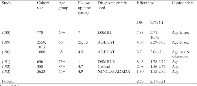
Table 2.2 Prevalence (%) and Incidence (%) of Alzheimer’s Disease (AD), Vascular Dementia (VaD) and Mixed Dementia (MD) and undifferentiated VaD/MD.
Population Source Age Follow -up (years)
All causes
MD VaD AD VaD
/MD
N
Prevalence proportion
London, United Kingdom (90) ≥65 n/a n/a 0.7 (5) 0.1 (1) 3.1 (22) n/a 705 Stockholm, Sweden (91) ≥75 n/a n/a 0.2 (3) 2.9 (52) 6.4 (116) n/a 1,810 Honolulu, Hawaii (92) 71-93* n/a n/a 1.4 (53) 1.8 (68) 2.1 (77) n/a 3,734 Framingham, United States (93) 61-93 n/a n/a 0.2 (5) 0.4 (8) 2.3 (50) n/a 2,180 Gothenburg, Sweden (94) ≥85 n/a n/a n/a n/a 13.0 (64) 14.0 (69) 494 New York, United States (95) 75-89 n/a n/a n/a n/a 8.4 (37) 5.7 (25) 442 Aichi Prefecture, Japan (96) ≥65 n/a n/a n/a n/a 2.4 (75) 2.8 (87) 3,106 Kanagawa Prefecture, Japan (97) ≥65 n/a n/a n/a n/a 1.2 (22) 1.6 (29) 1,800 Shanghai, China (98) ≥65 n/a n/a n/a n/a 2.6 (103) 1.1 (43) 3,888
Incidence proportion
Gothenburg, Sweden (94) 85-88 3 9.0 0.4 3.7 n/a n/a 494 Stockholm, Sweden (91) ≥75 3.5 4.3 0.2 0.7 n/a n/a 1,810 New York, United States (95) 75-85 8 3.4 1.0 ** n/a n/a 442 Multiple communities,
United States
(99) ≥65 5.7 14.0 n/a n/a n/a 6.3 3,375
Sources: (62, 68); * Men only; ** Rate not given.
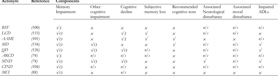
However, historically speaking, MCI has been plagued by the issue of alternative conceptualization as well as multiple operationalization. In fact, in 1962, Kral defined ‘Benign
Senescent Forgetfulness’ (BSF) as the inability to recall on certain occasions relatively unimportant
parts of past experiences (100). Kral characterized BSF as an age-related problem that did not
cross the boundary of disease, although it was suspected to be a transitional stage towards
Between 1968 and 1971, the word senility was rejected and replaced by dementia, to avoid the
redundancy of the term with normal aging. In 1986, Crook and colleagues (101) developed specific
criteria for ‘Age-Associated Memory Impairment’ (AAMI): age ≥ 50 with gradual onset memory complaints substantiated by relatively poor performance on neuropsychological tests (1.0 SD
below mean of test value, normed on young adults). During the late 1980s and the 1990s, the
AAMI criteria were criticized for the following reasons: First, they were thought to be
over-inclusive; second, they were difficult to operationalize in epidemiologic studies; third, they lacked
both construct and predictive validity; and finally, they did not necessarily represent decline. In
1989, Blackford and LaRue (102) altered the Crook criteria by adding the upper age limit of 79
years, requiring standardized self-report memory questionnaires, and using results on a battery of
four or more tests of secondary memory to define categories of impairment based on both young
adult and age-matched norms. They also required preserved general intelligence and revised the
exclusion criteria. Between 1994 and 1997, Ebly and colleagues (103) and Graham et al. (104)
coined the term ‘Cognitive Impairment, No Dementia’ which includes a classification proposed in
the Canadian Study of Health and Aging (105). The CIND category was meant to encompass a
variety of conditions which, while giving rise to cognitive impairment according the DSM-IIIR
criteria following clinical assessment and neuropsychological testing, did not meet the criteria for
dementia. Between 2000 and 2003, CIND was found to be a heterogenous group even though it
was found to increase the risk of progression towards dementia. Hogan and Ebly (106) found that
such progression was fastest in rate among a more narrowly defined group of CIND to whom the
name MCI was given. MCI was defined as memory impairment, intellectual decline and a
The risk of progression to dementia among MCI group was found to be 55%. Fisk and colleagues
(107) evaluated outcomes for a variety of definitions of MCI that either incorporated or did not
include a subject memory complaint and the presence of functional impairment. Although the
prevalence of MCI changed significantly with changing criteria (from 1% to 3%), rate of
progression to dementia did not change appreciably per year (8-10% per year over 5 years). Table
2.3 gives the detailed criteria used for differential diagnosis of “Mild Cognitive Impairment” along
the changing descriptive terminology and classification systems.
Although many of the above terms remain in use, none have received more attention than the
term MCI. Petersen and colleagues (108) was the first to give MCI a formal definition: “Having a
complaint of defective memory, normal activities of daily living, normal general cognitive
functioning, abnormal memory function for age, and absence of dementia.” A consensus
conference on MCI concluded that while MCI represents a high-risk stage for the development of
AD, its heterogeneity requires sub classification: amnestic MCI focuses on memory loss and may
progress to AD; MCI with slight impairment in multiple domains may represent normal aging or
may progress to AD or vascular dementia; and MCI with impairment of a single non-memory
domain may have a wide variety of outcomes. In addition to the clinical, functional and
psychosocial implications, it has been noted that if patients indentified with MCI were successfully
treated so as to delay progression to AD by only one year, the dollar cost savings would be quite
substantial (109). Given this, increased understanding as to the clinical and neurobiological aspects
of MCI, as well as the current status of potential therapeutic interventions will allow clinicians to
Prior studies indicate that the prevalence rates for MCI and other related conditions could range
between 3.2% and 53.8% depending on the characteristics of the cohort as well as the screening
instruments used (104, 110-112). Many potential factors for disease progression have been
identified, including informant report of functional deficits of which the patient is unaware (113),
EEG pattern changes (114), brain MRI imaging for volumetric measurements (115) and
magnetization ratios (116), cerebral glucose metabolism (117) and cerebrospinal fluid markers
(118, 119). No one factor or combination of factors has yet emerged as a clear predictor. Applying
the current criteria for MCI, individuals diagnosed with MCI converted to AD at an annual rate of
12-15%, compared with normally aging individuals who convert to AD at lower rates of 1-2% per
year. For longer periods of time of follow-up, MCI individuals convert to AD at a rate of 50%
after 3-4 years, and a rate of 80% after 6 years (88, 109). Given the high rates of conversion
between MCI and AD, some researchers contend that MCI is not nosologically separate from AD,
but rather a prodrome of it (120).However, others suggest that there is a heterogeneity within MCI
and that not all necessarily progress to AD when followed up for a sufficient period of time (121).
However the issue of whether MCI is a prodrome or a risk factor will continue to be a source of
continuous debate. Prevalence of dementia in epidemiological studies is reported at 0.3 to 1.0 per
100 people in individuals aged 60 to 64 years, and increases to a range from 42.3 to 68.3 per 100
people in individuals 95 years and older (122). Incidence rises from 0.7% per year in subjects aged
65 to 69 years to 6.6% per year in populations older than 90 years (123). Alzheimer pathology is
the underlying cause of 50-70% of clinically diagnosed senile dementias (124).