Coupled Electron Proton Transfer in Biological Redox Substrates
Christopher J. Gagliardi
A dissertation submitted to the faculty of the University of North Carolina at Chapel Hill in partial fulfillment of the requirements for the degree of Doctor of Philosophy in the Department of Chemistry
Chapel Hill 2012
Approved By:
Dr. Thomas J. Meyer Dr. H. Holden Thorp Dr. Gary Pielak
ABSTRACT
Christopher J. Gagliardi: Coupled Electron Proton Transfer in Biological Redox Substrates
(Under the direction of H. Holden Thorp and Thomas J. Meyer)
The amino acids tyrosine, tryptophan and cysteine are used to carryout electron
transfer within enzymes and proteins. The chemistry and oxidation mechanisms vary
considerably between these molecules. Proton coupled electron transfer (PCET), where
electrons and protons travel in sequence, or in concert is used by these amino acids in
both proteins as well as free in aqueous solution in order to minimize energy expenditure
and prevent build up of high-energy intermediates. The oxidation of these amino acids by
a series of metal M(bpy)33+ M = Os, Fe, Ru complexes in aqueous solution at ITO electrodes as well as spectroscopically in stopped-flow mixing studies is reported. A
common mechanism involving the pre-association of a base to form a hydrogen bound
adduct prior to electron transfer was observed in all cases. Each of the three amino acids
displayed various degrees of hydrogen bonding with bases, linked to their pKa values
(cysteine 8.2, tyrosine, 10.1 and tryptophan 16.0). Base assisted oxidation pathways
occur through two competing mechanisms; either proton transfer to a base followed
electron transfer (PT-ET), or concerted electron proton transfer (EPT). The kinetics of
these pathways as well as additional pathways such as outer-sphere electron transfer were
studied using electrochemical, and spectroscopic methods, giving rate information for
molecules behave in enzymatic electron transfer reactions. We have found that for PCET
reactions the driving force is the dominate factor dictating reactivity and can be
influenced by both the pKa of the proton acceptor and the oxidation potential of the
M(bpy)3III/II couple. This insight has provided a better understanding of how tyrosine, cysteine, and tryptophan are used to mediate biological electron transfer and allowed us
to observe commonalities in redox mechanisms with respect to common bases and
Acknowledgements
First and foremost, thank you Holden and Dr. Meyer for taking me on as a
graduate student. I knew when I told you I was a Student of Cliff’s, that it may not have
bode well for me, but you took a chance anyway. Holden, I would also like to specifically
thank you for taking me as a student as your administrative career was taking off, I never
would have imagined being the last graduate student of the Chancellor. I hope I have
been able to keep science in your busy life at an interesting level. I cannot thank you
enough for the guidance you have given my research and me when I needed it the most.
Dr. Meyer, thank you for pushing me when I needed it, and making me question myself
as a scientist. Without that kind of motivation I never would have made it to the finish
line. As someone who entered graduate school as a synthetic inorganic chemist, I never
would of imagined that kineticist, electrochemist and physical chemist would be the first
things to come to my mind when describing my time here at UNC.
I owe an immense debt of gratitude to my Alma Mater Roger Williams
University, and above all my mentor at RWU, Professor Cliff Timpson, who is also
another graduate of the Meyer group. Without you I never would have found my love for
chemistry. Without your mentorship, kind words, tough words and above all friendship, I
never would have found myself in the position that I am in today. From someone who
I would also like to thank Chrissie Murphy, or when I met her, Chrissie Fecenko,
without whose guidance and mentorship during my first two years of graduate school I
never would have stuck with or even come close to understanding my projects. Your
help, guidance and translation during meetings were more than I could have ever hoped
for. It was more of a help than you can possibly imagine, Thank you. I would also like to
thank Robert Binstead, whom without I would have been lost hopelessly trying to figure
out how tryptophan oxidation worked. With your help, not only did this project move
forward, but turned into a wonderful publication. Thank you so very much for all of your
time and patience. I would also like to thank all of the other members of the Meyer
group, especially Jonah Jurss, one of my best friends and collaborators during my time
here, David Weinberg and Javier Concepcion who helped me countless times studying
for my orals and for many other scientific questions and pursuits, including PCET
review, which I am so thankful is over. Finally, Aaron Vannucci, probably the best lab
mate someone could have, it is not easy spending countless 10-hour days back to back to
someone and still have room for a friendship, thank you for yours.
Lastly, I would like to thank my family and my friends for the loving support you
have given me in embarking on this endeavor, never once did I get a word of
discouragement. To my wife, who was Stefanie Haas when I started graduate school, and
is now Stefanie Gagliardi as I end it, your patience and comforting companionship has
kept me waking up day after day to pursue knowledge. This would have been an
intolerable herculean task without you by my side. Finally to my mother and father, I am
sorry high school did not workout as it perhaps would have for an ideal son and student,
have undergone. Thank you for your acceptance of my decisions, even if I didn’t really
know where I was going or why. I guess in the end, everything really works out.
Table of Contents
List of Figures ...…...……….……….……….………..………….…xii
List of Tables.……….……….……….………..…………..…xvii
List of Symbols and Abbreviations...………..………..…….………..……….xviii
Chapter 1: Introduction to Proton-Coupled Electron Transfer...………1
1.1 Introduction..……….……....………...2
1.2 Biological Redox Cofactors………...………..2
1.3 Proton Coupled Electron Transfer………...………5
1.4 A Short History and Nomenclature Background of PCET……….………...10
1.5 The Difference between MS-EPT, EPT and Hydrogen Atom Transfer (HAT).……….………....…12
1.5.1 The Thermodynamics of a Proton Acceptor H2O Verses Base..…...13
1.6 The Fundamentals of Proton Coupled Electron Transfer ………...………...17
1.6.1 Classical Electron Transfer………..…………...18
1.6.2 Proton Transfer …………..………..………...20
1.6.3 Proton Coupled Electron Transfer ……….……….…….…...20
1.6.3 Kinetic Isotope Effect in EPT..…….……….………..………...23
1.7 The Role of Tyrosine, Tryptophan and Cysteine in Biological PCET …..……....24
1.7.1 Ribonucleotide Reductase (RNR)...…..………..……….………...24
1.7.2 DNA Photolyase ……...………..……..………..………....25
1.8 Experimental Methods………….………...………..28
1.8.1 Electrochemical Methods………….…..……….………....29
1.8.2 Cyclic Voltammetry on Metal Oxide Electrodes.……...…..……...…...29
1.8.3 Mixing and Spectroscopic Methods....….….………..………….……...31
1.9 Concluding Remarks………...……….…………..32
1.10 References………..………..33
Chapter 2: Surface Activation of Electrocatalysis at Oxide Electrodes. Concerted Electron-Proton Transfer.…….……….………41
2.1 Abstract…….………...41
2.2 Introduction.…..………...41
2.3 Methods and Materials..……….44
2.3.1 General……….……..……….44
2.3.2 Reaction Solutions …..………...…………...…….….44
2.3.3 Deuterated Reaction Solutions and Kinetic Isotope Experiments.…….45
2.3.4 Sample and Electrode Preparation….……….………....46
2.3.5 Electrochemistry……….47
2.3.6 Thermodynamics……….………...……….47
2.4 Results and Discussion .……….…...………...47
2.4.1 Measurement of Rate Constants .……...….………....47
2.4.2 Catalytic Buffer Effects ...…….………..………50
2.5 Concluding Remarks...….………..58
Chapter 3: Concerted Electron–Proton Transfer (EPT) in the Oxidation of
Tryptophan with Hydroxide as a Base ………....………63
3.1 Abstract……….………...63
3.2 Introduction..……….………...63
3.3 Methods and Materials.……….………...66
3.3.1 Reaction Solutions….……….…………..……….…….66
3.3.2 Stopped-Flow kinetics...………..………..…….67
3.3.3 Electrochemistry ……….………...……….70
3.4 Results and discussion ………...……….……….70
3.4.1 Analysis of Stopped-Flow Data using SPECFIT/32…..……..…...……70
3.4.2 Modeling pH Dependence in SPECFIT/32……….………78
3.4.3 Validating Model at Low pH.……...………..….……….79
3.4.4 Testing Kinetic Model for Effects of Oxygen ..……….……….80
3.4.5 Hydroxide Dependence……...……….……..……….81
3.5 Electrochemistry……….………...86
3.5.1 Fitting Electrochemical Data Using Digital Simulations..………...…...91
3.6 Marcus Treatment for Lambda ………...….………..95
3.7 Concluding Remarks………..…..………..96
3.8 References..………….………..…..……...………98
Chapter 4: The Role of Concerted Electron–Proton Transfer in the Oxidation of Cysteine …………...……….………..………102
4.1 Abstract………...……….……….………... 102
4.2 Introduction..……….………... 102
4.3.1 Reaction Solutions….……….…………..……….…...104
4.3.2 Deuterated Solutions…………..……….……..……105
4.3.3 Stopped-Flow Kinetics.………..……….……..……106
4.3.4 Electrochemistry……..……….………...……….108
4.4 Results and Discussion ...………...…...……….…………109
4.4.1 Stopped-Flow Evaluation of Electron Transfer………109
4.4.2 Stopped-Flow Evaluation of PCET with Added Bases ...……....…….114
4.4.3 Kinetic Isotope Effects Measured by Stopped-Flow …….………...…116
4.4.4 Stopped-Flow Stoichiometry Measurements……..………..116
4.5 Dipicolininc Acid Inhibition of Cu2+ Catalysis …….…….………….…………118
4.5.1 Stopped-Flow Evaluation of DPA Effects.………...……119
4.5.2 Electrochemical Evaluation of DPA Effects.……….……...……120
4.6 Electrochemical Evaluation of Cysteine Oxidation Kinetic Pathways...……...121
4.6.1 Digital Simulation of Electrochemical Data.……..………..…….…...123
4.6.2 Limiting Conditions of the Rate Law ……….……..……126
4.7 Isolating EPT and PT-ET using Limiting Conditions of the Rate Law...………131
4.7.1 Pre-Association between [CysSH] and [B-]; Determination of KA and kEPTKA′.……….…131
4.7.2 Pre-Association between [CysSH---HPO2-] and [M(bpy)33+]; Determination of KA and kEPTKA′ .………...………..…132
4.7.3 Rapid Pre-equilibrium Formation of CysS- Followed by Rate Limiting Oxidation or Rate Limiting Proton Transfer; Determination of k1, k-1 and k2….….….….….….….….….….….….….….….…..………...133
4.7.4 Electrochemical Kinetic Isotope Effects……..……….……...……….135
4.9 References.…...…….…...…….…..…….…...…….…...…….…...……..…...138 Appendix 1: Derivation of Full and Limiting Forms of The Rate Law for
List of Figures
Chapter 1: Introduction to Proton-Coupled Electron Transfer
Figure 1: Biological redox cofactors……….……….3
Figure 2: Oxidative phosphorylation...….…...……….……….5
Figure 3: Simplified photosystem II PCET square scheme………...………6
Figure 4: MS-EPT with Yz……….………….………...………7
Figure 5: Full photosystem II PCET square scheme ……..………..………...…..8
Figure 6: Structure of photosystem II reaction center and electron transfer events induced by light absorption………...………9
Figure 7: Comproportionation of (RuIV =O22+ ) and (RuII =O22+ )………10
Figure 8: Differentiation of EPT and HAT with (RuIV =O22+ ) and (RuII =O22+ )……….13
Figure 9: Rhenium-tyrosine complex ……….……….……….……….……….…….16
Figure 10: Energy wells for electron transfer reactions, and illustration of vibrational overlap..……….……….……….……….……….……….……...21
Figure 11: Wave function overlap for OD versus OH complex………..23
Figure 12: The reaction carried out by ribonucleotide reductase………..…..25
Figure 13: Proposed ribonucleotide reductase electron transfer pathway…………...25
Figure 14: Full structure of the FAD/FADH2 mobile cofactor...……….………26
Figure 15: Proposed DNA photolyase electron transfer pathway.……...…………...27
Figure 16: Soybean lipoxygenase active site………...28
Figure 17: Cyclic voltammograms on an ITO electrode with tyrosine.……....……...30
Figure 2: Cyclic voltammograms of ITO-RuII
+ tyrosine on ITO.…….………43
Figure 3: Cyclic voltammograms of ITO-RuII
on ITO + (A) tyrosine-methyl-ester (B) 4-methylphenol and (C) N-acetyl-tyrosine....………43 Figure 4: Scan rate dependence cyclic voltammograms of tyrosine
methyl ester at ITO-RuII in 0.1M HClO
4 normalized to (A) scan rate (υ)
(B) square root of scan rate (υ1/2)…………...…….…..…………...…..…….……...44
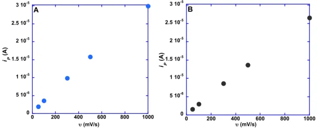
Figure 5: Scan rate dependence plots of (A) ITO-RuII
(B) ITO-OsII
..………...46
Figure 6: Cyclic voltammograms at ITO-RuII
varying A) tyrosine concentration B) ITO-RuII
surface loading (Γ).……….….48
Figure 7: icat/nFAΓ vs [TyrOH] (M) for tyrosine concentration dependence at ITO-RuII
...………..……….49
Figure 8: Base dependence cyclic voltammograms at ITO-RuII………….….……..51
Figure 9: Cyclic voltammograms at ITO-RuII
at constant [B
-] varying pH..………..51
Figure 10: Cyclic voltammograms at ITO-RuII
with pH 5.4 citrate buffer
and 0.1 and 1 mM HClO4……..……….…..52
Figure 11: Illustration of surface PCET with phosphonates as proton acceptor…..…52
Figure 12: Illustration of surface PCET with [B
-] as proton acceptor……….53
Figure 13: Scan rate dependence cyclic voltammograms of tyrosine methyl ester at ITO-RuII
in pH 5.4 citrate buffer normalized to (A) scan rate (υ) (B) square root of scan rate (υ1/2) .……..………..…….54
Figure 14: Cyclic voltammograms at ITO-OsII
in (A) pH 5.4 citrate
(B) pH 7.1 Tris...…………...………..………..………..………..………..………...55
Figure 15: Scan rate dependence cyclic voltammograms of tyrosine methyl ester at ITO-OsII
in pH 5.4 citrate buffer normalized to
(A) scan rate (υ) (B) square root of scan rate (υ1/2)..………...…………56
Figure 16: Scan rate dependence cyclic voltammograms of tyrosine methyl ester at ITO-OsII
in pH 7.1 Tris buffer normalized to (A) scan rate (υ) (B) square root of scan rate (υ1/2
).………..……….….56
Figure 17: Tris buffer dependence at ITO-OsII
(A) cyclic voltammograms
Chapter 3: Concerted Electron–Proton Transfer (EPT) in the Oxidation of Tryptophan with Hydroxide as a Base
Figure 1: Structures of tryptophan, N-acetyl-tryptophan and Tris.………..66
Figure 2: UV-Vis spectra of Os(bpy)3 2+
(A) 190-800 nm (B) 375-775 nm....……….67
Figure 3: Stopped-flow kinetic trace for Os3+→ Os2+ at pH 10.7
…...…………..…..69
Figure 4: OH
calibration curve ……….………..69
Figure 5: SPECFIT/32 diode array kinetic scans and representative kinetic trace...71
Figure 6: SPECFIT/32 SVD analysis of diode array scans at pH 7.1……….……….71
Figure 7: SPECFIT/32 calculated concentration profiles...………...……..72
Figure 8: Typical kinetic trace for reaction of Os(bpy)33+ with
N-acetyl-tryptophan at pH 7.1...……….……..74
Figure 9: Calculated concentration profiles for Os3+
and Os2+
from Figure 8..…...….74
Figure 10: Oxidation products of tryptophan……….………..75
Figure 11: SPECFIT/32 SVD analysis of diode array scans at pH 4.0.……...……..80
Figure 12: Rates of oxidation of N-acetyl-tryptophan as a function of (A) [OH
-] (B) [NAceTrp]...………...…...82
Figure 13: Typical equal concentration kinetic trace for reaction of Os(bpy)33+
with N-acetyl-tryptophan ………...83
Figure 14: Rates of oxidation of N-acetyl-tryptophan as a function of [OD
-]…...85
Figure 15: Mole fraction dependence for oxidation of N-acetyl-tryptophan…….…..85
Figure 16: Cyclic voltammograms on ITO of A) Os(bpy)3III/II
+ N-acetyl
tryptophan B) N-acetyl-tryptophan alone..……….……….…………87
Figure 17: Cyclic voltammograms of Ru(dmb)3
III/II + N-acetyl tryptophan
on ITO (A) 5 and 50 mM Tris Buffer (B) 5 and 50 mM phosphate buffer…...……..89
Figure 18: Cyclic voltammograms of Fe(dmb)3III/II
+ N-acetyl tryptophan
Figure 19: Cyclic voltammogram of N-acetyl tryptophan on ITO
sweep width 0.2-1.4V...………...91
Figure 20: DigiSim cyclic voltammogram simulation fit..…..……...………...93
Figure 21: Marcus plot, variation of RTln(kET) vs -∆G°′(eV)...……….…..96
Chapter 4: The Role of Concerted Electron–Proton Transfer in the Oxidation of Cysteine
Figure 1: Structures of cysteine, N-acetyl-cysteine and dipicolinic acid...…………105 Figure 2: Stopped-flow kinetic trace for Os3+→Os2+ at pH 5.76..…...…………..…107 Figure 3: Typical kinetic trace for the reaction of Ru(dmb)33+ with
N-acetyl-cysteine at pH 5.76, 2:1, [CysSH]:[Ru] ..……...……….……108
Figure 4: Typical kinetic traces for the reaction of Os(bpy)33+ with
N-acetyl-cysteine at pHs (A) 6.0 and (B) 4.5 with no buffer………....……110 Figure 5: Kinetic data for stopped flow oxidation of N-acetyl-cysteine
by Os(bpy)33+ as a function of [H+], pH 4-6, kobs/[Cys] vs [H+]-1………..113 Figure 6: Rates of oxidation of N-acetyl-cysteine by Os(bpy)33+ as a
function of (A) [HPO42-] and (B) [AcO-], kobs/[Cys] vs [B-]……...………...…115 Figure 7: Cyclic voltammograms of Os(bpy)3III/II + L-cysteine on ITO
(A) 1 mM DPA (B) No DPA.………...……….…121
Figure 8: Cyclic voltammograms of L-cysteine on ITO with and without
metal mediator Os(bpy)3III/II………...…122 Figure 9: Cyclic voltammograms of Os(bpy)3III/II + L-cysteine on ITO
with 300 mM [H2PO4-/HPO42-] at pH 6.2, 7.2 and 8.2 …..………...…….…123 Figure 10: Simulation of cyclic voltammograms of Os(bpy)3III/II + L-cysteine
on ITO with 300 mM [H2PO4-/HPO42-] at pH (A) 6.2, (B) 7.2 and (C) 8.2 .…..…...130 Figure 11: Cyclic voltammograms of Os(bpy)3III/II + L-cysteine on ITO
with 50 and 300 mM [H2PO4-/HPO42-] at pH 7.2 ……...………..…….…131 Figure 12: (A) Plot of kobs vs [HPO4-] for the oxidation of L-cysteine
by Os(bpy)33+ (B) kobs-1 vs [HPO4-]-1…....……….………..…...132 Figure 13: (A) Plot of kobs vs [CysSH] for the oxidation of L-cysteine
Figure 14: Plot of kobs vs [H2PO4-]-1 for the oxidation of L-cysteine
by Os(bpy)33+…...………..135
List of Tables
Chapter 1: Introduction to Proton-Coupled Electron Transfer
Chapter 2: Surface Activation of Electrocatalysis at Oxide Electrodes. Concerted Electron-Proton Transfer
Chapter 3: Concerted Electron–Proton Transfer (EPT) in the Oxidation of Tryptophan with Hydroxide as a Base
Table 1: Rate constants for redox pre-equilibrium/disproportionation
mechanism at various buffer concentrations.…….………..……76
Table 2: Rate constants for redox pre-equilibrium/tryptophan anion mechanism at various buffer concentrations..………..………77
Table 3: Rate constants for redox pre-equilibrium/disproportionation mechanism at various pHs……….…..………79
Table 4: Rate constants for oxygen sensitivity…………..………...81
Table 5: Rate constants for hydroxide dependence………..………...82
Table 6: Experimental electrochemical parameters……….………87
Table 7: Simulation parameters imported into DigiSim………..92
Table 8: Rate constants obtained from digital simulations……….……….94
Chapter 4: The Role of Concerted Electron-Proton Transfer in the Oxidation of Cysteine Table 1: Rate constants for N-acetyl-cysteine oxidation by Ru(dmb)33+ and Os(bpy)33+ at 5:1, 2:1 and 1:1 [Cys]:[M3+] …………...………...…117
Table 2: Rate constants for N-acetyl-cysteine oxidation by Os(bpy)33+ at pH 4.0 and 5.0, at 50 and 500 mM acetate buffer, with and without DPA..……….119
Table 3: Rate constants for N-acetyl-cysteine oxidation by Os(bpy)33+ at pH 4.0 and 50 mM acetate buffer, with varied concentrations of DPA ………....…....……120
Table 4 Experimental electrochemical parameters……….……….………..122
Table 5: Simulation parameters imported into DigiSim……….…………...…124
List of Symbols and Abbreviations
[TyrOH]T total concentration of tyrosine [CysSH]T total concentration of cysteine
°C degrees Celsius
α symmetry factor
∆E change in reduction potential
∆GEPT free energy change for electron proton transfer
∆GET free energy change for electron transfer
∆GPT free energy change for proton transfer
∆Qμ,ν change in equilibrium normal coordinate
Γ surface coverage
Γo maximum surface coverage
ε molar absorptivity
λ reorganizational energy
λmax maximum absorbance
µ initial state
µM micromolar
ν scan rate
ν vibrational mode
ν final state
π pi (3.1415926535898)
ρfc Frank-Condon density of states term
χD mole fraction deuterium
ψ wave function
ψµ electronic wave function initial state
ψν electronic wave function final state
ϕµ proton wave function initial state
ϕν proton wave function final state
ω angular frequency
A ampere
A electrode area
A absorbance
A
-/A acceptor
A∞ absorbance at time infinity
Abs absorbance
ADP adenosine diphosphate
Ag/AgCl silver/silver chloride reference
ATP adenosine triphosphate
B
-base form of buffer
bpy 2,2′-bipyridine
C coulomb
ClO4
perchlorate anion
CoQ10 ubiquinone
CV cyclic voltammograms
CysSH cysteine (protonated form) CysS-
cysteine (deprotonated anion) CysS•
cysteine (deprotonated radical) CysS•+
cysteine (protonated radical cation) CysSH---B cysteine (hydrogen bound base adduct)
d distance
D+
deuteron D+
/D donor
D2O deuterium oxide
DCl deuterochloric acid
dmb 4,4′-dimethyl-2,2′-bipyridine DNA deoxyribonucleic acid
Dop optical frequency dielectric constant Ds zero frequency dielectric constant
DPA dipicolinic acid (pyridine-2,6-dicarboxylic acid)
e-
electron
E°′ formal potential
E1/2 midpoint potential
EP peak potential
EPT electron proton Transfer
ET-PT step-wise electron transfer followed by proton transfer
eV electron volt
F Faraday constant
FAD flavin adenine dinucleotide (oxidized)
FADH- flavin adenine dinucleotide (mono protonated anion) FADH•- flavin adenine dinucleotide (protonated radical anion) FADH2 flavin adenine dinucleotide (reduced)
Fe iron
FEP fluorinated ethylene propylene
fj force constant
FTO fluorine doped tin oxide
H Hamiltonian
H+ proton
H190 histidine 190 in photosystem II
H2PO4- monobasic hydrogen phosphate
H3O+ hydronium ion
HB+ acid form of buffer
HCl hydrochloric acid
HClO4 perchloric acid
His histidine
HPO42- dibasic hydrogen phosphate
i electrode current
icat catalytic peak current
In indium
ip peak electrode current
KA acid disassociation constant
kB Boltzmann constant
kD rate constant for diffusion
kEPT electron proton transfer rate constant
KIE kinetic isotope effect
kobs observed rate constant
ks heterogeneous electron transfer rate constant
LiOH lithium hydroxide
Mµν initial and final state mixing matrix element
M molar
M(bpy)3III/II metal tris(2,2′-bypyridine) complex, couple
MeCN acetonitrile
Mj reduced mass
MLCT metal-ligand charge transfer
mM millimolar
MS-EPT multi-site electron proton transfer
MTHF 5,10- methylenetetrahydrofolylpolyglutamate
N number of replicates
n number of electrons
NA Avogadro’s number
NAceTrp N-acetyl-tryptophan
NAceTyr N-acetyl-tyrosine
NaCl sodium chloride
NAD+ nicotinamide adenine dinucleotide (oxidized)
NADP+ nicotinamide adenine dinucleotide phosphate (oxidized) NADH nicotinamide adenine dinucleotide (reduced)
NADPH nicotinamide adenine dinucleotide phosphate (reduced) NaOD sodium deuteroxide
NaOH sodium hydroxide
NHE normal hydrogen electrode AcO-
AcOH acetic acid OH-
hydroxide anion
Os osmium
P680 photosystem II primary electron donor P680+ oxidized form of P680
PCET proton coupled electron transfer
pD -Log[D+
]
pH -Log[H+
]
Pi inorganic phosphate
pKa -Log[Ka]
PMT photomultiplier tube
PSII photosystem II
PT-ET stepwise proton transfer electron transfer Q integrated peak area of the oxidative peak RNR ribonucleotide reductase
Ru ruthenium
s second
Sj Huang-Rhys factor
SLO soybean lipoxygenase
SVD singular value decomposition
T temperature
Tris tris base
Trp tryptophan
TrpNH tryptophan
TrpN-
deprotonated tryptophan anion TrpN● deprotonated tryptophan radical TrpNH●+ tryptophan radical cation
TrpNH---B tryptophan hydrogen bound base adduct
TyrOH tyrosine
TyrO- tyrosine anion
TyrO● deprotonated tyrosine radical
TyrOH---B tyrosine hydrogen bound base adduct
UV ultraviolet
V volts
VET electron transfer matrix element
Vis visible
Vµν energy from initial and final state mixing
W tryptophan amino acid residue
XRD X-ray diffraction
Y tyrosine amino acid residue
a
Chapter 1
Introduction to Biological Redox Substrates and Proton Coupled Electron Transfera
1.1 Introduction: Almost all process, whether chemical or biological, that produce and use
energy or move electrons employ proton-coupled electron transfer (PCET) as a means to do
so. PCET reactions involve the coupled transfer of protons and electrons and are critical for
many energy conversion processes including natural and artificial photosynthesis, catalytic
water oxidation and production of molecular hydrogen as well as many enzymatic reactions.
Whether it is electron transfer pathways through photosystem II to produce oxygen and
carbohydrates from sunlight, water and CO2 or in molecular water oxidation catalysts,
proton-coupled electron transfer is utilized for the purpose of charge leveling in the buildup
of oxidative equivalents and avoiding high energy intermediates that can be biologically
damaging.
1.2 Biological Redox Cofactors: In biology, electrons are the energy currency of all living
things. Shuttling these oxidative and reductive equivalents from site to site allows for living
things to grow as well as defend and heal themselves. Whether this energy comes from the
sun or from food, biological redox cofactors form the road upon which these redox
equivalents travel, enabling essential chemical reactions.
a Reproduced in parts with permission from the American Chemical Society; Christopher J.
Remarkably, the number of biological electron transfer shuttles, whether membrane
bound, protein based, or mobile, is relatively few given the immense diversity of life on this
planet. From the smallest prokaryotic cyanobacteria, to complex life such as higher
mammals, some of the same chemistry is, and to a further extent, the same redox cofactors
are utilized to perform a variety of reductive and oxidative chemical reactions. Figure 1
Amino Acids:
Moble Organic Cofactors:
Inorganic Cofactors:
Figure 1. Amino acid functional groups (R), and pKa values are shown with a zwitterionic
backbone. Amino acids are found as electron transfer mediators within proteins. For the
mobile cofactors, only the functional groups are shown. Mobile organic cofactors shuttle electrons and protons between immobile membrane bound proteins as shown in Figure 2. The inorganic cofactors play a large role in electron transfer reactions, with Fe-S clusters carrying out electron transfer reactions occurring in the protein subunits of mitochondrial respiration as well as other enzymes such as hydrogenases and photosystem I. In many cases, the active sites of these enzymatic reactions are metal centers, a few of which are also listed.
H3N+ CH C
H2C
O -O
R
HN
R = Tryptophan pKa = 16
OH
R = Tyrosine pKa = 10.1
SH
R = Cysteine pKa = 8.3
N H
NH
O
N HN O
R1
Fully Reduced isoalloxazine FAD/FADH2
N H2N
O
R2 Nicotinamide NAD+,NADP+/NADH, NADPH
H3C
H3C R3
O O 2,3-dimethyl-1,4-benzoquinone plastoquinone/plastoquinol Fe S S S Fe Fe Fe S Iron-Sulfur Clusters
Additional Metal Ion Cofactors: Cu1+/2+(azurin, cytochrome oxidase) Fe2+/3+ (nitrogenase, hemoglobin) Mn2+/3+ (PSII, arginase)
Ni2+/3+ (Ni-Fe hydrogenase, urease )
Proteins are folded chains of amino acids. Three of these amino acids; tyrosine,
cysteine and tryptophan mediate enzymatic electron-proton transfer reactions. Photosystem II
(PSII) in green plants1-7, ribonucleotide reductase (RNR),8-10 DNA photolyase11,12,
superoxide dismutase (SOD)13,14 and soybean lipoxygenase (SLO)15-18 have all been
highlighted for their utility of a PCET mechanism. Within a protein, these three chemical
functionalities - a phenol, an indole and a thiol - perform a wide variety of chemistry, and
individually on the molecular level, demonstrate distinctive chemical behavior.8-10,19-25 This
highlights why the residues cannot be used interchangeably and why mutagenesis studies
show a dramatic change in the overall functionality of a protein in the absence of even a
single residue.5-7,14
The role of amino acids in the diverse field of biochemistry is the subject of this work
and is further expanded upon below in Section 1.8. This section reviews some examples of
amino acids participating in biological PCET reactions. Knowledge in this area, especially on
PCET reactions involving the other mobile organic redox cofactors, is still in its adolescence.
We have much to gain from further experimentation on how these other cofactors work given
their biological ubiquity. A testament to this is the fact that there is a whole classification of
proteins called ferredoxins, which contain iron-sulfur clusters. The mobile cofactor,
ubiquinone, is named after its own biological prevalence.
All of these cofactors work in concert in biology. The next step in understanding
PCET on a macroscopic scale is looking at the interfaces where the different classes of
aforementioned cofactors interact. A mobile cofactor is able to pass on its redox equivalent(s)
at an interface between the cofactor and the first amino acid, or iron-sulfur cluster in an
respiration, shown in Figure 2. Oxidative phosphorylation employs two mobile electron
shuttle molecules, Coenzyme Q (CoQ), otherwise known as ubiquinone, and cytochrome c
(Cyt c) as well as iron-sulfur clusters and other metal centers for the transport of electrons
through each subunit.
Figure 2. Oxidative phosphorylation in the cellular respiratory chain occurs in the mitochondrial membrane, and consists of over 80 peptides, five enzymatic units (ATP synthase not shown) and produces ATP from the reduction of oxygen to generate energy for cellular function.
1.3 Proton Coupled Electron Transfer: According to the broad definition, proton-coupled
electron transfer encompasses any process involving the movement of both electrons and
protons.1,26,27
Within the context of this work, it will refer to a single 1e
-/1H+
reaction. PCET
is a general classification given to a broad set of electron-proton transfer reaction
mechanisms. A PCET reaction can occur through three distinct pathways:
i. Stepwise electron transfer followed by proton transfer (ET-PT)
ii. Stepwise proton transfer followed by electron transfer (PT-ET)
iii. Concerted electron proton transfer.
CoQ
Complex I NADH-ubiquinone
oxidoreductase
Complex II Succinate-ubiquinone
oxidoreductase
Complex III
Ubiquinol-Cytochrome c
oxidoreductase
Complex IV
Cytochrome c
oxidase
Oxidative Phosphorylation
Cyt c
= moble electron shuttles
Intramembrane Space
Mitochondrial Matrix
NAD+
NADH
Fumarate Succinate
1/2 O2 + 2H+
H2O
Within the context of concerted electron proton transfer there is another sub-set of
categorical nomenclature based qualitatively on donor (D) and acceptor (A) orbitals and
further defined by quantum mechanics. These are: electron proton transfer (EPT), multi-site
electron proton transfer (MS-EPT), hydrogen atom transfer (HAT) and hydride transfer.
a. EPT e-/H+, different D, A orbitals, same molecule
b. MS-EPT, e-/H+, different D, A orbitals on separate molecules
c. HAT, e-/H+, same D, A orbital
d. Hydride transfer (simultaneous transfer of 2e-/1H+)
To illustrate some key points in understanding PCET, PSII in green plants will be
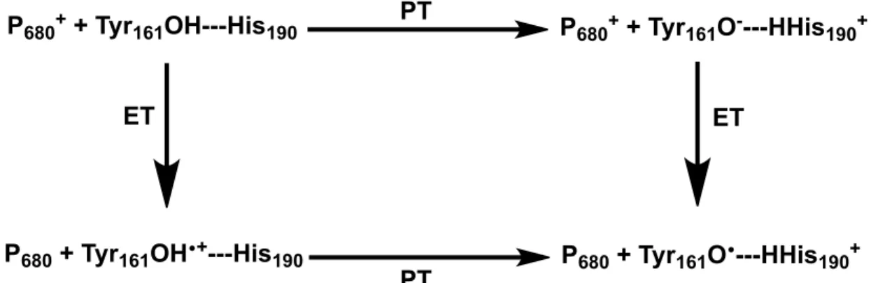
used as an example with the oxidation of the tyrosine (Tyr161) histidine (His190) pair (Yz)
by the reaction center, photooxidized P680+. The stepwise pathways PT-ET and ET-PT,
involve initial proton or electron transfer. Initial proton transfer (PT) produces the anionic
species before being oxidized to the radical and in the case of initial electron transfer (ET),
the tyrosine radical cation is produced before losing its proton to become the tyrosine radical,
as illustrated in Figure 3. Each of these initial stepwise products are high-energy
intermediates that can become damaging even within with the polypeptide architecture of a
complex set of proteins such as PSII.
Figure 3. Simplified illustration of the stepwise ET-PT, PT-ET pathways in the oxidation of
the Y pair in PSII.
P680+ + Tyr
161OH---His190 P680+ + Tyr161O----HHis190+
P680 + Tyr161OH•+---His190 P680 + Tyr161O•---HHis190+ PT
PT
The third mechanistic alternative for this reaction is concerted electron proton transfer
(EPT), where the movement of protons and electrons is in concert and does not produce any
radical cation or anionic intermediates. EPT is mechanistically more complex than either of
the stepwise mechanisms because of the simultaneous transfer of two particles: the proton
and electron. Thermodynamically, however, EPT conserves energy.
Within the context of PSII, the nomenclature MS-EPT is appropriate. This is because
the electron and proton go to acceptors on different molecules, as illustrated in Figure 4,
where the electron transfers to the photooxidised reaction center, P680+ and the proton
transfers to a nearby hydrogen bound histidine (His190).28,29 Figure 5 illustrates the
thermodynamic advantage of MS-EPT. In PSII, the concerted process is highly favored and,
as suggested by Babcock and coworkers and by Krishtalik, both the formation of Yz● and the
subsequent oxidation of the oxygen evolving complex (OEC) utilize concerted pathways in
order to avoid highly energetic intermediates.1,2,26,30-38
Figure 4. MS-EPT mechanism employed by the Yz Pair in PSII. Therein the proton is
transferred to the putative hydrogen bound histidine and the electron is transferred to the photooxidized PSII reaction center chlorophyll P680+.
O H
N
NH
His
190P
680+, Tyr
161O
H N
NH
His
190P
680, Tyr
161•
ET
Figure 5. Square scheme for the potential PCET reactions involving oxidation of the hydrogen bound Yz pair by the PSII reaction center chlorophyll, P680+. Either initial electron
or proton transfer in the stepwise pathways is thermodynamically unfavored and produce reactive, high-energy intermediates. The initial uphill thermodynamic cost of producing these intermediates is regained upon formation of the tyrosine radical product.
The initial steps for each of the stepwise PCET mechanisms are energetically uphill.
The oxidation of Yz by P680+ by simple outer sphere electron transfer occurs with a ∆G° =
+0.24 eV based on E°′ (P680+/●) = 1.26 V (vs NHE).36 Similarly, initial deprotonation of the
Tyr161, by His190 is uphill by ∆G° = +0.27 eV based on solution values for pKa(H+-His) =
5.5 and pKa(TyrOH) = 10.1. When these values are compared to the concerted reaction with
electron transfer from Yz to P680+ and proton transfer to His190 the reaction is highly favored
with ∆G° = -0.20 eV.1,26∆G° was calculated from ∆G° = F [(E°′(P680+/●) - (E°′(TyrOH●+/0) -
0.059(pKa(His) - pKa(TyrOH●+)] with pKa(TyrOH●+) = -2 and E°′(TyrOH●+/0) ~ 1.5 V. These
values are only approximations to the membrane potentials and neglect free energy (∆G°)
differences for the formation of the TyrO●---+H-His and TyrO-H---His H-bonded complexes.
The second step for each of the stepwise pathways is the difference between the ∆G° for the
first step and the ∆G° for the EPT step, because it is an overall thermodynamically adiabatic
process where the amount of work required to reach the final state is the same irrespective of
how the work is performed. P680+ + Tyr
161OH---His190 P680+ + Tyr161O----HHis190+
P680 + Tyr161OH•+---His190 P
680 + Tyr161O•---HHis190+ PT
PT
ET ET
!G°"= 0.24 eV
!G°"= -0.44 eV
!G°" = -0.47 eV
!G°"= 0.27 eV
!G
°" = -0
.20 eV
MS-EPT and the role of energetics appear in dramatic fashion in Figure 6: (1) light
absorption by an antenna apparatus sensitizes the singlet excited state of chlorophyll P680
(ChlD1), 1P680*; (2) 1P680* undergoes oxidative quenching with electron transfer to pheophytin
and then to quinone QA;(3) oxidation gives the powerful oxidant P680+ with E°′ (P680+/0) ~
1.26 V; (4) water oxidation at the CaMn4 cluster of the Oxygen Evolving Complex (OEC)
where the oxidative activation of the OEC occurs through intervening YZ as an electron
transfer relay; and (5) the semiquinone anion formed is further stabilized by a lateral electron
transfer step to a second quinone, QB. YZ consists of a tyrosine (Tyr161) and an associated
histidine, His190. Removal of His190 by mutagenesis shuts down photosynthesis. Loss of a
single base is sufficient to disrupt an enormously complex apparatus with multiple linked
functional elements and hundreds of thousands of atoms, highlighting the importance of the
MS-EPT mechanism.
Figure 6. Structure of the Reaction Center of Photosystem II illustrating terminal chlorophyll
P680, pheophytinD1,quinone acceptor QA, YZ (Tyr161-His190), the Oxygen Evolving Complex
(OEC), and the sequence of electron transfer events induced by light absorption and sensitization. The critical energetic role proposed for His190 as EPT acceptor base is also shown.3
Reproduced in part with permission from ref. 39. © 2008 Elsevier.
ET, PT, EPT, MS-EPT, HAT, and hydride transfer are all elementary steps available
for carrying out PCET reactions. In general, PCET reactions occur by more than one multi-Q
-A-Pheo-P680+ TyrZ
e- e
-~7x10³ s-1 ~1x10⁷ s-1
TyrO-H---His(190)
e
-~1x10⁷ s-1 !G°' ~ -0.24 eV
P680+
H+
TyrO---+H-His(190) P680
TyrO-H
e
-!G°' ~ +0.20 eV
P680+ P TyrO-H+
step mechanism with a competition between mechanisms with their relative importance
dictated by reaction conditions, temperature, pH and other factors.
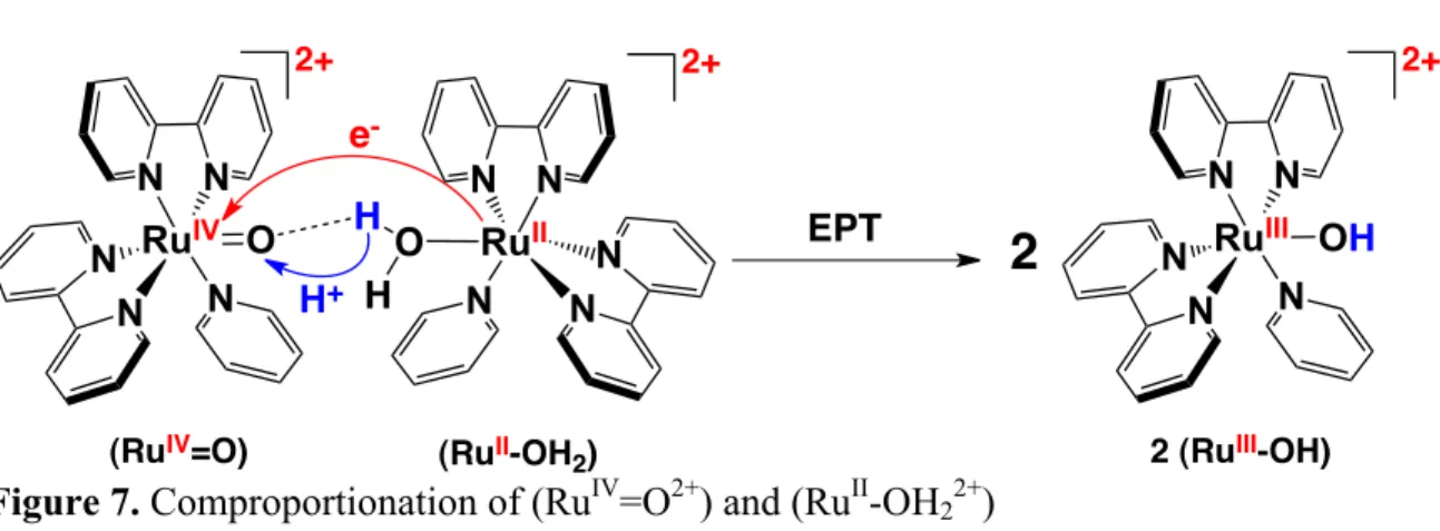
1.4 A Short History and Nomenclature Background of PCET: In 1981 the term proton
coupled electron transfer (PCET) was introduced to describe an elementary step, like electron
transfer or proton transfer but in which electrons and protons transfer together. The term was
coined to describe the concerted e-/H+ transfer process that occurs in the comproportionation
reaction between [RuIV(bpy)2(py)(O)]2+ (RuIV=O2+) and [RuII(bpy)2(py)(OH2)]2+(RuII-OH22+)
shown in Figure 7 (bpy = 2,2′-bipyridine, py = pyridine). In this reaction an electron and
proton are transferred simultaneously from RuII-OH22+ to RuIV=O2+ to give 2 equivalents of
RuIII-OH2+.40
Figure 7. Comproportionation of (RuIV=O2+) and (RuII-OH22+)
The term PCET has come to be used more broadly to describe reactions and half
reactions in which both electrons and protons are transferred without regard to mechanism.
Water splitting into H2 and O2 is a pH dependent reaction having separate PCET half
reactions, one for water oxidation, eq 1, and one for proton/water reduction, (eqs 2a, b) both
of which are pH dependent. RuIV
N N
N
N N
O e
-RuII
N N
N N N
O H H
2+
H+
EPT RuIII
N N
N
N N
OH
2+ 2+
2
(RuIV=O) (RuII-OH
2 H2O O2 + 2 H2 (1)
O2 + 4 H+ + 4 e- H2O (2a)
2 H+ + 2 e- H2 (2b)
Multi-electron, multi-proton PCET half reactions are universal in energy conversion
and storage reactions in chemistry and biology. In biology, key half reactions for energy
storage and production exploit PCET and the high-energy content in the C-H bonds of
hydrocarbons, sugars, or other oxygenates by indirect reactions with oxygen at physically
separated half reactions. Quintessential examples include carbohydrate formation by light
driven reduction of CO2 by water in photosynthesis and the reverse, oxidation of glucose by
oxygen which releases energy in respiration. In photosynthesis, CO2 reduction, coupled with
water oxidation, occurs in a reaction that stores 29.1 eV, 1.22 eV per redox equivalent. The
stored energy is released in respiration (Fig. 2) with the indirect oxidation of glucose by
oxygen used to drive oxidative phosphorylation in mitochondria. This combines inorganic
phosphate, Pi, and ADP to give ATP. ATP and the energy released by phosphate hydrolysis
to give ADP that is used to power cells for biosynthesis, motion, and signaling.
Unfortunately, the nomenclature used to describe concerted electron-proton transfer
has not been standardized, with different terms used to describe the same elementary step.
Alternate terms from the literature include concerted proton-electron transfer (CPET),41
electron transfer proton transfer (ETPT)42 and concerted electron-proton transfer (CEP).43
The use of EPT herein follows straightforwardly from ET to describe electron transfer and
PT to describe proton transfer as fundamental elementary reactions. It is descriptive,
nomenclature for a family of reactions that differ considerably in microscopic detail but in all
of which concerted electron-proton transfer is the defining redox event.
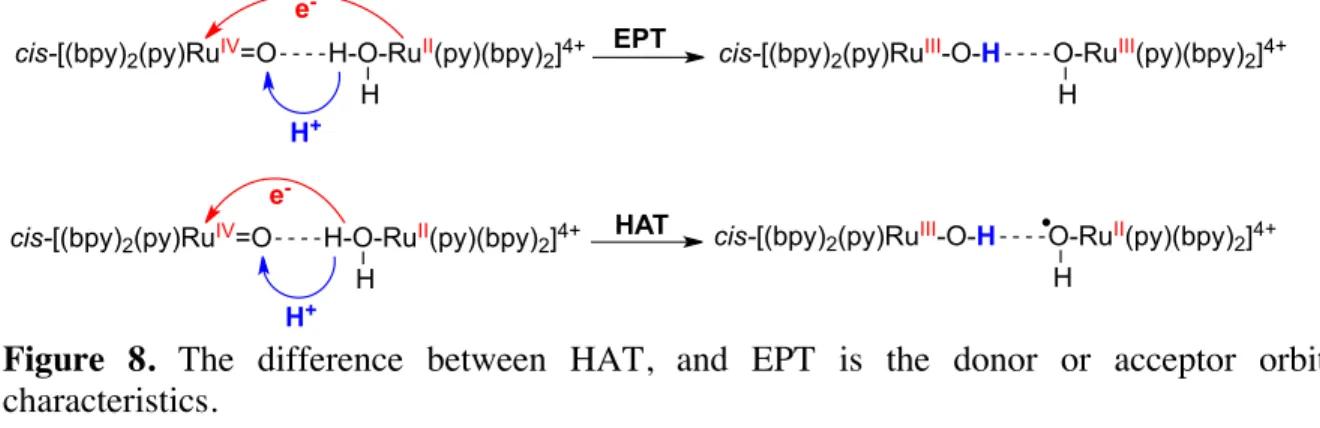
1.5 The Difference between MS-EPT, EPT and Hydrogen Atom Transfer (HAT): Concerted
proton-coupled electron transfer can be separated into three different sub-classes: multi-site
electron proton transfer (MS-EPT), electron proton transfer (EPT) and hydrogen atom
transfer (HAT), excluding hydride transfer because, unlike the aforementioned, it is by
definition, not a 1e-/1H+ reaction. The categorization of these concerted reactions is
dependent on the donor-acceptor orbital nature of the reaction. EPT is categorized as a
reaction that occurs between different donors or acceptors for the electron and the proton,
requiring charge redistribution because of this movement. HAT on the other hand is a
reaction in which the electron and proton are transferred to the same orbital, and thus does
not include a large amount of charge redistribution. This definition is not quantum
mechanically rigorous, however, these sub-classes can be quantitatively distinguished based
upon the degree of proton non-adiabaticity, which is reflective of the charge redistribution.44
In EPT, the e-/H+ donor orbitals and e-/H+ acceptor orbitals interact electronically,
enabling simultaneous transfer. Simultaneous means rapid relative to coupled vibrations (tens
of femtoseconds) and solvent modes (~1 picosecond).26 In EPT there is no discrete ET or PT
intermediate that is thermally equilibrated with its surroundings. If there were, the underlying
thermodynamics would be those of the intermediate and not those of the final EPT products.
Figure 8. The difference between HAT, and EPT is the donor or acceptor orbital characteristics.
In Figures 7 and 8 EPT leads to the final, energetically stable RuIII-OH2+ product and
is thermodynamically favored with ΔG°′= -0.11 eV.40,45 For the HAT pathway, e-/H+ transfer
occurs from a σ(O-H) orbital at RuII-OH22+ to electron (dπRu) and proton (pO) acceptor
orbitals at RuIV=O2+ giving RuIII-OH2+ and the high energy (~2.1 eV) charge transfer
intermediate, RuII-O●-H2+. There are different qualitative explanations for the differentiation
of EPT and HAT, however, a more accurate categorization comes from distinctions of
adiabaticity.
There is another class of EPT reactions, multiple site electron-proton transfer
(MS-EPT) as described in the context of PSII above. In MS-EPT an electron-proton donor
transfers electrons and protons to spatially separated acceptors or an electron-proton acceptor
accepts electrons and protons from spatially separated donors. MS-EPT is microscopically
more complex than electron or proton transfer. It shares with electron transfer requirements
for medium and intramolecular reorganization explained below in Section 1.6.1. However, it
has the additional complexity of a coupled proton transfer.
1.5.1 The Thermodynamics of a Proton Acceptor H2O versus Base: The rates of PCET
reactions and especially concerted PCET reactions are closely tied to the tunneling proton as
a result of the intrinsic nature of the reaction. As a product of this connection, it is a
cis-[(bpy)2(py)RuIV=O H-O-RuII(py)(bpy)2]4+
H
e
-H+
cis-[(bpy)2(py)RuIV=O H-O-RuII(py)(bpy)2]4+
H
e
-H+
EPT
HAT
cis-[(bpy)2(py)RuIII-O-H O-RuIII(py)(bpy)2]4+
H
cis-[(bpy)2(py)RuIII-O-H O-RuII(py)(bpy)2]4+
H
proton acceptors in PCET reactions. The nature of the proton acceptor is defined by its
conjugate acid pKa. For example, for H3O+→H2O, the pKa is -1.74, whereas for H2O→HO-,
the pKa is 15.74. Similarly, for phosphate H2PO4-→ HPO42- the pKa is 7.2. The robustness of
a proton acceptor in PCET can be calculated simply by the free energy equations for EPT, PT
and ET in eqs 3 – 7 below. From this treatment it can be seen that water is intrinsically and
microscopically a poor proton acceptor, even at its concentration of ~ 55M in neutral
aqueous solution. This is well stated by Costentin and coworkers which explain that EPT
oxidations involving water as a proton acceptor may compete with stepwise ET-PT reactions.
However, a necessary but perhaps not sufficient, prerequisite for this to occur is that the pKa
of the reduced form of the substrate be smaller than 0, where the excess driving force has to
be large enough to overcome pre-exponential and reorganizational factors that are unfavored
in the concerted reaction.46
For EPT with PT to an associated base:
ΔGEPT°′=-F{E°′(OxidantIII/II)-E°′(Substrate●+/0)}+0.059{pKa(Substrate●+)-pKa(HB)} (3)
For EPT with PT to “water”:
ΔGEPT°′= -F{E°′(OxidantIII/II)-E°′(Substrate●+/0)}+0.059{pKa(Substrate●+)-pH} (4)
For PT to an associated base:
ΔGPT°′= 0.059{pKa(Substrate●+)-pKa(HB)} (5)
For PT to “water”:
ΔGPT°′ = 0.059{pKa(Substrate●+)-pH} (6)
For ET:
The importance of water as a proton acceptor has been stressed in biological model
reactions involving tyrosine or tryptophan like molecules citing a Grotthaus or Eigen type
proton equilibration mechanism.41,43,47,48 However, this treatment is unsatisfactory on the
microscopic level because the proton must first release to a water molecule before
equilibrating to bulk solvent. In the case of tyrosine, the driving force for proton release to
water is ~ 0 eV given the difference in pKa values between pKa(TyrOH●+) = -2 and pKa(H2O)
= -1.74. In the case of tryptophan, the thermodynamics are even more uphill with
pKa(TrpNH●+) = 4.3.49
An example of the ongoing discussion about water as a proton acceptor is a
computational study conducted by Hummer and coworkers, on base assisted (direct) and
water mediated PCET between two stacked tyrosine molecules (TyrO●+ TyrOH → TyrOH +
TyrO●) mimicking a key step in the catalytic reaction in class Ia ribonucleotide reductase.50
Structures with and without a water molecule intervening between the phenol oxygen atoms
served as models for water mediated and direct PCET respectively. Results showed an
increase in reaction free energy barrier going from ∆G‡ = 6.5 kcal/mol (0.28 eV) for direct
PCET, and ∆G‡ = 11.0 kcal/mol (0.47 eV) for water mediated PCET, illustrating the less
favored nature of the reaction upon inclusion of water.50
Another example of the discussion on proton acceptors in PCET reactions is a set of
experiments carried out by Hammarström and coworkers.43,48,51 In these studies, tyrosine
appended metal-tris bipyridine complexes (Figure 9) were synthesized and studied via laser
flash-quench. In these studies the excited state of the metal complex is quenched by an
external quencher, followed by electron transfer from the tyrosine to a photo-oxidized metal
Figure 9. Structure of the rhenium-tyrosine complex that was the subject of reference 51, and 52 with a hydrogen bound phosphate (HPO42-) proton acceptor. The MS-EPT reaction occurs
via a proton transfer to the phosphate and electron transfer to the Rhenium.
A theoretical study by Hammes-Schiffer and coworkers found that a model, in which
the proton acceptor is the phosphate species, HPO42-, could successfully reproduce the
experimentally observed pH dependence of the overall rate as well as H/D kinetic isotope
effects.52 The model proposed by Hammarström and coworkers with water as a proton
acceptor was not physically reasonable for the system. Dibasic phosphate was found to be
favored over water as a proton acceptor in part because the proton donor-acceptor distance is
~0.2Å smaller for phosphate because of its negative charge.
Other theoretical treatments have addressed the pH dependence reported by
Hammarstöm and coworkers. Savéant and coworkers explored whether or not the driving
force and rate constants were pH dependent when H2O was acting as a proton acceptor.46 In
this study, the pH dependence was attributed to the participation of OH-, where the
concentration of H2O should not change significantly over the pH range, therefore, failing to
explain H2O as a proton acceptor coupled to pH as a driving force for the reaction.46 The use
of hydroxide as a stoichiometric proton acceptor has been shown in EPT reactions involving
have experimentally shown increased favorability of PCET reactions in the presence of a
base as opposed to just bulk water as a proton acceptor. 19-22
From Savéant and coworkers’ treatment it is plausible that the proton acceptor in
aqueous solution must be either hydroxide or a cluster of water. For the former, the OH
-acceptor is at such low concentrations except in very basic solution requiring a wavefunction
overlap between donor and acceptor through the solvent matrix. Hydroxide is the only
species that changes significantly over the pH range, suggesting its role as a proton acceptor
coupled to pH as a driving force. For the latter, however, it is microscopically challenging to
calculate a pKa for a solvent matrix proton acceptor and thus quantitatively assess
thermodynamically relevant values such as pKa. The state of the proton in water is complex
with two different water cluster structures proposed. One is the Zundel cation H5O2 +
(H2
O---H+
---OH2) 53
and the other the Eigen cation H9O4 +
(H3O +●
3H2O) 54,55
which undergo rapid
dynamical interchange.56-58
There is also experimental evidence for proton transfer through
individual solvent molecules acting as bridges.59,60
This has also drawn interest in biological
studies exploring the role of water molecules as proton relays as reported by Voth and
coworkers in Cytochrome c proton channels.61,62
1.6 The Fundamentals of Proton Coupled Electron Transfer: In summarizing the theory of
concerted electron proton transfer, it is useful to first consider simple electron transfer. Below
it is assumed that electronic coupling between electron donor and acceptor sites is relatively
weak. In this limit, the resonance energy arising from initial and final state wave function
1.6.1 Classical Electron Transfer: In diffusional electron transfer, pre-association between
donor D and acceptor A (eqs 8, 9) is followed by electron transfer and separation of products,
D+ and A-. Pre-association and close contact enhance electronic wave function mixing
between the electron donor and acceptor and increase the probability, given a perturbation, in
a barrier crossing or tunneling event. Within the classical limit for generic intramolecular
electron transfer in a pre-formed structure with electron transfer donor and acceptor spatially
arrayed as in a protein. There is no pre-association but changes in local orientation can be
influenced kET by their impact on electronic coupling.
DH-A→D-HA kobs = kET (8)
DH,A→D, HA kobs = KAkET (9)
Transfer of an initial Eigen state (µ) of one energy to a final Eigen state (ν) of another
energy, such as the case for election transfer, as well as proton coupled election transfer, is
best described using Fermi’s Golden Rule as a starting point. This mathematically defines the
probability of a transition as the result of a perturbation. The general format for this transition
probability is shown in equation 10 below.
(10)
Here the probability of the transition of initial (µ) to a final state (ν) is dictated by the
strength of the coupling between these states, represented by the matrix element Mµν as well
as the number of ways this transition can occur, which is represented by the Frank-Condon,
density of states term, ρfc.
This formalism can be expanded to define rates of election transfer as outlined by the
Marcus-Hush theory for election transfer.63,64 The equation for the Marcus non-adiabatic Tµ! =2"
! Mµ!
2
(11)
VET is the electron transfer matrix element discussed in detail below. In the
Frank-Condon density of states term, (4πλµνkBT)-1/2 is a Boltzmann distribution defining the thermal
population of vibrational states. The last exponential term defines the classical
thermodynamic barrier crossing. Within these terms λµν represents intramolecular and solvent
reorganizational energies in going from the initial (µ) and final (ν) states in the form of:
!
µ" =!
i+!
o (12)The lambda term is a summation of the inner-sphere (λi) and outer-sphere (λo)
reorganizational energies further broken down in equations 13 and 14.
(13)
Here a1, a2, d, Dop and Ds are the radii of the donor and acceptor, the distance between
their centers and the optical frequency and zero frequency dielectric constants of the solvent
respectively. The e2 term represents the amount of charge transferred. This treatment assumes
that the geometries of the reactants are simple spherical reactants. The innersphere
reorganizational energy is defined by:
(14)
Here the summation is over the coupled intramolecular vibrations. The contribution
of the jth normal mode to the reorganization energy is given in terms of the force constant fj
and the change in equilibrium position between the reactants and the products, represented by
∆Qµ,ν or the change in equilibrium normal coordinate for the quantum mode between initial
k
ET=
2
!
!
V
ET2
4
!"
µ#k
BT
(
)
!1/2exp
!
(
"
G
ET+
!
µ")
2
4
!
µ"RT
#
$
%
%
&
'
(
(
!o=e
2 1
2a1
+ 1
2a2
!1 d " # $ % & ' 1 Dop ! 1 Ds " # $$ % & ''
!i =
1 2 fj
j
!
(
"Qe,j)
2
= Sj
j
(reactant) and final (product) states. The only contributors to λiare those that have ∆Qµ,ν≠ 0.
Sj is the dimensionless quantity called the Huang-Rhys factor which is the
electron-vibrational coupling constant and is given by:
(15)
Here Mj, is the reduced mass and ωj the angular frequency. VET in eq 16 represents the
electron transfer matrix element which is the Hamiltonian for the wave function overlap of
the initial and final state the form of:
(16)
In this case the wave functions ψµ and ψν describe the initial and final states of the
transition. When the reaction is the sum of all contribution states the wave functions ψµ and
ψν are the total vibrational wave functions for the initial and final states. They are the
products of wave functions for all normal modes including collective solvent vibrations.
1.6.2 Proton Transfer: Proton motion along the proton transfer coordinate is described by a
linear combination of the high frequency (2000-4000 cm-1
) ν(O-H) vibrational modes before
and after proton transfer occurs.65-67
Because of the high quantum spacings between these
vibrational levels, proton transfer occurs by quantum mechanical tunneling not classical
barrier crossing.
1.6.3 Proton Coupled Electron Transfer: The coupling of a proton to the above quantum
mechanical treatment for pure electron transfer requires the inclusion of terms into equation
16 that take into account the tunneling of a proton in conjunction with the electron. The
following will initially define kEPT for a single vibrational transition, between an initial and
final state, and then define kEPT as a sum of all states, taking into consideration all possible Sj =
1 2
Mj!j ! ! "
# $
%
& '
(
Qe,j)
2
modes of transition from any vibrational level of the initial state to any vibrational level of
the final state. The complexity of this single state transition treatment in comparison to the
summation of states treatment can be illustrated by Figure 10 below.68
Figure 10.A) Schematic one-dimensional representation of the intersection between reactant and product vibrational levels. The optimal overlap of the ν = 0 initial vibrational level and the ν′ = 9 product level.68,69B) Two-dimensional vibronic free energy surface as functions of two collective solvent coordinates for a PCET reaction with the lowest energy reactant and product free energy surfaces shown. The notation of ΔG°µν represents the free energy difference between initial and final states and λ µν represents the outer-sphere reorganization energy. 4,70 Reproduced with permission from refs. 68 and 70. © 1996 and 2006 American
Chemical Society.
In the limit of strong electronic coupling across the hydrogen bond and weak
electronic coupling, kEPT is given for a single ν→ν′ transition in eq 17. It describes a single
vibronic transition from the initial level μ = 0 in the proton transfer mode to a final level ν
in the EPT product state and is analogous to the electron transfer result in eq 11.
(17)
k
EPT(!!!')=
2
"
!
V
µ!2
4
"#
µ$k
BT
(
)
"1/2exp
"
(
#
G
µ!+
"
µ!)
2
4
"
µ!k
BT
$
%
&
&
'
Initial coupled DH-A vibrational levels (µ), final coupled D-HA vibrational levels (ν)
and λµν is the reorganizational energy from solvent and low frequency (µ→ν) modes. Vµν is
the product of the electron transfer matrix element (VET) and proton vibrational wave
functions for initial and final states. Equation 17 above includes a classical barrier crossing
term with a Boltzmann distribution of states as well as the exponential term defining the
classical thermodynamic barrier crossing as in eq 11.
(18)
(19)
The EPT matrix element (eq 19) Vµν consists of the ET matrix element VET (eq 16)
and the wave function integral of the proton initial (µ) and final (ν) states. The dynamics of
the barrier crossing will depend on the product of the matrix element and the initial and final
proton wavefuntion overlap integral. The extent to which the square of this term is large is
going to dictate, much like in classical electron transfer, the probability of the initial and final
states being along the same spatial coordinate.
Approaching the total equation for kEPT requires the inclusion of all initial and final
overlapping states (summation of states) instead of the simplified treatment outlined above
which is for a single ν →ν′ transition. Contributions also exist from vibrational levels above
µ = 0. Even though populations in these levels fall off rapidly with ħω/kBT, there can be a
considerable compensation due to enhanced vibrational wave function overlap in levels
above µ = 0. Inclusion of the fractional population above µ = 0, PIµ, gives the final result in
eq 20. In this equation, the first summation is over the vibrational levels in the initial state
and the second summation is over all vibrational level in the final state. The summation of
states takes into consideration all of the possible interactions that can occur between all
!Gµ! = !GEPT+
(
µ"")
!#Vµ! =VET "µ
I


![Figure 7. i cat /nFAΓ vs [TyrOH] (M) for tyrosine concentration dependence at ITO-Ru II , I = 0.8M LiClO 4 , 0.1M HClO 4 , υ = 300 mV/s, Γ/Γ o = 1, slope = k obs,ET = 8.0x10 4 M -1 s -1](https://thumb-us.123doks.com/thumbv2/123dok_us/8239657.2183861/72.918.278.665.450.827/figure-nfaγ-tyroh-tyrosine-concentration-dependence-liclo-slope.webp)

