Corticosteroids Are Essential for Maintaining
Cardiovascular Function in Male Mice
Diana Cruz-Topete, Page H. Myers, Julie F. Foley, Monte S. Willis, and John A. Cidlowski
Signal Transduction Laboratory (D.C.-T., J.A.C.), Comparative Medicine Branch (P.H.M.), and Cellular and Molecular Pathology Branch (J.F.F.), National Institute of Environmental Health Sciences, National Institutes of Health, Department of Health and Human Services, Bethesda, Maryland 20892; and McAllister Heart Institute (M.S.W.), University of North Carolina at Chapel Hill, Chapel Hill, North Carolina 27514
Activation of the hypothalamic-pituitary-adrenal axis results in the release of hormones from the adrenal glands, including glucocorticoids and mineralocorticoids. The physiological association be-tween corticosteroids and cardiac disease is becoming increasingly recognized; however, the mecha-nisms underlying this association are not well understood. To determine the biological effects of corticosteroids on the heart, we investigated the impact of adrenalectomy in C57BL/6 male mice. Animals were adrenalectomized (ADX) at 1 month of age and maintained for 3– 6 months after surgery to evaluate the effects of long-term adrenalectomy on cardiac function. Morphological evaluation suggested that ADX mice showed significantly enlarged hearts compared with age-matched intact controls. These changes in morphology correlated with deficits in left ventricular (LV) function and electrocardiogram (ECG) abnormalities in ADX mice. Correlating with these functional defects, gene expression analysis of ADX hearts revealed aberrant expression of a large cohort of genes associated with cardiac hypertrophy and arrhythmia. Combined corticosterone and aldosterone replacement treatment prevented the emergence of cardiac abnormalities in ADX mice, whereas corticosterone replacement prevented the effects of adrenalectomy on LV function but did not block the emergence of ECG alterations. Aldosterone replacement did not preserve the LV function but prevented ECG abnormalities. Together, the data indicate that adrenal glucocorticoids and mineralocorticoids either directly or indirectly have selective effects in the heart and their signaling pathways are essential in maintaining normal cardiac function.(Endocrinology157: 2759 –2771, 2016)
G
lucocorticoids and mineralocorticoids are secreted by the cortex of the adrenal glands. Glucocorticoids such as corticosterone (mice) and cortisol (humans) are critical hormones for life, and their secretion is regulated by the hypothalamic-pituitary-adrenal axis in a circadian manner and in response to stress (1, 2). Mineralocortico-ids (aldosterone) are released by the adrenal gland in re-sponse to low blood volume or sodium depletion to reg-ulate blood pressure (BP) (3). Both steroids act in a tissue-specific manner by binding to their receptors, the glucocorticoid receptor (GR) (Nuclear Receptor Subfam-ily 3, Group C, Member 1; NR3C1) or the mineralocor-ticoid receptor (MR) (Nuclear Receptor Subfamily 3,Group C, Member 2; NR3C2), respectively (1, 2, 4, 5). GR and MR are members of the nuclear receptor su-perfamily of ligand-dependent transcription factors and regulate the expression of numerous genes involved in maintaining whole body homeostasis in response to al-terations in metabolism and in stress (1, 4, 5). Conse-quently, glucocorticoid and/or mineralocorticoid im-balance, such as excess or deficiency, can result in a variety of immunological, metabolic, and cardiovascu-lar complications (6).
ISSN Print 0013-7227 ISSN Online 1945-7170 Printed in USA
Copyright © 2016 by the Endocrine Society Received July 10, 2015. Accepted May 16, 2016. First Published Online May 24, 2016
For News & Views see page 2578
Abbreviations: ADX, adrenalectomized; BNP, brain natriuretic peptide; BP, blood pressure; CACNA1C, calcium channel, voltage-dependent, L type,␣1C subunit; CACNA2D1, cal-cium channel, voltage-dependent,␣2/␦subunit 1; CACNA2D2, calcium channel, voltage-dependent,␣2/␦subunit 2; CACNB2, calcium channel, voltage-dependent,2 subunit; CV, coefficient of variation; ECG, electrocardiogram; FS, fractional shortening; GR, glu-cocorticoid receptor; IPA, Ingenuity Pathway Analysis; LV, left ventricle; LVD, LV dysfunc-tion; LVEDD, LV internal diameter at diastole; LVESD, LV internal diameter at systole;
MHC,-myosin heavy chain; MR, mineralocorticoid receptor; PR, P-R interval; RYR2, ryanodine receptor 2; SERCA2, SR calcium transport ATPase 2; SKA, skeletal muscle␣ -ac-tin; SLC8A1, solute carrier family 8 (sodium/calcium exchanger), member 1; SR, sarco(en-do)plasmic reticulum.
doi: 10.1210/en.2015-1604 Endocrinology, July 2016, 157(7):2759 –2771 press.endocrine.org/journal/endo 2759
Primary adrenal insufficiency is an endocrine disorder characterized by insufficient secretion of glucocorticoids, and often mineralocorticoids (7–9). Despite clinical data suggesting that patients with adrenal insufficiency can de-velop cardiac complications, limited information exists on the biological actions of glucocorticoids and mineralocor-ticoids in maintaining normal cardiac function (10). Ad-dison’s disease, the most common form of primary adrenal insufficiency, was characterized more than 160 years ago by Thomas Addison, who described that among the lead-ing features of patients sufferlead-ing from adrenal disease was a remarkable feebleness of the heart actions (11). Subse-quent investigations revealed that decreased production of corticosteroids by the adrenal gland can lead to cardiac complications, including systolic failure and significant reduction in stroke volume (10, 12). Studies performed on isolated cardiac papillary muscle from adrenalectomized (ADX) rats suggested that corticosteroids regulate the gen-eration of mechanical force in the heart (13, 14). In addi-tion, several clinical studies in patients with heart failure have shown the existence of a correlation between both cortisol and aldosterone plasma levels and mortality (15– 17), which are consistent with potential effects of corti-costeroids on myocardial function. Furthermore, in vitro studies have shown that glucocorticoids and mineralocor-ticoids exert direct effects in cardiomyocytes (18 –20). Re-cent evidence found in transgenic mouse models have shown that prenatal inactivation of GR signaling in the heart leads to major structural and functional alterations in the maturation of the fetal heart (21). In addition, tar-geted deletion of GR in cardiomyocytes has demonstrated that intact GR signaling is critical for normal cardiac func-tion and viability in adult mice (22). In contrast, under normal physiological conditions, cardiomyocyte MR ap-pears to be nonessential for cardiac hemodynamics, struc-ture or function (23, 24); however, blockade of MR pro-tects the heart from injury in mouse models (23, 25), and prevents heart failure associated with myocardial infarc-tion in humans (26). Although, these results suggest that
both GR and MR play significant roles in cardiac physi-ology and pathphysi-ology, the nature of the ligand mediating the observed phenotypes remains unclear. The goal of the present study was to investigate the role of endogenous corticosteroids in the modulation of cardiac function in vivo in ADX C57BL/6 male mice. Our findings show that glucocorticoids have a primary role in the modulation of cardiac gene expression and function, whereas mineralo-corticoids seem to be critical in the regulation of cardiac electrical function.
Materials and Methods
Animals
1-month-old male intact (control, no surgical intervention), sham-ADX, and ADX C57BL/6 mice were purchased from Charles River Laboratories. One week after surgery, ADX mice were shipped to our facility, and upon arrival, all ADX mice were placed on 0.154M sodium chloride solution. One set of control and sham-ADX mice were also maintained on regular drinking water, whereas a second set was maintained on 0.154M sodium chloride solution. All studies were performed on male mice. All animals were maintained in accordance with the National Institutes of Health directives for the care and use of laboratory animals. These studies were approved by the National Institute of Environmental Health Sciences’ Animal Care and Use Committee.
Corticosterone, epinephrine, and aldosterone plasma levels
Blood samples from experimental animals were obtained by retroorbital sinus puncture using EDTA microhematocrit tubes. Blood samples were collected between 9 and 11:30AM. Corti-costerone, epinephrine, and aldosterone plasma levels were mea-sured by commercially available ELISAs following the manufac-turer’s instructions (DetectX Corticosterone Enzyme Arbor assays; Aldosterone ELISA kit, Abcam; Eagle Biosciences). The intra- and interassay coefficients of variation (CVs) for the cor-ticosterone assay were 6.3% and 9.9%, 6.6% and 10.8% for the aldosterone assay, and 9.0% and 7.7% for the epinephrine as-say. All samples were assayed in duplicate. For the first cohort of mice used in this study (Figures 1–7 and Table 1), the mean values for corticosterone and aldosterone in intact mice (1– 6 mo) were
Figure 1. Plasma levels of corticosterone (A), aldosterone (B), and epinephrine (C) in control (intact adrenal glands) and ADX mice. Hormone
levels were measured at 1, 3, and 6 months after adrenalectomy. Data are mean⫾SEM (n⫽10 mice per group). *,P⬍.05; **,P⬍.01; ***,P⬍.001 vs control mice.
2760 Cruz-Topete et al Corticosterone and Heart Disease Endocrinology, July 2016, 157(7):2759 –2771
between 15–36 and 0.03– 0.08 ng/mL, respectively, whereas mean values for corticosterone and aldosterone in ADX mice was 3–9 and 0.0 – 0.005 ng/mL, respectively. During the interval between our initial studies and the hormone replacement studies (Figures 8 and 9 and Table 2) our animal facility underwent changes in animal housing, which likely influenced the baseline for corticosterone levels. In this second cohort of mice the mean values for corticosterone and aldosterone in intact mice were 126⫾ 15 and 0.04⫾ 0.01 ng/mL, respectively. For ADX mice in the second cohort of mice, the mean values for corticosterone and al-dosterone were 21⫾14 and 0.005⫾0.0001 ng/mL, respectively. Despite the observed increase in corticosterone levels in both intact and ADX mice, corticosterone levels remained statistically signifi-cantly decreased in ADX mice (P⬍.001) in both studies.
Plasma electrolytes levels and liver panel
Na⫹, K⫹, Cl⫺, alanine aminotransferase (ALT), aspartate aminotransferase (AST), total bile acids (TBA), and sorbitol dehydrogenase (SDH) plasma levels were determined with the Olympus AU400e clinical analyzer, Beckman Coulter, Inc fol-lowing the manufacturer’s instructions. All reagents were pur-chased from Beckman Coulter, Inc.
Echocardiographic analysis
Transthoracic echocardiography was performed on con-scious mice with a VisualSonics Vevo 770 ultrasound biomicros-copy system (VisualSonics, Inc) with a 30-MHx 707B scan head as previously described (22). Briefly, 2-dimensional guided M-mode analysis of the left ventricle (LV) was performed on intact (controls) and ADX mice from 1 to 6 months of age. The leading edges of the epicardium and endocardium were used to measure anterior wall thickness, posterior wall thickness, and LV internal diameters (LV internal diameter at diastole [LVEDD], LV inter-nal diameter at systole [LVESD]). LV volume in diastole was calculated from the equation LV volume in diastole⫽(7/2.4⫹ LVEDD)⫻LVEDD3⫻1000, and LV volume in systole was
calculated from the equation LV volume in systole ⫽ (7/2.4 ⫹ LVESD) ⫻ LVESD3⫻1000. LV systolic function was assessed by fractional shortening (FS), calculated from the equation FS% ⫽(LVEDD⫺LVESD)/LVEDD⫻100. M-mode measurements represent 3 av-erage consecutive cardiac cycles from each mouse.
Electrocardiographic analysis Electrocardiographs were recorded from conscious mice with the noninva-sive ECGenie recording platform (Mouse Specifics, Inc) as previously de-scribed (22, 27). Briefly, mice were po-sitioned on the electrocardiogram (ECG) recording platform. Data were analyzed with the EzCG ECG analysis software (Mouse Specifics, Inc).
BP measurements
BP was measured with a tail-cuff sys-tem (CODA-6; Kent Scientific) following the manufacturer’s instructions. Briefly, conscious animals were preheated in a heating cabinet for 20 –30 minutes at 32°C. At least 10 accurate measurements per animal were used for analysis of diastolic, mean, and systolic pressure.
Real-time PCR (RT-PCR)
Total RNA was isolated from the whole hearts of intact (con-trol) and ADX C57BL/6 mice with the RNeasy Mini kit and RNase-Free DNase kit (QIAGEN) according to the manufac-turer’s instructions. mRNA levels were determined with a CFX96 Real-Time System C1000 Touch Thermal Cycler (Bio-Rad). Predesigned primer/probe sets for GR, ACTA1 (skeletal muscle␣-actin [SKA]), MYH7 (-myosin heavy chain [MHC]), NPPB (brain natriuretic peptide [BNP]), ATP2A2 (sarco[endo-]plasmic reticulum [SR] calcium transport ATPase 2 [SERCA2]), solute carrier family 8 (sodium/calcium exchanger), member 1 (SLC8A1), ryanodine receptor 2 (RYR2), calcium channel, volt-age-dependent, L type,␣1C subunit (CACNA1C), calcium chan-nel, voltage-dependent,␣2/␦subunit 1 (CACNA2D1), calcium channel, voltage-dependent, ␣2/␦ subunit 2 (CACNA2D2), CACNA1G, and cyclophilin B (peptidylprolyl isomerase B) were obtained from Applied Biosystems. Values measured for each primer/probe set were normalized to cyclophilin B.
Histological analysis
Hearts from intact and ADX mice were perfused with PBS and fixed with freshly prepared 4% paraformaldehyde. Samples were processed, embedded in paraffin, cut in 5m sections, and stained with hematoxylin-eosin.
Cross-sectional area analysis of cardiomyocytes Paraffin sections were stained with fluorescently labeled lec-tin as previously described (28). For cross-sectional area analysis of cardiomyocytes, TRITC-conjugated lectin (Triticum vulgaris; Sigma-Aldrich) staining was performed and measured as previ-ously described and examined by fluorescence microscopy (28).
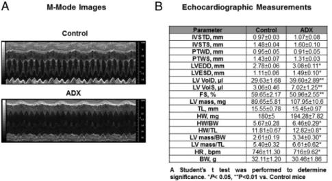
Figure 2. Long-term adrenalectomy leads to a significant decrease in cardiac performance. A,
Representative M-mode images from age-matched intact (control) and ADX mice at 6 months after surgery. B, Transthoracic echocardiography on conscious male C57BL/6 ADX and age-matched controls at 6 months after ADX. IVSTD, anterior wall thickness at diastole; IVSTS, anterior wall thickness at systole; PWTD, PWT at diastole; PWTS, PWT at systole; LV VolD, LV volume in diastole; LV VolS, LV volume in systole; TL, tibia length; HW, heart weight; BW, body weight; HR, heart rate. Data are⫾SEM (n⫽15 mice per group). A Student’sttest was performed to determine statistical significance (P⬍.05).
Microarray analysis
Gene expression analyses were performed on RNA from the hearts of long-term ADX mice (6 mo) and age matched controls with the Agilent Whole Mouse Genome oligo arrays (014868) (Agilent Technologies) following the Agilent 1-color microar-ray-based gene expression analysis protocol as described previ-ously (22). Briefly, data were obtained by the Agilent Feature Extraction software (v9.5), with the 1-color defaults for all pa-rameters. The Agilent Feature Extraction Software performed error modeling, adjusting for additive and multiplicative noise. The microarray data will be available in the Gene Expression Omnibus repository at the National Center for Biotechnology
Information ( http://www.ncbi.nlm.nih.gov/geo/info/link-ing.html). To further analyze the microarray datasets, the data were processed via Agi4x44PreProcess and Bioconductor for the R software environment (www.rproject.org). A single log scale normalized expression measure for each probe set was obtained after background correction and normalization between sam-ples. Box plots, density plots, MA plot, and spatial images of the raw and normalized data were examined in order to check the quality of the microarray data, and that no unusual results for any slide were observed. Principal component analysis was per-formed on all samples and all probes to reduce the dimensionality of the data while preserving the variation in the dataset. This
Figure 4. Dysregulation of genes associated to cardiac hypertrophy in long-term ADX mice. A–C, RT-PCR analysis ofMHC, SKA, and BNP mRNA
from hearts of control (intact) and long-term (6 mo) ADX mice. Values are normalized to cyclophilin (PPIB) mRNA. Data are mean⫾SEM (n⫽6 mice per group). **,P⬍.01 vs control mice. This PCR study is independent from the microarray data.
Figure 3. Adrenalectomy leads to changes in heart morphology and cardiomyocyte size. A, Representative images of intact hearts from
age-matched control (intact) and term ADX male mice (6 mo after surgery). B, Representative images of H&E-stained sections of control and long-term ADX mice. C, Representative images of TRITC-lectin staining of cardiomyocytes from control and long-long-term ADX mice. D, Morphometric analysis of cardiomyocyte cross-sectional area. Data are mean⫾SEM (n⫽10 mice per group). Scale bar, 50m. A Student’sttest was performed to determine statistical significance (*,P⬍.05).
2762 Cruz-Topete et al Corticosterone and Heart Disease Endocrinology, July 2016, 157(7):2759 –2771
allowed us to assess the similarities and differences of samples within a treatment group and between treatment groups. The significance of the log ratio for each probe was determined by calculating one modifiedtstatistic per probe using an empirical Bayesian approach. Probes with Benjamini-Hochberg multiple test correctedP⬍0.05 were considered to be differentially ex-pressed for comparison of the groups. The lists of probe sets generated were analyzed with the Pathway Analysis version 6.5 (Ingenuity Systems).
Corticosterone, aldosterone, and betamethasone treatments
Corticosterone, aldosterone, and betamethasone were given in 0.154M sodium chloride water at final concentrations of 25, 0.3, and 2.5g/mL, respectively, as previously described (29 – 32). Corticosterone, aldosterone, and betamethasone were pur-chased from Steraloids. Male C57BL/6 mice were ADX at 1 month of age and treated with corticosterone, betamethasone, aldosterone, or vehicle (PBS) in saline water for 3 months.
Statistical analysis
Statistics were performed with the GraphPad Prism software v6 (GraphPad Software, Inc). Student’st test (intact vs ADX mice) or one-way ANOVA with multiple comparisons analysis (intact vs ADX, corticosterone, aldosterone, ADX-corticosterone/aldosterone, and ADX-betamethasone) were used to evaluate whether differences between groups were sta-tistically significant. Differences were considered to be statisti-cally significant whenP⬍.05. For all studies we used 6 –15 mice per group, with the exception of ECG data on 6-month ADX mice (n⫽4).
Results
Plasma levels of corticosteroids, catecholamines, and electrolytes in controls (intact) and ADX male mice
In order to evaluate whether surgical removal of the adrenal glands leads to a sustained decrease in circulating corticosteroids and catecholamines, blood samples from controls and ADX mice were collected. Surgical adrenal ablation in ADX mice was confirmed during tissue har-vesting. Only mice with confirmed adrenal ablation were included in this study. Endogenous corticosterone, aldo-sterone, and epinephrine plasma levels were evaluated 1, 3, and 6 months after surgery. Our data show that corti-costerone, aldosterone, and epinephrine plasma levels were significantly diminished in ADX mice as compared with controls from 1 to 6 months after surgery (Figure 1, A–C).
Cardiac performance and heart morphology in long-term ADX mice
To understand the role of adrenal hormones in the eti-ology of heart disease, the chronic effects of adrenal in-sufficiency were evaluated in conscious ADX mice by transthoracic echocardiography. No major differences were observed between controls (intact) and ADX mice 1 month after adrenalectomy (Supplemental Figure 1). However, differences in cardiac function were detected between control and ADX mice 6 months after surgery (Figure 2A). Six-month ADX mice exhibited compro-mised LV function, as evidenced by a significant decrease in FS (59.65% control vs 50.96% ADX) (Figure 2B). In
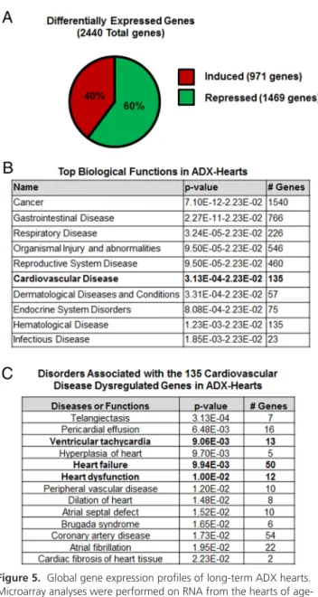
Figure 5. Global gene expression profiles of long-term ADX hearts.
Microarray analyses were performed on RNA from the hearts of age-matched male controls (intact) and ADX mice (6 mo after surgery). A, Total number of genes differentially expressed in the hearts of ADX mice compared with aged-matched controls: of the 2400 significant altered genes identified, 40% (971 genes) were induced, and 60% (1469 genes) were repressed in ADX hearts. Red and green colors correspond to induced and repressed expression, respectively. B, Differentially expressed genes were analyzed by IPA software for each age group. Shown are the top 10 biological functions most
significantly associated with the genes differentially regulated in the hearts of ADX mice. C, Most significant disorders or functions associated with the cardiovascular disease dysregulated genes in ADX hearts.
addition, we found that both heart weight and LV mass normalized to tibia length were significantly elevated in 6-month ADX mice (Figure 2B).
To evaluate the etiology of the LV dysfunction (LVD) observed in the long-term ADX mice, hearts were
col-lected and analyzed by histology. Gross morphological evaluation suggested that 6-month ADX mice had signif-icantly enlarged hearts compared with age-matched con-trols (Figure 3A). In agreement with these observations, histological evaluation of hematoxylin-eosin-stained
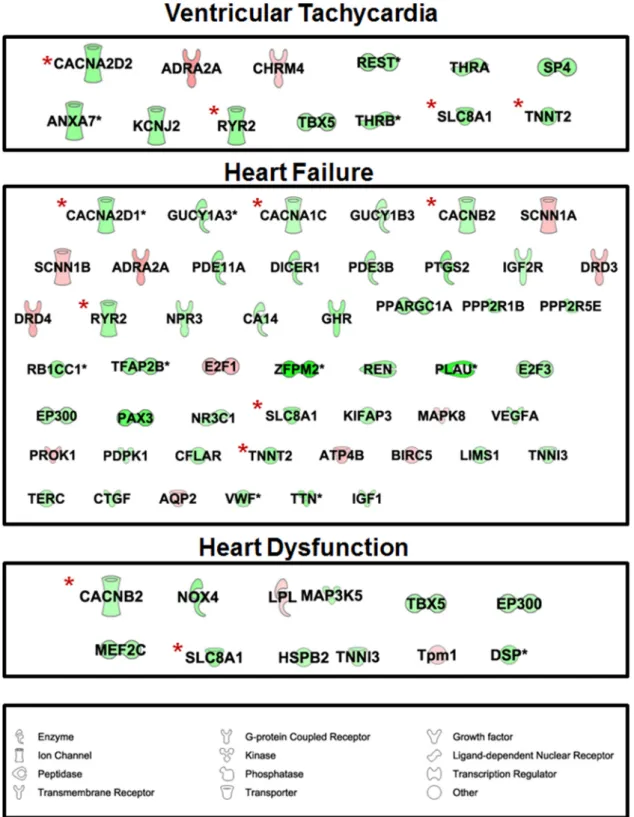
Figure 6. IPA identified that genes involved in cardiovascular disease were associated with ventricular tachycardia (13 genes), heart dysfunction
(50 genes), and failure (12 genes). Among the dysregulated genes in these 3 categories we found RYR2, CACNB2, CACNA1C, CACNA2D1, CACNA2D2, SLC8A1, and a Ca⫹⫹transporting, cardiac muscle, slow twitch ATPase (ATP2A2 or SERCA2). These genes are marked by a red asterisk (*). Red and green colors correspond to induced and repressed expression, respectively.
2764 Cruz-Topete et al Corticosterone and Heart Disease Endocrinology, July 2016, 157(7):2759 –2771
heart sections showed an increase in the thickness of the LV wall of the 6-month ADX mice (Figure 3B). Supporting these findings, a significant (P⬍.05) approximately 20% increase in cardiomyocyte cross-sectional area (Figure 3, C and D) was detected in TRITC-stained ADX heart sec-tions. These data suggest that long-term adrenalectomy leads to impairment of cardiac function associated with changes in heart morphology, as evidenced by a thickening of the myocardium of the LV and an increase in the cross-sectional area of the cardiomyocytes of ADX hearts. Be-cause control mice were maintained on regular drinking water, plasma electrolyte levels were measured to evaluate whether electrolyte abnormalities were secondary con-tributors to the cardiac phenotype observed in ADX mice. Plasma electrolyte levels did not differ between control
(regular drinking water) and ADX (saline solution) mice (Supplemental Figure 2). In addition, no significant ab-normalities in BP were observed in ADX mice compared with control mice (Supplemental Figure 3). Moreover, in-tact and sham-ADX groups of mice that were maintained on 0.154M sodium chloride showed no significant alter-ations in BP (Supplemental Figure 3), LV function ( Sup-plemental Figure 4), or heart electrical activity ( Supple-mental Table 1). Therefore, the cardiac phenotype observed in ADX mice is likely an effect of corticosteroid deficiency and is likely not associated with the 0.154M sodium chloride drinking water.
Effects of long-term adrenalectomy in the expression of cardiac stress marker genes
In order to dissect the molecular basis of the impaired heart function observed in the long-term ADX mice and to evaluate whether those changes corresponded to changes in gene expression observed in pathological cardiac hy-pertrophy, we evaluated mRNA levels of cardiacMHC, SKA, and BNP in the hearts of 6-month ADX and intact mice. A 4.32-fold increase inMHC and a 3.08-fold in-crease SKA (Figure 4, A and B) were found in ADX hearts. In addition, we measured a 2.84-fold increase in BNP (Fig-ure 4C). These data suggest that the compromised cardiac function and the increase in LV thickness in long-term ADX mice are pathological.
Figure 7. Down-regulation of genes involved in calcium signaling the
hearts of ADX mice. Total RNA was isolated from whole hearts of control and ADX mice (6 mo after surgery). CACNB2, CACNA1C1, CACNA2D1, CACNA2D2, RyR2, cardiac troponin T type 2, and SERCA2 mRNA levels were measured by qPCR. Data are mean⫾SEM (n⫽6 mice per group). *,P⬍.05; **,P⬍.01; ***,P⬍.001 vs control mice.
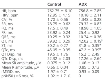
Table 1. Electrocardiographic Analysis of Conscious C57BL/6 ADX and Age-Matched Controls at 6 Months After ADX (⫾SEM)
Control ADX
HR, bpm 762.75⫾6.10 756.8⫾7.85 HRV, bpm 12.95⫾4.15 10.18⫾2.11 CV, % 1.70⫾0.56 1.348⫾0.28 RR, ms 78.75⫾0.62 79.32⫾0.83 PQ, ms 17.5⫾0.49 18.98⫾1.05 PR, ms 23.92⫾0.24 25.4⫾0.92 QRS, ms 10.25⫾0.32 10.74⫾0.36 QT, ms 39.92⫾0.29 42.02⫾0.19b
ST, ms 30.2⫾0.27 31.8⫾0.35a
QTc, ms 45.05⫾0.35 47.2⫾0.39b
QT Disp, ms 19.5⫾1.76 15.2⫾2.49 QTc Disp, ms 22.32⫾2.03 17.26⫾2.64 Mean SR amplitude,V 0.975⫾0.12 1.06⫾0.13 Mean R amplitude,V 0.66⫾0.04 0.85⫾0.13 rMSSD, ms 1.97⫾0.71 0.93⫾0.09 pNN50 (⬎6 ms), % 1.92⫾1.710 0
HR, heart rate; HRV, HR variability; RR, R-R interval; PQ, P-Q interval; QRS, QRS complex; QT, QT Disp, maximum QT interval minus minimum QT interval; ST, ST segment; QTc Disp, HR corrected QT interval; rMSSD, root mean square successive difference in heart period; pNN50, measure of HR variability. A Student’sttest was performed to determine significance. Values without asterisk were not significant different. n⫽4 mice per group.
aP⬍.05 vs control mice. b
P⬍.01 vs control mice.
Genome-wide cardiac gene expression profile of long-term ADX hearts
To define the molecular events and pathways underly-ing the effects of adrenalectomy in our model, genome-wide microarray analysis was performed on hearts from intact and ADX mice (6 mo). Adrenalectomy resulted in the differential expression of 2440 genes in the hearts of ADX mice (Supplemental Table 2). Interestingly, the ex-pression of most these genes was repressed in response to long-term adrenalectomy. Specifically, we discovered that 971 genes were induced, whereas 1469 genes were re-pressed in the hearts of ADX mice (Figure 5A). These find-ings suggest that adrenal hormones are either directly or indirectly essential in maintaining normal cardiac gene expression.
To determine the significance of the changes observed in the cardiac transcriptome of long-term ADX mice, 2440 genes were analyzed by literature-based Ingenuity Path-way Analysis (IPA) software. The analysis identified “car-diovascular disease” as one of the top 10 biological func-tions most significantly affected by the altered gene expression pattern in the heart of ADX mice (Figure 5B). A total of 135 genes associated with cardiovascular dis-ease were dysregulated in ADX hearts (Figure 5B and
Sup-plemental Table 3). Further analysis of these 135 genes showed that 13, 50, and 12 of these genes were associated with ventricular tachycardia, heart dysfunction, and heart failure, respectively (Figures 5C and 6 andSupplemental Table 4). Among the genes found in these categories were RYR2, calcium channel, voltage-dependent, 2 subunit (CACNB2), CACNA1C, CACNA2D1, CACNA2D2, SLC8A1, and cardiac troponin T type 2 (Figure 6).
The changes in gene expression were further validated by RT-PCR. For this purpose, we used hearts from addi-tional long-term ADX mice that were independent from those employed in the microarray studies. Consistent with the microarray data, the expression of all 7 genes was significantly repressed in the hearts of ADX mice (Figure 7). In addition, we found that the cardiac mRNA levels of SERCA2 were also significantly repressed in the hearts of ADX mice (Figure 7). These data indicate that long-term adrenal insufficiency leads to dysregulation of genes in-volved in regulating calcium handling and contractile function of the heart.
ECGs in long-term ADX mice
In order to assess whether the changes in gene expres-sion profiles displayed by long-term ADX mice translated
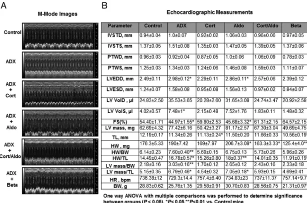
Figure 8. Hormone replacement effects on cardiac function in ADX mice. A, Representative M-mode images from age-matched intact (control),
ADX, and corticosterone-ADX (cort, 25g/mL)-treated mice, aldosterone-ADX-treated mice (aldo, 0.3g/mL), corticosterone/aldosterone-treated mice, and betamethasone-ADX (Beta, 2.5g/mL)-treated mice. B, Transthoracic echocardiography on conscious C57BL/6 hormone-treated ADX mice, ADX, and age-matched controls at 3 months after ADX. Data are mean⫾SEM (n⫽8 –12 mice per group). IVSTD, anterior wall thickness at diastole; IVSTS, anterior wall thickness at systole; PWTD, PWT at diastole; PWTS, PWT at systole; LV VolD, LV volume in diastole; LV VolS, LV volume in systole; TL, tibia length; HW, heart weight; BW, body weight; HR, heart rate. One-way ANOVA with multiple comparisons was performed to determine significance between groups (P⬍.05).
2766 Cruz-Topete et al Corticosterone and Heart Disease Endocrinology, July 2016, 157(7):2759 –2771
into abnormalities in the electrical conduction system of the heart, ECGs were performed on intact and ADX mice. We used a noninvasive system to record ECGs in ADX mice 6 months after surgery. All mice were conscious,
unsedated, and unrestrained. No differences were de-tected in average heart rates between control and ADX mice (controls 762.75⫾6.10 bpm vs ADX 756.8⫾7.85 bpm). In addition, adrenalectomy had no effects on P-R
Table 2. Electrocardiographic Analysis of Conscious C57BL/6 Hormone-Treated ADX, Age-Matched ADX, and Controls at 3 Months After ADX (⫾SEM)
Control ADX Cort Aldo Cort/Aldo Beta
HR, bpm 733.22⫾13.02 720.55⫾15.95 724.57⫾16.42 703.08⫾14.09 717.81⫾9.43 707.5⫾12.11 HRV, bpm 24.77⫾3.93 31.84⫾8.72 29.68⫾7.79 27.41⫾4.77 26.36⫾7.06 32.38⫾6.71 CV, % 3.41⫾0.54 4.48⫾1.26 4.15⫾1.08 3.97⫾0.75 3.60⫾0.92 4.67⫾0.9 RR, ms 82.17⫾1.62 83.86⫾1.96 83.37⫾1.97 85.93⫾1.82 83.94⫾1.08 85.23⫾1.49 PQ, ms 19.1⫾0.57 18.4⫾0.44 18.87⫾0.59 19.13⫾0.71 17.35⫾0.54 20.66⫾0.68 PR, ms 25.01⫾0.68 24.87⫾0.60 25.6⫾0.59 24.95⫾0.58 23.49⫾0.50 26.95⫾0.76 QRS, ms 10.05⫾0.29 11.61⫾0.29b 11.1⫾0.12b 10.20⫾0.27 10.19⫾0.27 10.77⫾0.34
QT, ms 39.86⫾0.36 43.26⫾0.97* 42.72⫾0.75a 41.79⫾0.66 41.56⫾0.81 42.43⫾0.34a
ST, ms 30.43⫾0.27 32.18⫾0.70 32.12⫾0.67 32.07⫾0.68 31.87⫾0.78 32.15⫾0.51 QTc, ms 44.46⫾0.43 47.18⫾0.81a 46.95⫾0.58a 45.1⫾0.53 45.37⫾0.77 46.06⫾0.44a
QT Disp, ms 22.25⫾1.51 25.56⫾3.13 21.85⫾4.41 25.29⫾1.04 23.68⫾2.02 21.31⫾2.83 QTc Disp, ms 25.41⫾1.76 27.32⫾3.57 24.01⫾4.84 26.67⫾0.84 25.27⫾2.01 22.55⫾2.81 Mean SR amplitude,V 0.87⫾0.10 0.73⫾0.10 0.88⫾0.11 0.91⫾0.10 1.00⫾0.09 1.11⫾0.11 Mean R amplitude,V 0.65⫾0.06 0.63⫾0.06 0.73⫾0.09 0.71⫾0.08 0.72⫾0.06 0.88⫾0.12 rMSSD, ms 2.62⫾0.40 4.15⫾1.56 4.0⫾1.87 2.61⫾0.52 2.59⫾0.45 2.52⫾0.42 pNN50 (⬎6 ms), % 4.04⫾1.27 1.23⫾1.06 5.67⫾2.29 4.78⫾2.13 7.62⫾4.97 8.91⫾3.36
Cort, corticosterone; Beta, betamethasone; Aldo, aldosterone; HR, heart rate; HRV, HR variability; RR, R-R interval; PQ, P-Q interval; QRS, QRS complex; QT, QT Disp, maximum QT interval minus minimum QT interval; ST, ST segment; QTc dispersion, HR corrected QT interval; rMSSD, root mean square successive difference in heart period; pNN50, measure of HR variability. One-way ANOVA with multiple comparisons was performed to determine significance between groups (P⬍.05). n⫽8 –12 mice per group.
a
P⬍.05 vs control mice.
bP⬍.01 vs control mice.
Figure 9. RT-PCR analysis ofMHC, SKA, CACNA1C1, CACNA2D1, and RYR2 mRNA from hearts of control, ADX, and corticosterone-treated
ADX mice. Values are normalized to cyclophilin (PPIB) mRNA. Data are mean⫾SEM (n⫽6 mice per group). One-way ANOVA was performed to determine significance between groups. *,P⬍.05; **,P⬍.01; ***,P⬍.001; n.s., not significant.
interval (PR) (controls 23.92⫾0.24 vs ADX 25.4⫾0.92 ms) and QRS interval (controls 10.25 ⫾ 0.32 vs ADX 10.74⫾0.36) (Table 1). Interestingly, QT (39.92⫾0.29 vs 42.02⫾0.19 ms), ST (30.2⫾0.27 vs 31.8⫾0.35 ms) and QTc intervals (45.05⫾0.35 vs 47.2⫾0.39 ms) were significantly prolonged in ADX mice (Table 1). These findings suggest that adrenal insufficiency also leads to alterations in the electrical depolarization and repolariza-tion of the ventricles, which increases the risk of develop-ing ventricular arrhythmias. Moreover, these results cor-relate with microarray data showing the dysregulation of several voltage-gated calcium channels and other genes involved in the regulation of cardiac action potential.
Effects of corticosterone treatment on cardiac gene expression, LV function, and electrical activity
To determine whether glucocorticoids were responsi-ble for the effects of adrenalectomy in cardiac gene ex-pression and function in our model, male C57BL/6 mice were ADX at 1 month of age and treated with corticoste-rone (25g/mL) in 0.154M NaCl for 3 months. Cortico-sterone-treated ADX mice reached similar corticosterone plasma levels to those observed in intact mice (126⫾15 control vs 153 ⫾ 9 ng/mL corticosterone-treated ADX) (Supplemental Figure 5A), and in ADX mice, corticoste-rone levels were statistically significantly decreased (P⬍ .0001) when compared with intact or corticosterone-treated ADX mice (Supplemental Figure 5). Cardiac func-tion was evaluated by conscious transthoracic echocardi-ography. Corticosterone treatment significantly improved LV function in ADX mice (Figure 8A). No major differ-ences were observed between control (intact) and corti-costerone-treated ADX mice (Figure 8B). Remarkably, corticosterone treatment also prevented increases in the gene expression of cardiacMHC and SKA in the heart of corticosterone-treated ADX mice (Figure 9A). Moreover, gene expression of CACNA1C, CACNA2D1, and RYR2 was significantly improved in corticosterone-ADX hearts as compared with untreated ADX mice (Figure 9B). These findings demonstrate that corticosterone play an impor-tant role in the regulation of cardiac gene regulation and are essential for the prevention of LVD in ADX mice. We also assessed whether corticosterone treatment prevented abnormalities in cardiac conduction displayed by long-term ADX mice. No differences in average heart rate, PR, QRS, or any other parameter were found between intact controls and ADX-corticosterone-treated mice (Table 2). However, QT and QTc remained significantly prolonged in corticosterone-treated ADX mice. Therefore, cortico-sterone treatment was not sufficient to prevent the emer-gence of ECG abnormalities in ADX mice.
Effects of aldosterone, corticosterone/aldosterone, and betamethasone treatment on LV function and electrical activity in ADX mice
Corticosterone has been reported to have a 10- to 30-fold lower affinity for GR than MR, thus to differentiate between glucocorticoid and mineralocorticoid effects in our model, ADX mice were treated with aldosterone (0.3
g/mL), corticosterone (25g/mL)/aldosterone (0.3g/ mL), or the GR selective ligand betamethasone (2.5g/ mL) in 0.154M NaCl for 3 months after surgery. Aldo-sterone plasma levels were also restored to comparable levels with those found in intact mice (0.044⫾0.01 con-trol vs 0.04⫾0.01 ng/mL aldosterone-treated ADX) ( Sup-plemental Figure 5B).
Cardiac function was evaluated by echocardiography. Despite betamethasone effects on growth and body weight, the treatment significantly improved LV function in ADX mice (Figure 8, A and B), and no major differences were observed between control (intact) and corticoste-rone/aldosterone or betamethasone-treated ADX mice (Figure 8B). Aldosterone alone did not restore LV function in ADX mice, whereas the combined treatment of corti-costerone and aldosterone prevented LVD in ADX mice (Figure 8, A and B). No significant effects in serum elec-trolyte levels were found in corticosterone- or betametha-sone-treated ADX mice (Supplemental Figure 2); how-ever, serum potassium levels were lower in the aldosterone-treated ADX mice (Supplemental Figure 2). Lower potassium levels (hypokalemia) have been reported to be associated with prolonged QT intervals (33). Despite the decrease in serum potassium levels, aldosterone re-placement treatment prevented the emergence of ECG normalities in ADX mice (Table 2). In addition, ECG ab-normalities were also blocked by the combined treatment of corticosterone and aldosterone (Table 2). In contrast, QT and QTc intervals remained prolonged in betametha-sone-treated ADX mice (Table 2). To investigate whether changes in BP contributed to the changes in LV function and ECG, BP was measured in hormone-treated ADX mice. Hormone treatments did not altered BP in this mouse model (Supplemental Figure 3). Thus, the increase in hypertrophy observed in aldosterone-treated mice is not likely a secondary effect of high BP.
Discussion
Corticosteroids can exert significant effects on heart func-tion; however, it is unclear whether their actions are direct or indirect and whether they are mediated through the GR or the closely related MR. The purpose of this study was to elucidate the role of endogenous corticosteroids in the 2768 Cruz-Topete et al Corticosterone and Heart Disease Endocrinology, July 2016, 157(7):2759 –2771
modulation of cardiac function. To achieve this goal we performed studies on long-term ADX male C57BL/6 mice. Our data demonstrate that glucocorticoids and mineralo-corticoids exert selective effects in the heart, with gluco-corticoid signaling playing a preponderant role in the reg-ulation of LV function and cardiac gene expression, whereas mineralocorticoid signaling has a predominant effect in the modulation of cardiac electrical activity.
We found that long-term adrenalectomy (6 mo) leads to cardiac dysfunction with a significant decrease in FS. Par-alleling the observed functional deficit, histological eval-uation of hearts from 6-month ADX mice showed an in-crease in thickness of the LV wall and in the cardiomyocyte cross-sectional area. These data are in agreement with re-cent studies by Oakley et al, which showed that mice lack-ing GR signallack-ing in cardiomyocytes (cardio GRKO) de-velop spontaneous cardiac hypertrophy and heart failure (22). Thus, removal of the hormones that activate both the GR and the MR leads to a similar phenotype in the heart. Our previous study showed that loss of cardiac GR signaling results in aberrant regulation of a large cohort of genes associated with abnormal calcium handling in car-diac hypertrophy and heart failure (22). Moreover, mouse models overexpressing MR in the heart have also demon-strated that mineralocorticoid signaling has effects on L-type calcium channel activity in cardiomyocytes (34). In vivo aldosterone infusion promotes the occurrence of widespread and long-lasting Ca⫹2 sparks due to an in-crease in the activity of cardiomyocyte RYRs (18). In ad-dition, recent studies by Messaoudi et al showed that MR activation in the heart leads to alterations in the gene ex-pression of ion channels, including potassium large con-ductance Ca2⫹-activated channel, subfamily M, -mem-ber 4, potassium channel, subfamily V, mem-mem-ber 2, and Ca2⫹ channel, voltage-dependent, l-type, ␣1S subunit (32). Consistently, hearts from ADX mice presented a sig-nificant dysregulation in a large cohort of cardiovascular disease genes associated with ventricular tachycardia and heart rhythm disorders, including Brugada syndrome, a disorder associated with mutations in genes involved in ion transport including sodium, potassium and calcium channels, with a characteristic prolongation of QT inter-vals (35). Thus, our genome wide gene expression results correlate well with the current literature (32).
In agreement with the changes in gene expression ob-served in ADX hearts, long-term adrenalectomy also led to ECG abnormalities characterized by prolonged QT inter-vals. These findings are consistent with in vivo studies showing that both GR and MR play a role in the etiology of atrio-ventricular alterations and ventricular arrhyth-mias in mice (34, 36).
To rule out the possibility that sodium in drinking wa-ter was contributing to the cardiac phenotype observed on the ADX hearts, functional studies were performed on control (intact) and sham-ADX mice drinking 0.154M NaCl. No significant alterations in BP, LV function, or ECG measurements were found associated with the 0.154M sodium chloride drinking water, suggesting that corticosteroid deficiency is important in the cardiovascu-lar phenotype observed in ADX mice.
In an attempt to differentiate the effects of glucocorti-coids and mineralocortiglucocorti-coids in our model, C57BL/6 male mice were ADX at 1 month of age and followed by re-placement with corticosterone, betamethasone (a gluco-corticoid lacking MR activity), and aldosterone (17, 29 – 31). Data from these studies revealed that, despite the effects of glucocorticoids on growth, body weight and liver chemistry (Supplemental Table 5), corticosterone and betamethasone replacement treatment prevented LVD in ADX mice. Similar results were obtained in mice treated with the combination of corticosterone and aldo-sterone. In contrast, treatment with aldosterone alone does not prevent LVD, suggesting that glucocorticoid sig-naling is playing a critical role in restoring cardiac function in our model. Provocatively, aldosterone was able to block the emergence ECG alterations in ADX mice either as a single treatment or in combination with corticosterone, despite the reduction in plasma potassium levels observed in aldosterone-treated ADX mice. Therefore, our data suggest that at the dose administered in our study, corti-costerone activates GR-dependent processes in the heart; however, it does not mimic aldosterone effects in terms of heart electrical conduction. Furthermore corticosterone neither activates nor antagonizes aldosterone actions at the sinoatrial nodal MR when they are coadministered. Together, our results demonstrate that glucocorticoids and mineralocorticoids have selective actions in the heart even in the context of the whole animal.
Given the fact that glucocorticoids bind GR and MR, the physiology underlying tissue-specific actions of gluco-corticoid is complex. Corticosterone has been reported to have 10- to 30-fold lower affinity for GR than MR, and in classic target tissues such as the distal nephron, MR se-lectivity is achieved by the coexpression of 11 -hydrox-ysteroid dehydrogenase type 2 (37). Cardiomyocytes do not express 11-hydroxysteroid dehydrogenase type 2; therefore glucocorticoids are not inactivated in the heart and can likely interact with MR (17). In addition, plasma levels of glucocorticoids are 1000 times greater than those of aldosterone suggesting that MR in cardiomyocytes are predominantly occupied by glucocorticoids (38). To gain insights into the role of GR and MR ligands in normal cardiac physiology, we used adrenalectomy as a model of
adrenal insufficiency. Clearly, this study does not differ-entiate between glucocorticoids and mineralocorticoids acting directly or indirectly in the heart. However, our data are consistent with data from transgenic mouse mod-els of GR and MR that have shown that GR is essential for normal cardiac function and survival (22), whereas MR signaling play an important role in the etiology of cardiac arrhythmias (34). Future studies are needed to decipher the molecular interplay between MR and GR signaling in the cardiovascular system of both male and female mice. In particular it will be essential to elucidate the direct and indirect actions of GR and MR signaling in cardiomyo-cytes in vivo.
Acknowledgments
We thank Ms Alyson Scoltock, Ms Chris Jewel, and Dr Robert H. Oakley and Dr Mahita Kadmiel for reading the present man-uscript and providing critical comments. We also thank Dr Ron Herbert and the Necropsy Support Group from the Pathology Core, Dr Kevin Gerrish from the Molecular Genomics Core, and Mr Ralph Wilson from the Clinical Pathology Core for their technical assistance. Finally, we thank National Institute of En-vironmental Health Sciences arts and photography, especially Lois Wyrick and Paul Cacioppo, for their assistance in the prep-aration of the figures for this manuscript.
Address all correspondence and requests for reprints to: John A. Cidlowski, PhD, Signal Transduction Laboratory, NIEHS, NIH, MD F3-07, 111 T. W. Alexander Drive, Research Triangle Park, NC 27709. E-mail:[email protected].
This work was supported by the Intramural Research Pro-gram of the National Institutes of Health (J.A.C.).
Disclosure Summary: The authors have nothing to disclose.
References
1. Kadmiel M, Cidlowski JA.Glucocorticoid receptor signaling in health and disease.Trends Pharmacol Sci. 2013;34:518 –530. 2. Cruz-Topete D, Cidlowski JA.One hormone, two actions: anti- and
pro-inflammatory effects of glucocorticoids. Neuroimmunomodu-lation. 2015;22:20 –32.
3. Funder JW, Pearce PT, Smith R, Smith AI.Mineralocorticoid action: target tissue specificity is enzyme, not receptor, mediated.Science. 1988;242:583–585.
4. Oakley RH, Cidlowski JA.Glucocorticoid signaling in the heart: a cardiomyocyte perspective.J Steroid Biochem Mol Biol. 2015;153: 27–34.
5. Meinel S, Gekle M, Grossmann C.Mineralocorticoid receptor sig-naling: crosstalk with membrane receptors and other modulators.
Steroids. 2014;91:3–10.
6. Rhee SS, Pearce EN.Update: systemic diseases and the cardiovas-cular system (II). The endocrine system and the heart: a review.Rev Esp Cardiol. 2011;64:220 –231.
7. Hsieh S, White PC.Presentation of primary adrenal insufficiency in childhood.J Clin Endocrinol Metab. 2011;96:E925–928.
8. Perry R, Kecha O, Paquette J, Huot C, Van Vliet G, Deal C.Primary adrenal insufficiency in children: twenty years experience at the Sainte-Justine Hospital, Montreal.J Clin Endocrinol Metab. 2005; 90:3243–3250.
9. Malikova J, Flück CE.Novel insight into etiology, diagnosis and management of primary adrenal insufficiency.Horm Res Paediatr. 2014;82:145–157.
10. Krug JJ.Cardiac arrest secondary to Addison’s disease.Ann Emerg Med. 1986;15:735–737.
11. Ten S, New M, Maclaren N.Clinical review 130: Addison’s disease 2001.J Clin Endocrinol Metab. 2001;86:2909 –2922.
12. Krishnamoorthy A, Mentz RJ, Hyland KA, et al.A crisis of the heart: an acute reversible cardiomyopathy bridged to recovery in a patient with Addison’s disease.Asaio J. 2013;59:668 – 670.
13. Lefer AM.Influence of corticosteroids on mechanical performance of isolated rat papillary muscles.Am J Physiol. 1968;214:518 –524. 14. Daw JC, Lefer AM, Berne RM.Influences of corticosteroids on cardiac glycogen concentration in the rat.Circ Res. 1968;22:639 – 647.
15. Swedberg K, Eneroth P, Kjekshus J, Wilhelmsen L.Hormones reg-ulating cardiovascular function in patients with severe congestive heart failure and their relation to mortality. CONSENSUS Trial Study Group.Circulation. 1990;82:1730 –1736.
16. Rossi GP, Cesari M, Sacchetto A.LVH in primary aldosteronism.
Hypertension. 1997;30:1297–1298.
17. Messaoudi S, Azibani F, Delcayre C, Jaisser F.Aldosterone, miner-alocorticoid receptor, and heart failure.Mol Cell Endocrinol. 2012; 350:266 –272.
18. Gómez AM, Rueda A, Sainte-Marie Y, et al.Mineralocorticoid modulation of cardiac ryanodine receptor activity is associated with downregulation of FK506-binding proteins.Circulation. 2009;119: 2179 –2187.
19. Rossier MF, Lenglet S, Vetterli L, Python M, Maturana A. Corti-costeroids and redox potential modulate spontaneous contractions in isolated rat ventricular cardiomyocytes.Hypertension. 2008;52: 721–728.
20. Ren R, Oakley RH, Cruz-Topete D, Cidlowski JA.Dual role for glucocorticoids in cardiomyocyte hypertrophy and apoptosis. En-docrinology. 2012;153:5346 –5360.
21. Rog-Zielinska EA, Thomson A, Kenyon CJ, et al.Glucocorticoid receptor is required for foetal heart maturation.Hum Mol Genet. 2013;22:3269 –3282.
22. Oakley RH, Ren R, Cruz-Topete D, et al.Essential role of stress hormone signaling in cardiomyocytes for the prevention of heart disease.Proc Natl Acad Sci USA. 2013;110:17035–17040. 23. Fraccarollo D, Berger S, Galuppo P, et al.Deletion of cardiomyocyte
mineralocorticoid receptor ameliorates adverse remodeling after myocardial infarction.Circulation. 2011;123:400 – 408.
24. Fraccarollo D, Bauersachs J.Cardiomyocyte mineralocorticoid re-ceptor function post myocardial infarction. Trends Cardiovasc Med. 2011;21:42– 47.
25. Lother A, Berger S, Gilsbach R, et al.Ablation of mineralocorticoid receptors in myocytes but not in fibroblasts preserves cardiac func-tion.Hypertension. 2011;57:746 –754.
26. Greenberg B, Zannad F, Pitt B.Role of aldosterone blockade for treatment of heart failure and post-acute myocardial infarction.
Am J Cardiol. 2006;97:34F– 40F
27. Chu V, Otero JM, Lopez O, Morgan JP, Amende I, Hampton TG.
Method for non-invasively recording electrocardiograms in con-scious mice.BMC Physiol. 2001;1:6.
28. Willis MS, Ike C, Li L, Wang DZ, Glass DJ, Patterson C.Muscle ring finger 1, but not muscle ring finger 2, regulates cardiac hypertrophy in vivo.Circ Res. 2007;100:456 – 459.
29. Coll AP, Fassnacht M, Klammer S, et al.Peripheral administration of the N-terminal pro-opiomelanocortin fragment 1–28 to Pomc-/-mice reduces food intake and weight but does not affect adrenal
2770 Cruz-Topete et al Corticosterone and Heart Disease Endocrinology, July 2016, 157(7):2759 –2771
growth or corticosterone production.J Endocrinol. 2006;190:515– 525.
30. Michailidou Z, Coll AP, Kenyon CJ, et al.Peripheral mechanisms contributing to the glucocorticoid hypersensitivity in proopiomel-anocortin null mice treated with corticosterone.J Endocrinol. 2007; 194:161–170.
31. Aiello R, Crupi R, Leo A, et al.Long-term betamethasone 21-phos-phate disodium treatment has distinct effects in CD1 and DBA/2 mice on animal behavior accompanied by opposite effects on neu-rogenesis.Behav Brain Res. 2015;278:155–166.
32. Messaoudi S, Gravez B, Tarjus A, et al.Aldosterone-specific acti-vation of cardiomyocyte mineralocorticoid receptor in vivo. Hyper-tension. 2013;61:361–367.
33. Kallergis EM, Goudis CA, Simantirakis EN, Kochiadakis GE, Var-das PE.Mechanisms, risk factors, and management of acquired long
QT syndrome: a comprehensive review. ScientificWorldJournal. 2012;2012:212178.
34. Ouvrard-Pascaud A, Sainte-Marie Y, Bénitah JP, et al.Conditional mineralocorticoid receptor expression in the heart leads to life-threatening arrhythmias.Circulation. 2005;111:3025–3033. 35. Grant AO, Carboni MP, Neplioueva V, et al.Long QT syndrome,
Brugada syndrome, and conduction system disease are linked to a single sodium channel mutation.J Clin Invest. 2002;110:1201– 1209.
36. Sainte-Marie Y, Nguyen Dinh Cat A, Perrier R, et al.Conditional glucocorticoid receptor expression in the heart induces atrio-ven-tricular block.FASEB J. 2007;21:3133–3141.
37. Funder JW.Mineralocorticoid receptors: distribution and activa-tion.Heart Fail Rev. 2005;10:15–22.
38. Young M, Funder JW.Aldosterone and the heart.Trends Endocri-nol Metab. 2000;11:224 –226.