primers used to analyse IkBatranscripts (þ2180 toþ2378) contained the sequences 50
-GATCCGCCAGGTGAAGGG-30 and 50
-GCAATTTCTGGCTGGTTGG-30 , the primers used to analyse IL-8 transcripts (þ1 toþ239) contained the sequences 50
-ATGACTTCCAAGCTGGCCGT-30 and 50
-TTACATAATTTCTGTGTTGGC-30 , and the primers used to analyse the 18S ribosomal RNA contained the sequences 50
-AGGAATTGACGGAAGGGCAC-30 and 50
-GGACATCTAAGGGCATCACA-30 .
ChIP assays
ChIP assays were performed with a previously described protocol (Upstate Biotechnology). In brief, chromatin from crosslinked cells was sheared by sonication (three times, 15 s each; one-third power) and incubated overnight with specific antibody followed by incubation with protein G–Sepharose saturated with salmon sperm DNA. Precipitated DNAs were analysed by quantitative PCR (34 cycles) with aTaqPCR Master mix kit (Qiagen) and primers for either the human 50
-GACGACCCCAATTCAAATCG-30 and 50
-TCAGGCTCGGGGAATTTCC-30
or murine 50
-GGACCCCAAACCAAAATCG-30 and 50
-TCAGGCGCGGGGAATTTCC-30
IkBapromoters (2316 to215), together with the human IL-8 promoter (2121 toþ61) 50
-GGGCCATCAGTTGCAAATC-30 and 50
-TTCCTTCCGGTGGTTTCTTC-30
and the humanb-actin promoter (2980 to2915) 50
-TGCACTGTGCGGCGAAGC-30 and 50
-TCGAGCCATAAAAGGCAA-30
. Quantitative real-time PCR was performed in triplicate to determine the association of IKK-a, p65 and CBP with the IkBaand IL-8 promoters by using 500 nM of the above oligonucleotide primers and input DNA standards diluted in threefold increments from 10% to 0.01% with SYBR Green Master Mix and the ABI Prism 7700 Sequence Detection System.
In vitrointeraction assay
Fragments of the CBP coding sequence were cloned into pGEX vector (Pharmacia). Purified GST–CBP fusion proteins were immobilized to glutathione–agarose and incubated overnight with cell lysates (100mg protein). After extensive washing with cold PBS, the protein complexes were analysed by immunoblotting.
IKK-akinase assay
Total cell lysates (100mg protein) prepared from cells transfected with expression vectors encoding Flag-tagged IKK-a, IKK-a(K/M) or IKK-a(SS/AA) were incubated for 1 h with anti-Flag antibody (Sigma) and with protein A–agarose for a further 1 h. After extensive washing of the immunoprecipitates, kinase assays were performed as described29with 5mg of histone H3 (Sigma) as a substrate.
Received 4 February; accepted 21 March 2003; doi:10.1038/nature01576.
1. Baldwin, A. S. The NF-kB and IkB proteins: new discoveries and insights.Annu. Rev. Immunol.14,
649–681 (1996).
2. Ghosh, S., May, M. J. & Kopp, E. B. NF-kB and Rel proteins: evolutionarily conserved mediators of immune responses.Annu. Rev. Immunol.16,225–260 (1998).
3. Silverman, N. & Maniatis, T. NF-kB signaling pathways in mammalian and insect innate immunity.
Genes Dev.15,2321–2342 (2001).
4. Zandi, E. & Karin, M. Bridging the gap: composition, regulation, and physiological function of the IkB kinase complex.Mol. Cell. Biol.19,4547–4551 (1999).
5. Takeda, K.et al.Limb and skin abnormalities in mice lacking IKKa.Science284,313–316 (1999). 6. Hu, Y.et al.Abnormal morphogenesis but intact IKK activation in mice lacking the IKKasubunit of
IkB kinase.Science284,316–320 (1999).
7. Li, Q.et al.IKK1-deficient mice exhibit abnormal development of skin and skeleton.Genes Dev.13,
1322–1328 (1999).
8. Sizemore, N., Lerner, N., Dombrowski, N., Sakurai, H. & Stark, G. R. Distinct roles of the IkB kinasea andbsubunits in liberating nuclear factorkB (NF-kB) from IkB and in phosphorylating the p65 subunit of NF-kB.J. Biol. Chem.277,3863–3869 (2002).
9. Li, X.et al.IKKa, IKKband NEMO/IKKgare each required for the NF-kB mediated inflammatory response program.J. Biol. Chem.277,45129–45140 (2002).
10. Birbach, A.et al.Signaling molecules of the NF-kB pathway shuttle constitutively between cytoplasm and nucleus.J. Biol. Chem.277,10842–10851 (2002).
11. Janknecht, R. & Hunter, T. Transcription. A growing coactivator network.Nature383,22–23 (1996). 12. Goodman, R. H. & Smolik, S. CBP/p300 in cell growth, transformation, and development.Genes Dev.
14,1553–1577 (2000).
13. Zhong, H., Voll, R. E. & Ghosh, S. Phosphorylation of NF-kB p65 by PKA stimulates transcriptional activity by promoting a novel bivalent interaction with the coactivator CBP/p300.Mol. Cell1,
661–671 (1998).
14. Zhong, H., May, M. J., Jimi, E. & Ghosh, S. The phosphorylation status of nuclear NF-kB determines its association with CBP/p300 or HDAC-1.Mol. Cell9,625–636 (2002).
15. Sakurai, H., Chiba, H., Miyoshi, H., Sugita, T. & Toriumi, W. IkB kinases phosphorylate NF-kB p65 subunit on serine 536 in the transactivation domain.J. Biol. Chem.274,30353–30356 (1999). 16. Madrid, L. V., Mayo, M. W., Reuther, J. Y. & Baldwin, A. S. Jr Akt stimulates the transactivation
potential of the RelA/p65 subunit of NF-kB through utilization of the IkB kinase and activation of the mitogen-activated protein kinase p38.J. Biol. Chem.276,18934–18940 (2001).
17. Gerritsen, M. E.et al.CREB-binding protein/p300 are transcriptional coactivators of p65.Proc. Natl Acad. Sci. USA94,2927–2932 (1997).
18. Wang, C. Y., Mayo, M. W. & Baldwin, A. S. J. TNFa- and cancer therapy-induced apoptosis: potentiation by inhibition of NF-kB.Science274,784–787 (1996).
19. Van Antwerp, D. J., Martin, S. J., Kafri, T., Green, D. R. & Verma, I. M. Suppression of TNF-a-induced apoptosis by NF-kB.Science274,787–789 (1996).
20. Strahl, B. D. & Allis, C. D. The language of covalent histone modifications.Nature403,41–45 (2000). 21. Cheung, P., Allis, C. D. & Sassone-Corsi, P. Signaling to chromatin through histone modifications.Cell
103,263–271 (2000).
22. Cheung, P.et al.Synergistic coupling of histone H3 phosphorylation and acetylation in response to epidermal growth factor stimulation.Mol. Cell5,905–915 (2000).
23. Lo, W.-S.et al.Phosphorylation of serine 10 in histone H3 is functionally linkedin vitroandin vivoto Gcn5-mediated acetylation at lysine 14.Mol. Cell5,917–926 (2000).
24. Sassone-Corsi, P.et al.Requirement of Rsk-2 for epidermal growth factor-activated phosphorylation of histone H3.Science285,886–891 (1999).
25. Thomson, S.et al.The nucleosomal response associated with immediate-early gene induction is mediated via alternative MAP kinase cascades: MSK1 as a potential histone H3/HMG-14 kinase.
EMBO J.18,4779–4793 (1999).
26. Saccani, S., Pantano, S. & Natoli, G. p38-dependent marking of inflammatory genes for increased NF-kB recruitment.Nature Immunol.3,69–75 (2002).
27. Anest, V.et al.A nucleosomal function for IkB kinase-ain NF-kB-dependent gene expression.Nature
423,659–663 (2003).
28. Senftleben, U.et al.Activation by IKKaof a second, evolutionary conserved, NF-kB signaling pathway.Science293,1495–1499 (2001).
29. Yamamoto, Y.et al.IKKg/NEMO facilitates the recruitment of the IkB proteins into the IkB kinase complex.J. Biol. Chem.276,36327–36336 (2001).
AcknowledgementsWe thank J. Guo for assistance, A. Herrera and M. Singh for preparation of the figures, J. Darrah for preparing the manuscript, and T. Collins for GAL4–CBP constructs.
Competing interests statementThe authors declare that they have no competing financial interests.
Correspondenceand requests for materials should be addressed to R.B.G. ([email protected]).
...
A nucleosomal function for IkB
kinase-a
in NF-kB-dependent
gene expression
Vasiliki Anest*†, Julie L. Hanson*†, Patricia C. Cogswell*, Kris A. Steinbrecher*, Brian D. Strahl*†‡& Albert S. Baldwin*†§
*Lineberger Comprehensive Cancer Center,
†Curriculum in Genetics and Molecular Biology,
‡Department of Biochemistry and Biophysics, and
§Department of Biology, University of North Carolina, Chapel Hill, North Carolina 27599, USA
... NF-kB is a principal transcriptional regulator of diverse cyto-kine-mediated processes and is tightly controlled by the IkB kinase complex (IKK-a/b/g). IKK-band IKK-gare critical for cytokine-induced NF-kB function, whereas IKK-ais thought to be involved in other regulatory pathways1–4. However, recent data
suggest a role for IKK-ain NF-kB-dependent gene expression in response to cytokine treatment1,5–7. Here we demonstrate nuclear
accumulation of IKK-a after cytokine exposure, suggesting a
nuclear function for this protein. Consistent with this, chromatin
immunoprecipitation (ChIP) assays reveal that IKK-a was
recruited to the promoter regions of NF-kB-regulated genes on stimulation with tumour-necrosis factor-a. Notably, NF-k B-regulated gene expression is suppressed by the loss of IKK-a
and this correlates with a complete loss of gene-specific phos-phorylation of histone H3 on serine 10, a modification previously associated with positive gene expression. Furthermore, we show
that IKK-a can directly phosphorylate histone H3 in vitro,
suggesting a new substrate for this kinase. We propose that
IKK-a is an essential regulator of NF-kB-dependent gene
expression through control of promoter-associated histone phos-phorylation after cytokine exposure. These findings provide additional insight into the role of the IKK complex in NF-k B-regulated gene expression.
Characteristic cytokine-mediated activation of the NF-kB path-way involves IKK-b-directed phosphorylation of and subsequent degradation of inhibitors of NF-kB (IkBs), resulting in rapid nuclear accumulation of NF-kB subunits1–3. It has been found that IKK-a,
but not IKK-b, constitutively shuttles between the cytoplasm and the nucleus, suggesting a nuclear function for this IKK subunit8. To
cytokine exposure, nuclear and cytoplasmic extracts from mouse embryonic fibroblasts (MEFs) stimulated with tumour-necrosis factor-a (TNF-a) were immunoblotted with antibodies against p65 and the different IKK subunits. Notably, stimulation of MEFs with TNF-aresults in marked nuclear accumulation of IKK-a(Fig. 1a). In contrast to IKK-a, both IKK-band IKK-gare present in the nucleus and the cytoplasm, and exhibit no appreciable changes on TNF-ainduction (Fig. 1a). We demonstrated the purity of nuclear and cytoplasmic fractions with antibodies specific for p105, an NF-kB precursor known to be cytoplasmic, and for TFIIB, a known nuclear protein (Fig. 1a). Immunofluorescence staining with an anti-IKK-aantibody confirms the western analysis. IKK-ais pre-dominantly cytoplasmic with low but detectable nuclear levels in unstimulated wild-type MEFs, whereas TNF-a-induced cells exhi-bit higher levels of nuclear IKK-a(Fig. 1b, left panel). Loss of immunoreactivity in IKK-a2/2cells confirms antibody specificity (Supplementary Information). Consistent with previous reports, TNF-a treatment induces nuclear translocation of p65 (Fig. 1b, right panel). Although the kinetics of induced IKK-a nuclear accumulation are similar to those of p65, TNF-ainduction leads to nuclear accumulation of IKK-ain p652/2cells (data not shown),
indicating that IKK-adoes not require p65 for nuclear entry. These data suggest that IKK-amay have a nuclear function distinct from the canonical NF-kB activation pathway.
Consistent with previous reports1, we observed normal TNF-a
-induced IkBadegradation and induced p65 nuclear translocation in MEFs lacking IKK-a(data not shown). However, IkBaresynthesis is delayed in IKK-a2/2cells compared with wild-type MEFs (Fig.
1c). The defect in resynthesis is consistent with a previous report implicating a role for IKK-a in TNF-a-induced IkBa gene expression5. Accordingly, we performed real-time polymerase
chain reaction (PCR) analysis to examine the expression profile of the NF-kB-regulated IkBagene (Nfkbia) in IKK-a2/2, IKK-b2/2 and wild-type MEFs. TNF-arapidly induces IkBagene expression in wild-type MEFs (Fig. 2a), whereas TNF-a-induced IkBagene expression is significantly decreased in both IKK-a2/2and IKK-b2/
2
cells. We next examined the kinetics of TNF-a-induced inter-leukin-6 (IL-6) gene expression by northern analysis. Both IKK-a2/
2and IKK-b2/2cells are deficient in IL-6 gene (Il6) activation at all
time points after TNF-a-induction (Fig. 2c). Previous reports have shown that loss of IKK-aand IKK-bresults in altered transcrip-tional activation of IkBa, IL-6 and other NF-kB-dependent genes5–7.
Thus, our results support a requirement for IKK-aand IKK-bin optimal IkBaand IL-6 gene expression.
The molecular mechanism(s) through which IKK-aregulates the expression of specific cytokine-induced NF-kB-dependent genes has not been determined previously. To investigate a nuclear role for IKK-a, we performed ChIP assays with antibodies directed against p65, IKK-a and IKK-b in TNF-a-stimulated wild-type MEFs. Furthermore, we used phosphorylated and acetylated histone H3 antibodies to examine histone H3 modifications that are known to correlate with active gene expression at the IkBapromoter9. The
results reveal a rapid recruitment of p65 to the IkBapromoter in response to TNF-a(Fig. 2b, left panel). Of note, both IKK-aand IKK-brecruitment are observed after TNF-a-induction, whereas we detect a low level of IKK-arecruitment in unstimulated cells.
Figure 1TNF-a-induced nuclear accumulation of endogenous IKK-a.a, MEFs were stimulated with TNF-a(10 ng ml21) and fractionated into nuclear and cytoplasmic fractions, and then analysed directly by western blotting with IKK-a, IKK-b, IKK-gand p65 antibodies. Antibodies specific for cytoplasmic p105/p50 and nuclear TFIIB demonstrate the purity of these fractions.b, MEFs were treated for 15 min with TNF-aas indicated,
and were processed for indirect immunofluorescence and stained with IKK-aor p65 antibodies.c, Kinetics of TNF-a-induced IkBadegradation in IKK wild-type and IKK-a2/2MEFs. Cells were treated with TNF-afor the indicated times and western
The kinetics of IKK-arecruitment and histone H3 Ser 10 phos-phorylation correlate in uninduced and TNF-a-induced cells. Consistent with previous reports and with evidence of basal IkBa gene expression9–11, we observed constitutive levels of acetylated
histone H3 at this promoter (Fig. 2b, left panel). Enrichment of promoter sequences is not detected at theb-actin promoter or when
the antibody is omitted from the immunoprecipitation reaction, confirming the specificity of the ChIP assay (data not shown). These results demonstrate a TNF-a-inducible association of IKK-aand IKK-bwith the IkBapromoter, and suggest a new function for these kinases in NF-kB-dependent gene regulation.
To address whether IKK-a has a unique nuclear function in modulating gene expression, we examined the recruitment profile of p65 and the status of histone modifications at the IkBapromoter in IKK-a2/2cells following TNF-atreatment. The kinetics of p65 recruitment in these cells is similar to those of wild-type MEFs, whereas IKK-b recruitment is delayed (Fig. 2b, right panel), indicating that p65 and IKK-bassociation with the IkBapromoter does not require IKK-a. The levels of TNF-a-induced p65 recruit-ment are reduced in IKK-a2/2cells. Notably TNF-a-induced H3 Ser 10 phosphorylation levels are completely abolished in the IKK-a2/2cells. Basal levels of H3 acetylation are decreased in the IKK-a2/2cells but normal levels are restored after TNF-a-induction. These data indicate that IKK-ais required for Ser 10 phosphoryl-ation of histone H3 at the IkBapromoter. The requirement for IKK-ain controlling H3 Ser 10 phosphorylation is also observed on another immediately accessible NF-kB-regulated gene,Mip2(also known asCxcl2; data not shown).
Next, we investigated recruitment of p65 and IKK-a, and the status of H3 Ser 10 phosphorylation at the IkBapromoter, using IKK-b2/2 MEFs. We detected reduced and transient levels of promoter-associated p65 and IKK-a after stimulation with TNF-a. The onset of Ser 10 phosphorylation matches the pattern of IKK-arecruitment in these cells after stimulation with TNF-a (Fig. 2b, lower panel). These data show that the loss of IKK-bdoes not affect Ser 10 phosphorylation at the IkBapromoter.
To examine further a requirement for IKK-ain regulating Ser 10 phosphorylation of histone H3, we evaluated the IL-6 gene, an NF-kB-regulated gene that requires prior chromatin modifications to ensure accessibility to NF-kB11. In wild-type MEFs, p65,
IKK-a and IKK-b all show a similar profile of promoter recruitment occurring 2 h after stimulation with TNF-a(Fig. 2d, left panel). Similar to the results obtained with the IkBapromoter, the presence of IKK-adirectly correlates with the onset of H3 Ser 10 phos-phorylation. Promoter-associated IKK-alevels decrease 4 h after treatment whereas Ser 10 levels remain detectable, suggesting that either additional H3 kinases may be involved in the maintenance of Ser 10 levels at the IL-6 promoter or that Ser 10 phosphorylation levels remain stable after transient recruitment of IKK-a. Notably, we did not detect TNF-a-induced Ser 10 phosphorylation in IKK-a2/2 cells at the IL-6 promoter (Fig. 2d, right panel). p65 recruitment in IKK-a2/2cells exhibits similar kinetics to wild-type cells, although overall levels appear to be lower. We did not detect IKK-bat this promoter in IKK-a2/2cells, suggesting a role for IKK-a in recruiting IKK-b to the IL-6 promoter. Collectively, these results indicate a requirement for IKK-ain mediating H3 Ser 10 phosphorylation at different NF-kB-dependent promoters.
The timing of TNF-a-induced expression of IL-6 messenger RNA does not coincide with recruitment of p65 to the IL-6 promoter (Fig. 2c, d). We examined IL-6 mRNA levels in wild-type and p652/2
MEFs to determine whether expression of IL-6 is strictly p65-dependent. Northern blotting experiments revealed decreased IL-6 expression in p652/2cells, although low-level induction of
IL-6 by TNF-ais observed (data not shown). This is consistent with p65 being necessary for efficient IL-6 transcription but suggests that other transcription factors, probably additional NF-kB subunits (J.L.H., unpublished observations), can initiate IL-6 transcription at a minimal level after TNF-astimulation.
The co-occupancy of p65 and IKK-a at both IkBa and IL-6 promoters in wild-type MEFs stimulated with TNF-aled us to examine whether p65 is required for the recruitment of IKK-aat these promoters. In p652/2MEFs, neither IKK-arecruitment nor Ser 10 phosphorylation is detected, indicating that p65 activation Figure 2Promoter-associated IKK-ais essential for TNF-a-induced NF-kB-dependent
gene expression and histone H3 phosphorylation.a,c, IKK-a2/2and IKK-b2/2cells
complexes are required for IKK-aassociation with NF-kB-regulated promoters (data not shown). Further investigation is needed to clarify whether p65 binding may function to alter the chromatin environment for IKK-arecruitment and subsequent Ser 10 phos-phorylation of histone H3, or whether p65 directly recruits IKK-a in this response.
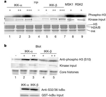
The role of IKK-a in controlling promoter-specific Ser 10 phosphorylation may be indirect through the regulation of other H3 kinases, or alternatively, IKK-a may directly phosphorylate histone H3. To address this latter point, we tested free core histones as substrates for recombinant IKK-abyin vitrokinase assays. IKK-a efficiently phosphorylates histone H3in vitroin a dose-dependent fashion (Fig. 3a). Furthermore, H3 phosphorylation catalysed by IKK-ais comparable to levels obtained with known H3 kinases, MSK1 and RSK2 (refs 12, 13; Fig. 3a). Site-specificity for histone H3 phosphorylation by IKK-awas confirmed by protein immunoblot-ting with a well-characterized anti-H3 (Ser 10) phospho-specific antibody (Fig. 3b). IKK-bfails to phosphorylate H3 on free core histones after incubation with [g-32P]ATP in vitro or using the Ser 10 phospho-specific antibody (Fig. 3a, b). As a positive control for IKK activity, phosphorylation of glutathione S-transferase (GST)–IkBafusion by IKK-aand IKK-bis shown (Fig. 3b, lower panel). The analysis of the histone H3 primary sequence does not reveal the IKK phosphorylation consensus sequence. These results demonstrate that IKK-adirectly phosphorylates histone H3 protein on Ser 10in vitro.
Our data demonstrate TNF-a-inducible IKK-arecruitment and subsequent Ser 10 phosphorylation on specific NF-kB-regulated promoters. To address the potential involvement of IKK-a in mechanisms associated with global levels of histone H3 phosphoryl-ation, we took two approaches. First, we acid-extracted core histones from wild-type, IKK-a2/2 and IKK-b2/2 MEFs and
examined H3 Ser 10 phosphorylation by western blotting. In wild-type and IKK-b2/2MEFs, TNF-atreatment leads to increased
Ser 10 phosphorylation (Fig. 4). However, both basal and TNF-a -induced levels of Ser 10 phosphorylation are significantly reduced in IKK-a2/2cells (Fig. 4) but are restored by the introduction of stably
expressed IKK-a(data not shown). Previous reports suggest that impaired recognition of phosphorylated Ser 10 by phospho-H3 antibodies may occur when adjacent lysine groups are acetyl-ated14,15. Therefore, we tested an antibody against dimodified H3
to examine H3 phosphorylated on Ser 10 and acetylated on Lys 14. Levels of phosphorylated and acetylated H3 increase in response to TNF-astimulation, with kinetics similar to that of Ser 10 phos-phorylation in wild-type and IKK-b2/2MEFs (Fig. 4). However, levels of phosphorylated and acetylated H3 remain reduced in the IKK-a2/2MEFs. This suggests that the reduced affinity for Ser 10 phosphorylation in IKK-a2/2MEFs is not due to antibody occlu-sion by adjacent acetylation on Lys 14. In addition, global levels of acetylated H3 are largely unaffected in IKK-a2/2MEFs (data not shown). Second, immunofluorescence staining of asynchronous wild-type and IKK-a2/2 cells with the same phospho-specific antibody demonstrates similar H3 Ser 10 phosphorylation levels in both cell types (data not shown). However, this probably represents mitotic-associated Ser 10 phosphorylation16, not
tran-scriptionally associated histone phosphorylation. Thus, although it is clear that IKK-a controls promoter-associated Ser 10 phos-phorylation on a set of NF-kB-regulated genes, current investigation is aimed towards examining the role of IKK-ain modulating global levels of histone H3 phosphorylation.
Although a role for IKK-ain controlling NF-kB-dependent gene expression has been suggested previously, a mechanism whereby IKK-amight facilitate such a response has remained unclear. Our results suggest that one role for IKK-a in controlling gene expression is through an unexpected nuclear mechanism involving histone H3 phosphorylation. Furthermore, the data indicate that IKK-adirectly phosphorylates histone H3 under TNF-a stimu-lation. However, we cannot rule out the possibility that IKK-ais involved in the regulation of other known H3 kinases. In this regard, we observe no TNF-a-induced changes in MSK1 or RSK2 protein levels in wild-type or IKK-a2/2 MEFs (data not shown). Also, MSK1 activity is minimally induced by TNF-astimulation in wild-type or IKK-a2/2MEFs using core histones as a substrate (data not shown). Therefore, our results suggest that IKK-a-mediated histone phosphorylation may provide one nucleosomal component in the overall mechanism required for optimal gene expression. Future experiments will be necessary to determine whether IKK-a -mediated Ser 10 phosphorylation regulates other histone modifi-cations associated with positive gene expression, such as acetylation of Lys 14 on histone H3 as predicted by the histone code hypoth-esis15,17–20.
Our observation that IKK-bis recruited to NF-kB-dependent promoters and is not required for histone phosphorylation (Fig. 2b, lower panel) indicates that IKK-a does not require IKK-b to control histone phosphorylation, and suggests a distinct chromatin-associated role for IKK-bin controlling NF-kB-dependent gene
Figure 4IKK-amodulates global levels of histone H3 phosphorylation. Acid-soluble proteins were extracted from asynchronous IKK wild-type, IKK-a2/2and IKK-b2/2
MEFs, and were immunoblotted with anti-phospho-specific Ser 10, anti-phosphorylated/ acetylated H3, or anti-H3 antibodies.
expression. Current experiments are directed towards investigating the role of promoter-localized IKK-b. Additionally, TNF-ainduces recruitment of IKK-gto the IkBapromoter, albeit at later time points than IKK-aand IKK-bin wild-type MEFs (data not shown). Collectively, these data support a function for IKK-athat is distinct from the classical role of the cytoplasmic IKK-a/b/gcomplex in controlling cytokine-induced NF-kB-regulated gene expression. Thus, we propose independent roles for IKK-a, IKK-band IKK-g in cytokine-induced NF-kB-dependent gene expression. Whereas IKK-bcontrols IkBa degradation and efficient DNA binding of NF-kB subunits1 and IKK-g acts as a structural or regulatory
mediator of this kinase complex1,3,21, IKK-afunctions as a
chro-matin modifier through histone phosphorylation. The data also raise the possibility of a role for IKK-ain NF-kB-independent gene
expression. A
Methods
Cells and reagents IKK wild-type, IKK-a2/2
and IKK-b2/2
MEFs were provided by I. Verma and M. Karin. Antibodies against IKK-a, IKK-b, IKK-g, p105/p50, actin and tubulin were obtained from Santa Cruz and Upstate Biotechnology. The p65/RelA-specific antibody was obtained from Rockland. Phosphorylated histone H3 (Ser 10) antibody was obtained from Cell Signal Technology. Acetylated (Lys 9/Lys 14) histone H3 antibody and phosphorylated/ acetylated (Ser 10/Lys 14) histone H3 antibody was obtained from Upstate Biotechnology. TFIIB antibody was obtained from Transduction Laboratories. TNF-a(Kamiya) was used at a final concentration of 10 ng ml21.
Immunofluorescence IKK wild-type, IKK-a2/2
or IKK-b2/2
cells were seeded onto chamber slides (Nalge Nunc International) and treated with TNF-a(10 ng ml21) for 15 min. After fixation with 4% paraformaldehyde, cells were permeabilized with 0.2% Triton-X and blocked with 10% goat serum, 1% bovine serum albumin in phosphate-buffered saline (PBS). Primary antibodies were incubated at 48C overnight. One-hour incubations with fluorescein isothiocyanate-conjugated secondary antibodies (Santa Cruz Biotechnology) were used to detect primary antibody and were visualized on a Zeiss Axioskop. All images are shown at
£200.
Real-time quantitative PCR
Five micrograms of total RNA was incubated with Moloney murine leukaemia virus-reverse transcriptase (Invitrogen) as recommended by the manufacturer. The resulting complementary DNA was analysed quantitatively for the expression of IkBaby fluorogenic 50
-nuclease PCR as described previously. Specific primers (forward 50 -AGGATGAGCTGCCCTATGATGA-30
and reverse 50 -TGCCACTTTCCACTTATAATGTCAGA-30
) and probe (50 -6-FAM
TGTGTGTTTGGAGGCCA-TAMRA) were designed to the IkBagene and PCR products were continuously measured by means of ABI Prism 7900 during 40 cycles. All data were normalized to 18S ribosomal RNA.
Northern blot analysis
Total RNA was isolated using Trizol (Invitrogen) as recommended by the manufacturer. A total of 10–20mg total cellular RNA was separated on 1.5% formaldehyde-agarose gels and transferred overnight to a nylon filter according to standard procedures. RNA was then crosslinked to the membrane by ultraviolet irradiation (Stratagene) and probed with randomly labelled IL-6 probe. Hybridization and wash was preformed using ExpressHyb (Stratagene) as described by the manufacturer.
ChIP assay
ChIP analysis was performed following a protocol provided by Upstate Biotechnology under modified conditions. After TNF-a(20 ng ml21) stimulation, 3£106cells were fixed with 1% formaldehyde. After 5 min, cells were washed extensively with ice-cold PBS and lysed for 10 min in lysis buffer (Upstate Biotechnology). Chromatin was sheared by sonication to an average size of approximately 1 kilobase and pre-cleared for 2 h at 48C with salmon sperm DNA-saturated protein G Sepharose. Chromatin solutions were precipitated overnight at 48C using 10ml anti-p65, 10mg anti-IKK-aor anti-IKK-b, 10ml anti-phospho-H3 (Ser 10), and 5ml acetylated H3-specific antibodies or beads alone. Immune complexes were collected with salmon sperm DNA-saturated protein G Sepharose for 1 h and washed extensively following the manufacturer’s protocol. Input and immunoprecipitated chromatin were incubated at 658C overnight to reverse crosslinks. After proteinase K digestion, DNA was extracted with phenol/chloroform and precipitated with ethanol. Precipitated DNAs were analysed by PCR (30–35 cycles) using Platinum Taq PCR Master Mix (Invitrogen). The following promoter-specific primers were used: primer pair 50
-TGGCGAGGTCTGACTGTTGTGG-30 and 50
-GCTCATCAAAAAGTTCCCTGTGC-30
was used to amplify a 230-base-pair (bp) region in the mouse IkBapromoter; primer pair 50
-TGTGTGTGTGTGTATGTGTGTGTCG-30 and 50
-TCGTTCTTGGTGGGCTCCAG-30
was used to amplify a 440-bp region in the mouse IL-6 promoter or theb-actin promoter (50
-TGCACTGTGCGGCGAAGC-30 and 50
-TCGAGCCATAAAAGGCAA-30 ).
In vitrokinase assay
Kinase assays were performed following a previously described protocol (Upstate Biotechnology). Kinase activity was determined by incubating purified chicken core histones (10 mg ml21) with increasing amounts of recombinant IKK-aor IKK-b, or MSK1 (20 mU) and RSK2 (77 U) in the presence of 1mCi ml21[g-32P]ATP or cold ATP (100mM) for 30 min at 308C. Reactions were resolved by SDS–polyacrylamide gel electrophoresis (PAGE; 15%) and processed for autoradiography or protein immunoblotting. Recombinant IKK-aand IKK-bwere provided by L. Dang.
Western blot analysis
Extractions of acid-soluble proteins were done according to the protocol described by Upstate Biotechnology and resolved on 10–20% Tris-tricine SDS–PAGE gels.
Received 6 March; accepted 28 March 2003; doi:10.1038/01648.
1. Ghosh, S. & Karin, M. Missing pieces in the NF-kB puzzle.Cell109,S81–S96 (2002).
2. Silverman, N. & Maniatis, T. NF-kB signaling pathways in mammalian and insect innate immunity.
Genes Dev.15,2321–2342 (2001).
3. Courtois, A., Smahi, A. & Israel, A. NEMO/IKKg: linking NF-kB to human disease.Trends Mol. Med.
7,427–430 (2001).
4. Senftleben, U.et al.Activation by IKKaof a second, evolutionarily conserved, NF-kB signaling pathway.Science293,1495–1499 (2001).
5. Li, Q.et al.IKK1-deficient mice exhibit abnormal development of skin and skeleton.Genes Dev.13,
1322–1328 (1999).
6. Li, Q.et al.Severe liver degeneration in mice lacking the IkB kinase 2 gene.Science284,321–325 (1999).
7. Li, X.et al.IKKa, IKKb, and NEMO/IKKgare each required for the NF-kB mediated inflammatory response program.J. Biol. Chem.277,45129–45140 (2002).
8. Birbach, A.et al.Signaling molecules of the NF-kB pathway shuttle constitutively between cytoplasm and nucleus.J. Biol. Chem.277,10842–10851 (2002).
9. Saccani, S., Pantano, S. & Natoli, G. p38-dependent marking of inflammatory genes for increased NF-kB recruitment.Nature Immunol.3,69–75 (2002).
10. Ainbinder, E.et al.Mechanism of rapid transcriptional induction of TNFa-responsive genes by NF-kB.Mol. Cell. Biol.22,6354–6362 (2002).
11. Saccani, S., Pantano, S. & Natoli, G. Two waves of NF-kB recruitment to target promoters.J. Exp. Med.
193,1351–1359 (2001).
12. Sassone-Corsi, P.et al.Requirement of Rsk-2 for epidermal growth factor-activated phosphorylation of histone H3.Science285,886–891 (1999).
13. Thomson, S.et al.The nucleosomal response associated with immediate-early gene induction is mediated via alternative MAP kinase cascades: MSK1 as a potential histone H3/HMG-14 kinase.
EMBO J.18,4779–4793 (1999).
14. Clayton, A., Rose, S., Barratt, M. & Mahadevan, L. Phosphoacetylation of histone H3 on c-fos and c-jun associated nucleosomes upon gene activation.EMBO J.19,3714–3726 (2000).
15. Thompson, S., Clayton, A. & Mahadevan, L. Independent dynamic regulation of histone phosphorylation and acetylation during immediate early gene induction.Mol. Cell8,1231–1241 (2001).
16. Crosio, C.et al.Mitotic phosphorylation of histone H3: spatio-temporal regulation by mammalian Aurora kinases.Mol. Cell. Biol.22,874–885 (2002).
17. Berger, S. L. An embarrassment of niches: the many covalent modifications of histones in transcriptional regulation.Oncogene20,3007–3013 (2001).
18. Strahl, B. D. & Allis, C. D. The language of covalent histone modifications.Nature403,41–45 (2001). 19. Cheung, P.et al.Synergistic coupling of histone H3 phosphorylation and acetylation in response to
epidermal growth factor stimulation.Mol. Cell5,905–915 (2000).
20. Lo, W.et al.Phosphorylation of serine 10 in histone H3 is functionally linkedin vitroandin vivoto Gcn5-mediated acetylation at lysine 14.Mol. Cell5,917–926 (2000).
21. Prajapati, S. & Gaynor, R. Regulation of IkB kinase (IKK)g/NEMO function by IKKb-mediated phosphorylation.J. Biol. Chem.277,24331–24339 (2002).
22. Yamamoto, Y., Verma, U. N., Prajapati, S., Kwak, Y.-T. & Gaynor, R. B. Histone H3 phosphorylation is critical for cytokine-induced gene expression.Nature423,655–659 (2003).
Supplementary Informationaccompanies the paper onwww.nature.com/nature.
AcknowledgementsWe thank D. Kashatus for critical reading of the manuscript. We appreciate scientific discussions with Y. Yamamoto and R. Gaynor. Support was provided by the NIH and by the UNC breast cancer SPORE program to A.S.B., and by the UNC Comprehensive Center for Inflammatory Disorders.
Competing interests statementThe authors declare that they have no competing financial interests.