Changes in antioxidative enzymes and isozyme pattern due to Exobasidium vexans infection in tea cultivars
Copyright © 2017 MJB 11
RESEARCH ARTICLE
Open Access Full Text Article
Changes in antioxidative enzymes and isozyme pattern due
to
Exobasidium vexans
(blister blight) infection in tea
cultivars
Shalini D
1*and Raj kumar R
21SNS College of Engineering, Coimbatore-641017.
2Department of Plant Physiology and Biotechnology, UPASI Tea Research Foundation, Tea Research Institute,
Valparai, Coimbatore-642127.
*For correspondence: [email protected]
Article Info: Received 18 Feb 2017; Revised: 27 Apr 2017; Accepted 16 May 2017.
ABSTRACT
Assessment of antioxidative enzymes and their isoenzyme pattern were studied with field grown tea plants having highly resistant (SA-6 & SMP-1) and highly susceptible (UPASI-14 & TES 31) traits to Exobasidium vexans. Antioxidative enzymes were assayed in healthy and infected leaves collected from selected tea clones. Peroxidase, ascorbate peroxidase and glutathione reductase have higher activity in resistant clones but catalase activity was very low while there was no trend in SOD activity. The zymograms of peroxidase and catalase in the resistant genotype showed amplified expression than susceptible tea clones. Induction and /or amplified expression of particular isoforms of peroxidase and catalase against blister blight infection in resistant tea clones led to the apparent conclusion that amplified isoenzyme expression linked with blight resistance. This might be considered as an important criterion in breeding programme to identify the Exobasidium resistant genotype in tea.
Keywords: Isoenzyme, antioxidative enzymes, blister blight
1.
INTRODUCTION
Tea is the most popular and inexpensive beverage produced from tender shoots of the commercially cultivated tea plants (Camellia sps.). Being perennial crop, tea is affected by various pest and diseases (1). The crop loss in tea due to pests and diseases is estimated around 10-20% according to severity in Southern India (2). Chen and Chen (3) described nearly 400 tea pathogens where the foliar diseases are important because they affect the crop yield as well as the quality of final produce (made tea) (4,5).
Among the biotic stress, blister blight caused by a
biotropic fungus, Exobasidium vexans Massee is the
most destructive foliar disease (6). When a plant recognizes an invading pathogen, active defence mechanism are induced which can include a
hypersensitive response, accumulation of
antimicrobial phytoalexins, synthesis and hydrolyses of pathogenesis related proteins, reinforcement of cell walls through callose deposition and lignification besides activation of defence related genes (7, 8). The production of reactive oxygen species (ROS) is an important defence mechanism in plants against
Malaya
Journal of
Biosciences
12 Changes in antioxidative enzymes and isozyme pattern due to Exobasidium vexans infection in tea cultivars
pathogens. In plant cells, ROS, mainly H2O2,
superoxide anion (O2–) and hydroxyl radical (OH)
are generated in the cytosol, chloroplasts,
mitochondria and the apoplasticspace (9, 10). A higher concentration of intracellular ROS has been shown to create cytotoxic conditions, including oxidative damage to lipids, proteins, and nucleic acids. Thus, excessive production of ROS may cause the disruption of cellular functions, finally leading to cell death (11). To overcome the negative consequence of ROS, plants have evolved various protective mechanisms either to reduce or to completely eliminate it (12) through antioxidative system of producing antioxidative enzymes and low molecular mass antioxidants (13). The enzymatic antioxidative system operates with a sequential and simultaneous action of many enzymes such as superoxide dismutase, peroxidase and catalase, which react with ROS and keep them at low levels while the low molecular weight antioxidants such as ascorbate
and glutathione scavenge H2O2 at the expense of
NADPH or NADH (14). There is no report on changes in antioxidative enzyme activity and their isozymes pattern among the resistant and susceptible tea cultivars in relation to leaf blister blight disease. Hence an attempt has been made to find out the changes in antioxidative enzyme activities, mainly those participating in the metabolism of ROS during pathogenesis.
2.
MATERIALS AND METHODS
2.1. Plant material
Tea leaf samples were collected randomly from susceptible (UPASI-14 & TES-34) and resistant tea clones (SA-6 & SMP-1) grown at experimental field of UPASI Tea Research Institute, Valparai, located alongside the western Ghats of Southern India (10º30’ N, 77º0’ E, altitude 1050 m above MSL). During the course of study period, five times sampling were done between June and October 2008.
2.2. Biochemical analysis
All the biochemical parameters were done with freshly collected tea shoots (two leaves and a bud) extracted with alcohol. Catechins were estimated adopting the method described by Swain and Hillis (15), while the polyphenols were quantified following the method described by Dev Choudhury and Goswami (16). Amino acids and reducing sugars were quantified following the methods described by Moore and Stein and Hedge and Hofreiter (17), respectively. All the results were expressed as per cent dry weight
2.3. Assay of enzyme activities
Peroxidase (POX) activity was determined using pyrogallol as the substrate with the amount of purpurogaline produced (18) and expressed as unit
mg-1 protein. Catalase (CAT) activity was determined
by involving the decomposition of hydrogen peroxide
(19) and expressed as µmol H2O2 oxidized min-1 mg-1
protein. Super oxide dismutase (SOD) activity was assayed by measuring its ability to inhibit the photochemical reduction of nitroblue tetrazolium chloride (NBT) (20). One unit of SOD activity was defined as the amount of enzyme required to cause 50% inhibition of the NBT photoreduction rate and the results were expressed as units per mg protein. Ascorbate peroxidase was determined according to Nakano and Asada (21). The decrease in ascorbate concentration was followed as a decline in optical
density at 290 nm [ԑ = 2.8 (mmol L-1)-1 cm-1] and
expressed as µmol ascorbic acid oxidized min-1 mg-1
protein. Glutathione reductase activity was assayed by monitoring the glutathione dependant oxidation of NADPH (22). GR activity was calculated using the
extinction coefficient for NADPH of 6.2 (mmol L-1)-1
cm-1 and expressed as µmol NADPH oxidized min-1
mg protein-1. Total protein content measured
spectrophotometrically at 595 nm where bovine serum albumin were used as a standard (23).
2.4. Native PAGE and visualization
The isoform profiles of peroxidase (POX) and catalase (CAT) were examined by discontinuous native polyacrylamide gel electrophoresis (native-PAGE) (24). Native PAGE was carried out by using 10% separating gel and 4.0% stacking gel. Equal amount of protein from each extract was loaded along with sample loading dye (Tris, pH 6.8; 0.1% bromophenol blue and 15% glycerol) in the wells. The electrophoresis was carried out at 20 mA constant current until the blue dye reached the bottom of the gel. Immediately after electrophoresis, staining activity was carried out by incubating the gel in substrate solution. All the steps were carried out at 4ºC. Staining of POD isoenzymes was achieved by incubating the gels in sodium acetate buffer (pH 4.5) containing 2 mM benzidine (dissolved in DMSO).
The reaction was initiated by adding 3 mM H2O2 and
the reaction was allowed to continue for 20 min. (28).
2.5. Statistical analysis
13 Changes in antioxidative enzymes and isozyme pattern due to Exobasidium vexans infection in tea cultivars
3.
RESULTS AND DISCUSSION
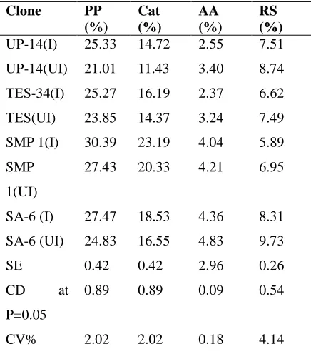
Due to pathogen infection polyphenol and catechin contents were significantly higher in infected shoots than healthy shoots but reverse trend was observed in amino acids and reducing sugars. Tea clones exhibited significantly wide variation in polyphenol content and the ranged from 30.39 to 21.01% (Table 1). Identical trend was noticed with catechins, as the total catechins are a part and parcel of polyphenols. Amino acids and reducing sugars were significantly higher in healthy leaves than infected leaves. Accumulation of certain biochemical constituents due to pathogen infection in tea leaves were reported earlier (29, 30). Reports were also available on prominent reduction in all the constituents in certain tea clones due to their susceptibility to the disease (31). The reduction in sugar content of diseased leaves might be due to the increase in the rate of utilization by the pathogen as respiratory substrate during pathogenesis (32).
Table 1.Analysis of biochemical constituents
Clone PP
(%)
Cat (%)
AA (%)
RS (%)
UP-14(I) 25.33 14.72 2.55 7.51
UP-14(UI) 21.01 11.43 3.40 8.74
TES-34(I) 25.27 16.19 2.37 6.62
TES(UI) 23.85 14.37 3.24 7.49
SMP 1(I) 30.39 23.19 4.04 5.89
SMP
1(UI)
27.43 20.33 4.21 6.95
SA-6 (I) 27.47 18.53 4.36 8.31
SA-6 (UI) 24.83 16.55 4.83 9.73
SE 0.42 0.42 2.96 0.26
CD at
P=0.05
0.89 0.89 0.09 0.54
CV% 2.02 2.02 0.18 4.14
UP- UPASI, PP-Polyphenol, Cat- Catachin, AA- Amino acid, RS- Reducing sugar
Quantitative assay of POD, APX and GR were higher in healthy shoots than infected leaves (Table 2) and two times enhanced activity was observed in resistant clones (SA-6 and SMP-1) when compared to that of susceptible tea clones (UPASI-14 and TES–34). The results indicated existence of diversity among the tested tea clones during blight stress conditions and marked variations between susceptible and resistant
clones. Higher activity of POD enzyme in the resistant cultivar may support the development of defence mechanism in the host to overcome infection. Similarly, peroxidases were associated with disease resistance/susceptibility in many crops (35, 36, 37 and 38). The key enzyme, APX enhanced to overcome the stress conditions. Peroxidase could serve as useful biochemical marker for the selection
of tea clones which resist the Exobasidum vexans
pathogen efficiently. When accumulation of
intracellular concentrations of H2O2 due to the
depression of the CAT scavenger are induced to
detoxify H2O2. Because of these conditions, sensible
clone have higher activity of CAT. However there was no trend in SOD activity.
Table 2. Antioxdative enzyme activity
Clone Pox APX GR SOD CAT
UP-14(I) 22.85 4.70 1.31 0.83 1.60
UP-14(UI) 32.31 6.19 2.42 0.46 0.87
TES-34(I) 23.44 2.73 0.89 0.50 1.35
TES(UI) 28.93 6.43 2.47 0.33 0.75
SMP 1(I) 38.29 9.22 1.96 0.82 0.83
SMP 1(UI) 45.19 11.13 2.04 0.52 0.49
SA-6 (I) 53.15 9.34 1.87 0.89 0.80
SA-6 (UI) 69.54 8.75 2.98 0.74 0.38
CD 2.96 0.11 0.28 0.02 0.06
CV 9.91 3.02 22.09 6.36 36.07
SE 1.41 0.05 0.13 0.01 0.03
*POD-Peroxidase activity (µ moles product formed/min/mg protein), APX –Ascorbate peroxidise activity (µ moles product formed /mg protein), GR- Glutathione activity (µ moles of NADPH oxidised /min/ mg protein),- SOD – Superoxide dismutase (U/ mg protein),Cat-Catalase (μmole H2O2 Oxi/min/mg
protein)
Differential response exhibited by the tea clones against leaf blister blight disease with particular reference to the expression of POX and CAT. Peroxidases are homoproteins, present as multiple isozymes in plant tissues and are distributed throughout the cell and catalyze the reduction of
H2O2 to H2O. The results of the isozyme profile
14 Changes in antioxidative enzymes and isozyme pattern due to Exobasidium vexans infection in tea cultivars
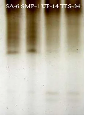
electrophoresed in a non-denaturing gel, the band intensity equates to enzyme activity.
Figure 1. Isoforms of catalase native gel
Figure 2. Isoforms of peroxidase native gel
The resistant clones expressed high intensity bands while this isoforms disappeared and seemed to be absent in susceptible clones. However, blister blight susceptible clones have faint bands at the end of regions and it was absent in resistant clones. Thus,
there is an apparent correlation of higher POD activity (intense bands) with blister blight resistance, while lower activity with susceptibility. (Fig 2).The activity of CAT and POD isoforms indicated that induction and/or increased expression of either of these in the studied clones might be associated with leaf blister blight disease resistance.
Conflicts of Interest
There are no conflicts of interest.
References
1. Hajra G. N. (2001) .Tea Cultivation.
Comprehensive Treatise. International Book Distributing Company, Lucknow .
2. Sathyanarayana, G., and Barua G.C.S.
(1983).Leaf and stem diseases of tea in north east India with reference to recent advances
in control measures, Journal of Plantation
Crops 11: 27-31.
3. Chen. Z.M. and Chen. J.F. (1990). The
Diagnosis of Tea Diseases and Their Control. [In Chinese] Shanghai science tech. Publ., Shanghal.
4. Muraleedharan,N. and Chen.Z.M. (1997).
Pests and diseases of tea and their
management. Journal of Plantation Crops,
25: 15-43.
5. Baby.U. (2001). Diseases of tea and their
management – a review, In: P.C. Trivedi,
(ed.) Plant Pathology, pp. 315-327, Pointer
Publication, Jaipur.
6. Baby.U.I, Ravichandran.R, Ganesan V,
Parthiban. B. and Sukumar, S (1998). Effect of blister blight on the biochemical and quality constituents of green leaf and CTC tea, Tropical agriculture (Trinidad) 75: 452-456.
7. Bruce, R.J. and West, C.A. (1989)
Elicitation of lignin biosynthesis and isoperoxidase activity by pectic fragments in
suspension cultures of castor bean. Plant
Physiology, 91:889–897.
8. Kleczkowska, H.E. (2001) hMSH3 and
hMSH6 interact with PCNA and colocalize with it to replication foci. Genes & Development, 15(6):724-36
9. Bowler, C. and Fluhr, R. (2005). The role of
calcium and activated oxygen as signals for
controlling cross-tolerance. Trends in Plant
Science, 10:241–6.
10. Mittler, R. (2002). Oxidative stress,
antioxidants and stress tolerance. Trends in
15 Changes in antioxidative enzymes and isozyme pattern due to Exobasidium vexans infection in tea cultivars
11. Halliwell B and Gutteridge J M C (1984).
Oxygen toxicity, oxygen radicals, transition
metals and disease. Biochemistry Journal,
219:1–14.
12. Mehdy M C (1994). Active oxygen species
in plant defense against pathogens. Plant
Physiology. 105, 467-472.
13. Gao, S., Ouyang. C., Wang, S.,Xu, Tang,L
and Chen.F (2008). Effects of salt stress on
growth, antioxidant enzyme and
phenylalanine ammonia-lyase activities in Jatropha curcas L. seedlings Plant Soil and Environment, 54, (9): 374–381.
14. Noctor G, Arisi A.C.M., Jouanin L, Valadier
M.H, Roux Y, Foyer C.H (1997). The role of
glycine in determining the rate of
glutathione synthesis in poplars. Possible implications for glutathione production
during stress. Physiologia
Plantarum.100:255–63.
15. Asada K. (1994) Production and action of
active oxygen species in photosynthetic tissues. In Foyer, c.h., and Mullineaux,P.M. (eds.),Causes of Photooxidative Stress and Amelioraton of Defense Systems in Plants, CRC Press, Boca Raton, 77-104
16. Premkumar R, Nepolean, P, Vidhya Pallavi,
R, Balamurugan A and Jayanthi, R. (2012). Integrated disease management of grey blight in tea. Two and A Bud, 59:27-30.
17. Swain T and Hillis, W.E. (1959). The
phenolic constituents of Prunus domestica.
I. The quantitative analysis of phenolic
constituents. Journal of the science of food
and agriculture, 10: 63-68.
18. Dev Choudhury. M.N and Goswami,M.R
(1983). A rapid method for the estimation of total polyphenolic matters in tea (Camellia sinensis L.), Two and A Bud 30: 59-61.
19. Moore.S and Stein.W.H (1984). In: Methods
in Enzymology, Eds. S.P. Colowick and N.D. Kaplan, Academic Press, New York.
20. Hedges.J.E. and Hofreiter. B.T (1962).
Determination of reducing sugar method in Carbohydarate Chemistry M.L. Academic Press, New York 1:388-389.
21. Chance. B and Maehly, A.C. (1955). Assay
catalases and peroxidases, Methods in Enzymology 2: 764–768.
22. Luck, H. (1974). Catalase. In Methods in
Enzymatic Analysis, éd. H.W. Bergmeyer. Academic Press, New York.
23. Giannopolitis, C.N. and Ries, S.K. (1977).
Superoxide dismutase 1. Occurrence in higher plants. Plant Physiology 59, 309–314.
24. Nakano Y and Asada K (1981). Hydrogen
peroxide is scavenged by ascorbate-specific
peroxidase in spinach chloroplasts. Plant
Cell Physiology. 22: 867-880.
25. Carlberg, I and Mannervik, B (1985).
Glutathione reductase assay. Methods in
Enzymology. Academic Press, Orlando FL., 113: 484-495.
26. Bradford M M (1976). A rapid and sensitive
method for the quantitation of microgram quantities of protein utilizing the principle of
protein-dye binding. Analytical
Biochemistry, 72: 248-254.
27. Rao M V, Paliyath G. and Ormrod, D.P
(1996). Ultra violet B and ozone induced biochemical changes in antioxidant enzymes of Arobidopsis thaliana, Plant Physiology. 110: 125-136.
28. Dhillon S.S. (1992). Dual inoculation of
pretransplant stage Oryza sativaL.plañís
with indigenous vesicular-arbuscular
mycorrhizal fungi and íluoresccnt
Pseudomonas spp. Biology and Fertility,
Soils 13:147-151.
29. Ponmurugan P, Baby U I and Raj Kumar R.
(2007). Growth, photosynthetic and
biochemical responses of tea cultivars
infected with various diseases.
Photosynthetica 45(1): 143-146.
30. Ponmurugan, P, Deepa M.A. and
Damodaran, P.N (2002). Effect of
environmental factors on lichens factors
distribution. Journal of Ecology and
Environment.147-150.
31. Gao S, Ouyang C, Wang S, Xu Y, Tang L
and Chen, F (2008). Effects of salt stress on
growth, antioxidant enzyme and
phenylalanine ammonia-lyase activities in
Jatropha curcas L. seedlings. Plant Soil
Environment, 54(9): 374–381
32. Retig N (1974). Changes in peroxidase and
polyphenol oxidase associated with natural and induced resistance of tomato to
Fusarium wilt, Physiological Plant
Pathology 4:145–50.
33. Alcazar,M D, Egea,C, Espin A and Candela
E (1995). Peroxidase isoenzymes in the defense response of Capsicum annum to
Phytophthora capsicum, Physiolgica
Plantarum, 94:736–42.
34. Melger J.C, Abney TS and Vierling, R.A
(2006). Peroxidase activity in soybeans following inoculation with Phytophthora sojae. Mycopathologia, 161:37-42.
35. Dalisay R.F. and Kuc,J.A.(1995).
16 Changes in antioxidative enzymes and isozyme pattern due to Exobasidium vexans infection in tea cultivars
Colletotrichum lagenarium into cucumber leaves with induced systemic resistance and its relation to enhanced peroxidase and
chitinase activities, Physiolgical Molecular
Plant Pathology, 47:329-38.
36. Thurman R.G, Ley H.G and Scholz Eur R.
(1972). Hepatic microsomal ethanol
oxidation. Hydrogen peroxide formation and
the role of catalase. The Journal of