Refractive Features and Diffraction Scattering Patterns
Observed in the Elastic Scattering of
12
C from
12
C at
Various Energies
Sh. Hamada1, N. Burtebayev2
1Faculty of Science, Tanta University, Tanta, Egypt 2Institute of Nuclear Physics, Almaty, Kazakhstan
Email: [email protected]
Received March 25, 2013; revised April 24, 2013; accepted May 17,2013
Copyright © 2013 Sh. Hamada, N. Burtebayev. This is an open access article distributed under the Creative Commons Attribution License, which permits unrestricted use, distribution, and reproduction in any medium, provided the original work is properly cited.
ABSTRACT
We have measured the angular distributions for 12C ion beam elastically scattered from 12C target of thickness 17.4
μg/cm2 at energies 15, 18 and 21 MeV which is close to the Coulomb barrier energy for 12C + 12C nuclear system. The
elastic scattering of 12C beam on 12C was analysed also at different energies (139.5, 158.8, 180, 240, 288.6, 300, 360
and 420 MeV) from literature in order to obtain the global optical potential parameters, which could fairly reproduce the experimental data. The experimental results were analysed within the framework of both the optical model and the dou- ble folding potential obtained with different density-dependent NN interactions which give the corresponding values of the nuclear incompressibility K in the Hartree-Fock calculation of nuclear matter. The agreement between the experi- mental results and the theoretical predictions in the whole angular range is fairly good.
Keywords: Scattering Patterns; Refractive Features; Elastic Scattering; Optical Model; Double Folding; Density
Distribution
1. Introduction
There have been rather extensive differential cross sec- tion studies for 12C + 12C nuclear system at different en-
ergies [1-7] which have been attempted to be explained theoretically by using both phenomenological and mi- croscopic potentials [6-12]. It is commonly known that
12C + 12C nuclear system is one of the key reactions for
better understanding of the formation of heavier elements in nuclear burning process. In this work, we performed the experimental measurements for 12C ion beam elasti-
cally scattered from 12C target at low energies close to
the Coulomb barrier energy for this nuclear system, and the theoretical calculations were performed using both optical model (OM) and double folding potential (DF). In comparison with the various studies concerned 12C + 12C
elastic scattering at high and intermediate energies, we found that, the experimental data at intermediate and high energies displaying the typical Fraunhofer oscilla-tions at forward angles and the symmetrization interfer- ence near 90˚, with gross structures in between which one could surmise are due to Airy maxima and minima. Our experimental data at low energies close to the
Cou-lomb barrier energy are displaying the typical Fresnel scattering pattern and the symmetrization interference near 90˚ is clearly observed as the experimental measurements were extended up to angle ~150˚.
2. Experimental Details
The experiments were performed in the cyclotron DC-60 INP NNC located at Institute of Nuclear Physics, Almaty, Kazakhstan. The 12C ion beam was accelerated up to en-
ergies 15, 18 and 21 MeV and then directed to 12Ctarget
of thickness 17.4 μg/cm2. The dead time was monitored
and kept as constant as possible by changing the spec-trometer entrance slits and/or the beam intensity. The angular distributions for 12C(12C,12C)12C nuclear system
were measured in the angular range 20˚ - 155˚ in the centre of mass system with an increment Δθ = 2˚. The registration system of nuclear reaction products included electronic components firms ORTEC and CANBERRA
of 100 μm. The detector was located at a distance of 24 cm from the scattering region and had the opportunity to move in the angular range from 10˚ to 75˚ in the labora- tory system. The maximum voltage which could be ap- plied on the detector was 30 volt but, during the experi- ment it was raised up to 20 volt. The 12C beam passed
through three collimators of 1.5 mm diameter and fo- cused on the target to a spot diameter of ≈3.9 mm. In order to minimize the evaporation of the target, the beam current was 30 nA. Spectrum analysis has been done using the program MAESTRO [13]. Figure 1 shows the
spectrum for 12C(12C,12C)12C elastic scattering at angle
35˚ and at energy 18 MeV. Final normalization of the absolute cross sections was determined by comparing the measurements at the most forward angles, where Mott scattering dominates, with optical model predictions which in this angular region are only weakly dependent on potential parameters.
3. Preparation of
12C Target and Thickness
Measurements
The 12C target was prepared using the technique of
vac-uum evaporation by resistance heating of the specimen with a spot electron gun at the facility UVS-2 (universal vacuum system) see Figure 2. The preparation chamber
was connected with two pumps rotary pump and turbo molecular pump to achieve the desired vacuum (2 × 10⁻⁶
torr) inside the chamber. Firstly, we deposited layers of NaCl crystal salt onto glass plates with the aid of tanta- lum cradle. Then, electron gun is directed to a sample of graphite with 12C concentration of 98% and placed at 3
cm from the electron gun. The size of the beam spot (2 - 3 mm diameter). Under the influence of electrons, the sample is strongly heated, evaporated, and deposited onto glass plates with previously deposited layers of salt which acts as a release agent and facilitates the removal
400 500 600 700 800 900 1000 1100 1200 1300 100
200 300 400 500 600 700 800
12
C(12
C,12
C)12
C Elastic Scattering at E=18 MeV, lab=35o
C
ount
s
Channel
[image:2.595.322.531.320.481.2]12 C Peak
Figure 1. Spectrum for 12C(12C,12C)12C elastic scattering at angle 35˚ and at energy 18 MeV.
of carbon film. After annealing for 12 hours at 150˚C, the carbon films were removed from the glass plates and placed to specially prepared frames.
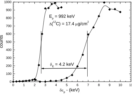
The target thickness was carried out using reactions that have narrow and well isolated resonances. For this purpose, the reaction 27Al(p,)28Si at E
p, lab. = 632, 773,
992, 1089 keV [14] was used. After the measurement of the target yield curves over the 27Al(p,γ)28Si resonance at
Ep = 992 keV for (an aluminum foil) and for (the 12C
target + the aluminum foil) is made, target thicknesses are taken from the shift of the resonance energy obtained by comparing these yield curves as shown in Figure 3,
these measurements were performed using the resonance chamber in the accelerator UKP-2-1 INP NNC RK lo-cated in Almaty, Kazakhstan.
4. Theoretical Analysis
[image:2.595.59.285.538.708.2]Our experimental results for 12C + 12C nuclear system at
Figure 2. Block diagram of the vacuum system UVS-2; K1, K2, K3: Vacuum Valve; BB: Backing Balloon; S: Manual Vacuum Gate; BP: Backing Pump; VC: vacuum chamber; CH: Vent System; DP: a high-vacuum diffusion pump.
0 1 2 3 4 5 6 7 8 9 10 1
0 100 200 300 400 500 600 700 800 900 1000
1 E
p = 992 keV
(12C) = 17.4 g/cm2
cou
nt
s
p - (keV)
E = 4.2 keV
Figure 3. The shift of the 992 keV resonance energy of the
27
[image:2.595.310.533.539.698.2]energies 15, 18 and 21 MeV and the experimental data at energies (139.5, 158.8, 180, 240, 288.6, 300, 360 and 420 MeV) from literature [4,15] were analysed (phe-nomenologically) within the frame work of optical model using Code SPI-GENOA [16] and (semi-microscopically) within the frame work of double folding using code FRESCO [17].
In the phenomenological analyses, Woods-Saxon form factor was taken for both the real and imaginary parts of the potential.
1 0 1 0 , 1 exp 1 exp C v v w wU V V iW
V V r R a
W W r R a
(1)
where, V0 and W0, av and aw, Rv and Rw being the depth,
diffuseness and radii of the real and imaginary potentials respectively, VC is the Coulomb potential. The radii are
expressed in terms of the target and projectile mass numbers At and Ap of the nuclei involved given by
1 3 1 3, ,
v w v w t p
R r A A
s
(2)
In the double folding calculations, the effective NN
interaction potential is assumed to have a separable form
where D and EX are
the direct and exchange terms, respectively, derived from the M3Y interactions [18,19], and s is the inter-nucleon separation;
DEX , DEX ,
v s F v v v
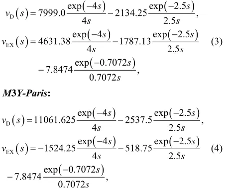
is the density of the surrounding nuclear medium in which the two nucleons are embedded. The radial shape of the M3Y-Paris interaction [18] used in the present folding calculation is given in terms of three Yu-kawas. The original M3Y interaction is density inde-pendent and given in terms of the Yukawa functions as follows:
M3Y-Reid:
D
EX
exp 4 exp 2.5
7999.0 2134.25 ,
4
exp 4 exp 2.5
4631.38 1787.13 4 2 exp 0.7072 7.8474 , 0.7072 2.5 .5 s s v s s s s s v s s s s s (3)
M3Y-Paris:
D
EX
exp 4 exp 2.5
11061.625 2537.5 ,
4 2
exp 4 exp 2.5
1524.25 518.75 4 2 exp 0.7072 7.8474 , 0.7072 .5 .5 s s v s s s s s v s s s s s (4)
These interactions, especially the M3Y-Reid version, have been used with some success in the double folding
model calculation of the HI optical potential at low ener- gies [20], with the elastic data usually limited to the for- ward scattering angles and, thus, sensitive to the optical potential (OP) only at the surface. However, in cases of refractive (rainbow) nucleus–nucleus scattering where the elastic data are sensitive to the nucleus-nucleus OP over a much wider radial domain, the density-independ- ent M3Y interactions failed to give a good description of the data. The inclusion of explicit density dependence was needed to account for a reduction in the strength of the nucleus-nucleus interaction that occurs at small R
where the overlap density of the nuclear collision in-creases. An early version of the density dependence of the M3Y-Reid interaction was constructed by Kobos et al. [21] based upon the G-matrix results obtained by Jeukenne et al. [22]. It was dubbed as the DDM3Y in-teraction and has been used to improve the folding model description of the elastic α-nucleus [22,23] and light HI [24] scattering. Hartree-Fock (HF) calculation of the nu- clear matter (NM) energy has shown that the original density-independent M3Y interaction Equations (3) and (4) failed to saturate NM, leading to a collapse. The HF method is the first order of nuclear many-body calcula-tion and the introduccalcula-tion of a density dependence into the original M3Y interaction accounts, therefore, for higher order NN correlations which lead to the NM saturation. Several versions of the density dependence of the M3Y- Reid and M3Y-Paris interactions have been introduced by scaling them with an explicit density-dependent func-tion F(ρ). In our present work the density-dependent function F(ρ) was taken in the following form:
1 exp
nF C , (5)
The parameters C,α,β,γand n given in Table 1 were
taken from ref. [25].
The nuclear density distribution for 12C was calculated
using Three-parameter Fermi model (3PF), where ρ(r) was calculated using the following formula
2
0 1 2 1 exp wr
r r
c
c z
where w = –0.149, Z = 0.5224 and c = 2.355.
5. Theoretical Analysis
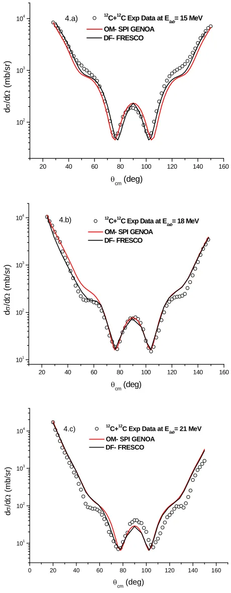
[image:3.595.64.291.514.703.2]The comparison between the experimental data and the theoretical predictions using both optical potential and double folding at energies 15, 18 and 21 MeV is shown in Figure 4. In the case of double folding calculations,
Table 1. Parameters of density dependence function F(ρ).
Model c α β(fm3) γ(fm3n) n K(MeV)
20 40 60 80 100 120 140 160 102
103
104 12
C+12C Exp Dataat Elab= 15 MeV OM- SPI GENOA
DF- FRESCO
d
/d
(mb/
sr)
cm (deg)
4.a)
20 40 60 80 100 120 140 160
101 102
103 104
4.b) 12 C+12
C Exp Dataat Elab= 18 MeV OM- SPI GENOA
DF- FRESCO
d
/d
(mb/s
r)
cm (deg)
0 20 40 60 80 100 120 140 160
101
102 103
104 4.c) 12C+12C Exp Dataat Elab= 21 MeV OM- SPI GENOA
DF- FRESCO
d
/d
(m
b/sr)
[image:4.595.57.287.86.675.2] cm (deg)
Figure 4. Comparison between experimental and calculated differential cross section within the framework of OM (SPI-GENOA) code and DF (FRESCO) code, for 12C elasti-cally scattered by 12C at energies 15, 18 and 21 MeV respec-tively.
the normalization factor (Nr) equals 1.2. The optical po-tential parameters for 12C + 12C nuclear system and also
those from double folding potential at different energies are listed in Table 2. The Coulomb radius parameter rc
was fixed at 0.95 fm, the radius parameter for the real part of the potential was fixed at 1.225 fm, the diffuse-ness parameter for the real part of the potential was fixed at 0.44 fm. While, the radius parameter for the imaginary part of the potential used in the optical model calcula-tions was fixed at 1.294 fm.
In identical particles elastic scattering such as 12C + 12C, due to symmetry under the interchange of spatial
coordinates of the two particles, the differential cross section is given by
2identical
d π
d f f
.
where f
fC
fN
,
π
C
π
N
π
f f f , with fC is the Cou-
lomb scattering amplitude and fN is the nuclear
scat-tering amplitude. As shown in our experimental results, the total symmetry between cross sections in forward and backward hemispheres tells that the experiments were done correctly.
The comparison between the experimental data and the theoretical predictions using both optical potential and double folding at energies 139.5, 158.8, 180, 240, 288.6, 300, 360 and 420 MeV is shown in Figures 5 and 6. The
optical potential parameters for 12C + 12C nuclear system
and also those from double folding potential at energies (139.5 - 420 MeV) are listed in Table 3.
The Coulomb radius parameter rc was fixed at 0.95 fm,
the radius parameter for the real part of the potential was
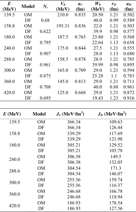
Table 2. The optimal potential parameters or 12C elastically scattered on 12C at energies 15, 18 and 21 MeV, R
1 3 1 3
0 t p
r A A .
E (MeV) Model Nr V0 (MeV) W0 (MeV) rW (fm) aw(fm)
15.0 OM DF 1.2
99.93 20.54 40.0
1.294 1.0
0.292 0.532 18.0 OM
DF 1.2
92.98 26.92 40.0
1.294 1.2
0.3 0.765 21.0 OM
DF 1.2
85.98 29.64 40.0
1.294 1.1
0.292 0.4
E (MeV) Model JV(MeV·fm3) JW(MeV·fm3)
15.0 OM
DF
544.7 544.7
127.25 126.58
18.0 OM
DF
528.07 528.07
198.02 182.53
21.0 OM
DF
468.54 468.54
[image:4.595.307.537.547.729.2]Table 3. The optimal potential parameters for 12C elastically scattered on 12C at different energies,
1 3 1 3
0 t p
R r A A . E
(MeV) Model Nr
V0 (MeV) aV (fm) W0 (MeV) rW (fm) aW (fm) 139.5 OM
DF 0.68
210.0 0.835 20.50 40.0
1.21 0.99
0.502 0.589 158.8 OM
DF 0.622
195.31 0.836 22.0 39.9
1.21 0.98
0.503 0.577 180.0 OM
DF 0.795
187.5 0.765 23.80 22.64
1.21 1.13
0.568 0.658 240.0 OM
DF 0.907
175.0 0.844 27.5 28.0
1.21 1.13
0.555 0.680 288.6 OM
DF 0.961 158.5 0.878 39.99 28.9 1.210.98 0.7850.895 300.0 OM
DF 0.875
165.0 0.709 29.0 25.28
1.21 1.1
0.594 0.783 360.0 OM
DF 0.708
145.0 0.813 29.0 40.0
1.21 0.88
0.711 0.961 420.0 OM
DF 0.695
125.0 0.668 29.0 19.43
1.21 1.23
0.872 0.916
E (MeV) Model JV(MeV·fm3) JW(MeV·fm3) 139.5 OM
DF
366.34 366.34
109.63 126.44 158.8 OM
DF
339.29 339.29
117.69 121.98 180.0 OM
DF
305.21 305.21
129.52 105.78 240.0 OM DF 306.38 306.38 132.05 149.5
288.6 OM DF 304.54 304.54 146.07 171.3
300.0 OM DF 255.56 255.56 159.74 116.37
360.0 OM DF 246.68 246.68 166.78 118.94
420.0 OM DF 186.93 186.93 178.54 127.36
fixed at 0.726 fm. At small angles, the cross section is dominated by diffraction and exhibits a typical Fraun-hofer diffraction pattern. Beyond this region the cross section exhibits a structureless exponential falloff. Dif-ferent experiments showed that, there is no evidence of rainbow scattering could be seen in the 12C + 12C system
studied at low incident energy [26]. At energies 140 and 159 MeV, the differential cross sections were measured up to 90˚, and it is possible to guess at the presence of broad Airy maxima (centred at ≈50˚ at 159 MeV and at ≈
62˚ at 140 MeV), in between the Fraunhofer pattern at forward angles and the oscillations near 90˚ due to sym-metrization. While the data at 240 and 289 MeV didn’t extend far enough to display the full extent of a nuclear rainbow, the measurements at 360 MeV show the begin-ning of the exponential falloff of the nuclear rainbow, distinctly evident in the angular distribution beyond θcm ≈
25˚. As E/A approaches 100 MeV and higher, the rain-bow moves forward faster than the Fraunhofer oscilla-tions, and the visual identification of its Airy minima in the angular distribution is less clear.
0 20 40 60 80 100
10-8 10-7 10-6 10-5 10-4 10-3 10-2 10-1 100 101 102 103 104
Elab= 240 MeV x 10
-6
Elab= 180 MeV x 10
-4
Elab= 158.8 MeV x 10-2
Elab= 139.5 MeV Exp Data DF- FRESCO OM SPI-GENOA d /d (mb /sr)
[image:5.595.59.284.119.470.2]cm (deg)
Figure 5. Comparison between experimental and calculated differential cross section for 12C + 12C elastic scattering at energies 139.5, 158.8, 180 and 240 MeV using both OM and DF potentials.
0 5 10 15 20 25 30 35 40
10-6 10-5 10-4 10-3 10-2 10-1 100 101 102 103 104 105
Elab= 420 MeV x 10-6
Elab= 360 MeV x 10-4
Elab= 300 MeV x 10-2
Elab= 288.6 MeV Exp Data DF- FRESCO OM- SPI-GENOA d /d (mb /sr)
cm (deg)
Figure 6. Comparison between experimental and calculated differential cross section for 12C + 12C elastic scattering at energies 288.6, 300, 360 and 420 MeV using both OM and DF potentials.
It has been found that, at incident energies of a few MeV per nucleon, several light heavy-ion systems, among which 12C + 12C display more transparency than
[image:5.595.308.533.319.492.2]observa-tion in the elastic scattering data of distinctive refractive effects, like strong Airy minima, superimposed on more classic diffractive features. This refractive behavior is clear in the systematic analyses carried out for the 16O + 16O system by Nicoli et al. [28] at incident energies
be-tween 75 and 124 MeV and by Khoa et al. [29] between 124 and 1120 MeV, and for the 12C + 16O system by
Nicoli et al. [30] between 62 and 124 MeV and by Ogloblin et al. [31] at 132 MeV. In particular, we want to clarify the transition between the region of relatively high incident energies where rainbow scattering has set in and lower energies close to Coulomb barrier energy where rainbow scattering is not yet observed. In our pre-sent work, it is clearly shown that, refractive features such as; nuclear rainbow phenomenon is not observed in
12C + 12C nuclear system at low energies close to
Cou-lomb barrier energy. The optical model analysis for this nuclear system at low energies near the Coulomb barrier energy doesn’t require deep real potential as at high en- ergies.
6. Summary
The angular distributions for 12C ion beam elastically
scattered by 12C at energies 15, 18 and 21 MeV have
been measured at the cyclotron DC-60. At these energies which are close to the Coulomb barrier energy for 12C + 12C nuclear system (V
CB = 17.44 MeV), the 12C + 12C
nuclear system doesn’t show any refractive features such as rainbow and Airy minimum which is clearly observed for this nuclear system at high energies and the optical model analysis requires deep real potential and shallow imaginary potential. At low energies close to Coulomb barrier energy, rainbow scattering is not observed and the optical model analysis at such low energies doesn’t re- quire deep real potential.
The experimental data were analysed within the frame work of both optical model and double folding. The ob-tained normalization coefficient Nr equals 1.2. The
ex-perimental data showed the typical Fresnel scattering pattern and the symmetrization interference near 90˚ is clearly observed, while at high energies, the 12C + 12C
nuclear system displaying typical Fraunhofer scattering pattern.
REFERENCES
[1] S. M. Lenzi, A. Vitturi and F. Zardi, Nuclear Physics A, Vol. 536, 1992, pp. 168-178.
doi:10.1016/0375-9474(92)90252-F
[2] S. M. Lenzi, A. Vitturi and F. Zardi, Physical Review C, Vol. 40, 1989, pp. 2114-2123.
doi:10.1103/PhysRevC.40.2114
[3] J. Y. Hostachy, et al., Nuclear Physics A, Vol. 490, 1988, 441-470. doi:10.1016/0375-9474(88)90514-3
[4] C.-C. Sahm, T. Murakami, J. G. Cramer, A. J. Lazzarini, D. D. Leach, D. R. Tieger , R. A. Loveman, W. G. Lynch, M. B. Tsang and J. Van der Plicht, Physical Review C, Vol. 34, 1986, pp. 2165-2170.
doi:10.1103/PhysRevC.34.2165
[5] M. E. Brandan, Physical Review Letters, Vol. 60, 1988, pp. 784-787. doi:10.1103/PhysRevLett.60.784
[6] H. Emling, R. Nowotny, D. Pelte, G. Schrieder and W. Weidenmeier, Nuclear Physics A, Vol. 239, 1975, pp. 172-188. doi:10.1016/0375-9474(75)91141-0
[7] W. Treu, H. Fröhlich, W. Galster, P. Dück and H. Voit, Physical Review C, Vol. 22, 1980, pp. 2462-2464.
doi:10.1103/PhysRevC.22.2462
[8] I. Boztosun and W. D. M. Rae, Physical Review C, Vol. 63, 2001, Article ID: 054607.
doi:10.1103/PhysRevC.63.054607
[9] F. Michel and S. Ohkubo, The European Physical Jour- nal A, Vol. 19, 2004, pp. 333-339.
doi:10.1140/epja/i2003-10133-0
[10] M. Freer, M. P. Nicoli, S. M. Singer, C. A. Bremner, S. P. G. Chappell, W. D. M. Rae, I. Boztosun, B. R. Fulton, D. L. Watson, B. J. Greenhalgh, G. K. Dillon, R. L. Cowin and D. C. Weisser, Physical Review C, Vol. 70, 2004, Ar- ticle ID: 064311. doi:10.1103/PhysRevC.70.064311
[11] D. L. Watson, B. J. Greenhalgh, G. K. Dillon, R. L. Cow- in and D. C. Weisser, Physical Review C, Vol. 70, 2004, Article ID: 064311. doi:10.1103/PhysRevC.70.064311
[12] C. A. Bremner, S. P. G. Chappell, W. D. M. Rae, I. Boz- tosun, M. Freer, M. P. Nicoli, S. M. Singer, B. Fulton, D. L. Wat-son, B. J. Greenhalgh, G. K. Dillon and R. L. Co- win, Physical Review C, Vol. 66, 2002, Article ID: 034 605. doi:10.1103/PhysRevC.66.034605
[13] “MAESTRO®-32 MCA Emulator for Microsoft® Win-
dows® 98, 2000, NT®, and XP® A65-B32 Software User’s
Manual Software Version 6.”
[14] J. W. Bulter, US Naval Research Laboratory, “NRL Re-port 5282,” 1959.
[15] M. E. Brandan, Physical Review Letters, Vol. 60, 1988, pp. 784-787. doi:10.1103/PhysRevLett.60.784
[16] F. Perey, “SPI-GENOA: An Optical Model Code,” Un- published, 1975.
[17] I. J. Thompson, Computer Physics Reports, Vol. 7, 1988. [18] G. Bertsch, J. Borysowicz, H. McManus and W. G. Love,
Nuclear Physics A, Vol. 284, 1977, pp. 399-419.
doi:10.1016/0375-9474(77)90392-X
[19] N. Anantaraman, H. Toki, G. F. Bertsch, Nuclear Physics A, Vol. 398, 1983, pp. 269-278.
doi:10.1016/0375-9474(83)90487-6
[20] G. R. Satchler, W. G. Love, Physics Reports, Vol. 55, 1979, pp. 183-254. doi:10.1016/0370-1573(79)90081-4
[21] A. M. Kobos, B. A. Brown, P. E. Hodgson, G. R. Satchler and A. Budzanowski, Nuclear Physics A, Vol. 384, 1982, pp. 65-87. doi:10.1016/0375-9474(82)90305-0
[22] J. P. Jeukenne, A. Lejeune and C. Mahaux, Physical Re- view C, Vol. 16, 1977, pp. 80-96.
doi:10.1103/PhysRevC.16.80
chler, Nuclear Physics A, Vol. 425, 1984, pp. 205-232.
doi:10.1016/0375-9474(84)90073-3
[24] M. E. Brandan and G. R. Satchler, Nuclear Physics A, Vol. 487, 1988, pp. 477-492.
doi:10.1016/0375-9474(88)90625-2
[25] Dao T. Khoa, W. von Oertzen, H. G. Bohlen and S. Oh-kubo,Journal of Physics G: Nuclear and Particle Physics, Vol. 34, 2007, pp. R111-R116.
[26] R. M. Wieland, R. G. Stokstad, G. R. Satchler and L. D. Rickertsen, Physical Review Letters, Vol. 37, 1976, pp. 1458-1461. doi:10.1103/PhysRevLett.37.1458
[27] D. A. Goldberg and S. M. Smith, Physical Review Letters, Vol. 33, 1974, pp. 715-718.
doi:10.1103/PhysRevLett.33.715
[28] M. P. Nicoli, F. Haas, R. M. Freeman, N. Aissaoui, C. Beck, A. Elanique, R. Nouicer, A. Morsad, S. Szilner, Z.
Basrak, M. E. Brandan and G. R. Satchler, Physical Re- view C, Vol. 60, 1999, Article ID: 064608.
doi:10.1103/PhysRevC.60.064608
[29] D. T. Khoa, W. von Oertzen, H. G. Bohlen and F. Nuof-fer, Nuclear Physics A, Vol. 672, 2000, pp. 387-416. [30] M. P. Nicoli, F. Haas, R. M. Freeman, S. Szilner, Z. Bas-
rak, A. Morsad, G. R. Satchler and M. E. Brandan, Phy- sical Review C, Vol. 61, 2000, Article ID: 034609.
doi:10.1103/PhysRevC.61.034609