Back to the future? The fallopian tube, precursor escape and a dualistic model of high-grade serous carcinogenesis
Thing R. Soong, MD, PhD, MPH 1
David L. Kolin, MD, PhD 2
Nathan J. Teschan, DO 2
Christopher P Crum, MD 2
1 Department of Pathology, University of Washington Medical Center, Seattle, WA
2 Department of Pathology, Division of Women's and Perinatal Pathology, Brigham and Women's
Hospital, Boston, MA 02115
Address correspondence to Christopher P Crum, MD, Department of Pathology, Brigham and Women's Hospital, 75 Francis Street, Boston MA 02115
Tel: 617-732-5481
FAX: 617-264-5125
Email: [email protected]
Abstract
Beginning with the discovery of the BRCA ovarian cancer susceptibility genes and subsequent detailed examination of risk reduction salpingo-oophorectomy (RRSO) specimens, a new paradigm of ovarian carcinogenesis has unfolded with attention to the distal fallopian tube. The primary focus has been an early cancer in the fallopian tube which is seen in virtually all incidentally discovered high-grade serous cancers in asymptomatic women. This high-frequency of tubal involvement in early serous cancer - serous tubal intraepithelial carcinoma or STIC - has galvanized attention to this organ as a primary source of this disease. However, an enduring mystery has been the relatively low frequency of STIC in fallopian tubes of women with advanced malignancy. This paradox, a high-frequency of tubal
involvement early and a low-frequency late in the disease process has spurred interest in other potential sources, such as the ovarian surface or secondary Mullerian system. However, because essentially all high-grade serous carcinomas are linked by TP53 mutations, and because fallopian tubes frequently contain early serous proliferations (ESPs) with these mutations, attention has turned to the possibility that nonmalignant but TP53 mutated tubal epithelium could be responsible for an eventual malignancy. Recent data have shown evidence of lineage continuity between ESPs and concurrent serous carcinomas prompting the concept of "precursor escape". This creates a 2nd component of the paradigm by which cells from early lesions can escape the fallopian tube and undergo future malignant transformation later, emerging suddenly as widespread malignancy. This dualistic model thus explains the paradox and opens new questions pertaining to the challenge of both early detection and prevention of this lethal malignancy.
The past
difficulty in detecting the tumor at a curable stage, combined with eventual resistance to chemotherapy. 1 Unlike endometrial and cervical malignancies, which are preceded by recognizable precursor lesions in
the respective site, the majority of ovarian carcinomas – high-grade serous carcinomas (HGSC) – have been assigned to the ovary by default. Systems for estimating origin have been primarily dependent upon location of tumor rather than identification of a specific carcinogenic sequence.2 Because most of these carcinomas do involve the ovarian surface, an origin in the ovarian surface epithelium was
presumed.2 For those tumors that did not involve the ovarian surface or demonstrated scant evidence of ovarian involvement a source in the peritoneal cavity was presumed. These so-called "primary
peritoneal carcinomas" were generally assumed to arise within epithelial rests such as endosalpingiosis, endometriosis or other components of the "secondary Mullerian system". 2
The paradigm shift
Several unrelated but temporally linked observations facilitated the emergence of a potential new source for HGSC. The first was the discovery of the BRCA cancer susceptibility genes.3 The ability to identify patients at risk by testing for germline BRCA1 and BRCA2 mutations accelerated the adoptions of risk reduction salpingo-oophorectomy. This in turn increased the likelihood that early cancers would be discovered in the ovary or fallopian tubes. A second was a concurrent study that underscored the rarity of early HGSC of the ovary.4 A third was the progressive realization that serologic screening or ultrasound demonstrated very little efficacy in detecting these HGSCs at a curable stage. 5
fallopian tube, where the majority of early malignancies were being found.9 This was followed by studies of earlier precursor lesions in the fallopian tube ranging from small stretches of epithelium with TP53 mutations and proliferations termed serous tubal intraepithelial lesions.7 1011 Based on these
observations a serous carcinogenic sequence was assembled in the distal tube which began with a p53 signature and terminated in STIC, with serous tubal intraepithelial lesions signifying a transition.
Application of the SEE-FIM protocols to carefully examine the tubes of women at risk for HGSC accelerated the percentage of early cancers attributed to the distal tube, approaching 100% in some studies.12 The tubal theory of high-grade serous carcinogenesis was thus superimposed upon the prior literature and like most new models began as a simple paradigm in which precursor to cancer evolution occurred in the tube, followed by dissemination to the peritoneal surfaces.13 This explained the rather rapid emergence of a malignancy which began as an occult carcinoma in the fallopian tube and then rapidly became advanced once the tumor is disseminated to the peritoneum.
Unanswered questions
The above serous carcinogenic model required transition from precursor to cancer in the fallopian tube, which led investigators to multiple conclusions. The first was the assumption that the metastatic
carcinoma was launched from a primary malignancy in the fallopian tube. Encouraging this were
observations that up to 75% or more of HGSC involved the fallopian tube in some manner.14 This has led to consensus opinions concluding - based on circumstantial evidence - that any significant tubal
salpingectomy should be practice whenever possible to reduce the risk of eventual HGSC.16
Salpingectomy alone was also proposed for managing women with BRCA mutations. The latter strategy is currently under study, but with careful guidelines to minimize risk to the patients who insist on and interval of ovarian preservation.17
A second and entirely different viewpoint coming from this work was informed by critical pathologic observation. Based on multiple studies, the frequency of an intramucosal carcinoma in the fallopian tube in patients with a symptomatic or advanced HGSC ranged from as low as 10% to as high as 60%.18 Either estimate left a large percentage of HGSC in which a clear-cut early malignancy in the fallopian tube could not be identified. Proponents of other potential sites of origin pointed to the ovarian surface epithelium or the secondary Mullerian system.192021These proposals did not exclude the fallopian tube as the ultimate source of the tumor but theorized that benign cells derived from either the fallopian tubes or embryonic Mullerian rests (endosalpingiosis) in the pelvis (second Mullerian system) could undergo malignant transformation and explain the absence of a concurrent serous tubal intraepithelial carcinoma. Even STICs were viewed with suspicion as launch pads for HGSC when studies revealed that STICs could be secondary deposits from a widespread cancer rather than a primary site.22
associated with a STIC.10 What these studies did show was that the overall frequency of a STIC carcinoma in cases of advanced serous cancer cases from women with BRCA mutations individual was low, further emphasizing the likelihood that another pathway must be considered.25 Another piece of information suggesting more than one mechanism of carcinogenesis was the similarity in mean age between women with BRCA mutations who presented with isolated serous tubal intraepithelial carcinoma and symptomatic widespread malignancy.26 Given the assumption that there must be a lag period between the onset of early cancer and later disseminated malignancy, the similarities in age between early and advanced BRCA-mutation associated HGSC was at odds with the traditional
precursor- cancer models, such as seen in the uterine cervix.27 Finally, molecular genetic studies of HGSC with and without an associated STIC failed to show any noticeable difference between the two groups.28 In all of these tumors a TP53 mutation was a requirement and in contrast to the fallopian tube, there was no evidence of early or occult TP53 mutations within alternate sites of origin, including
endosalpingosis, endometriosis, or ovarian inclusion cysts.
In summary it was clear that a very high percentage of early serous cancers discovered in asymptomatic women arose from within the fallopian tube. At the same time a rather low percentage of advanced cancers were associated with a tubal malignancy. However, both cancer groups were genetically similar and there was no compelling evidence for an alternate origin containing a cancer precursor with a TP53 mutation. This raised the fundamental question of how the fallopian tube could be involved in the development of HGSC in the absence of a STIC.
Serous cancer precursors in the fallopian tube
fallopian tubes. In one study, fallopian tubes from 50% of controls and 70% of women with BRCA mutations contained at least one small stretch of 12 or more cells with a TP53 mutation.29 The small foci - p53 signatures - like their malignant counterparts localized to the distal fallopian tube, occurred in non-ciliated cells, and were associated with evidence of DNA damage in the form of foci of gamma H2AX immunostaining.30 This and other studies showed examples where these early lesions could be found in continuity with intraepithelial carcinomas or occasionally malignancies in the fallopian tube.31 Given the frequency of these lesions, they were presumed to be at very low risk for giving rise to an eventual serous cancer. Proliferative lesions, termed either tubal intraepithelial lesions in transition or serous tubal intraepithelial lesions (STIL) were less common and were considered an intermediate step
between p53 signature and STIC. 32 For the purposes of this discussion, this spectrum of tubal precursors containing Tp53 mutations will be designated early serous proliferations or ESPs.33
Early serous proliferations and "precursor escape"
Until recently, ESPs were not considered to play a major role in the development of HGSC. Given that they are found in up to 50% of the general population and in view of the low risk of HGSC, the overall risk for an individual with one of these lesions would seem to be very low. Moreover, is well known that women with BRCA mutations who present with an asymptomatic STIC carry only an approximately 5% risk of ever developing a metastatic serous cancer.34 Nevertheless the only recognizable precursor lesion to date with a TP53 mutation has been found in the fallopian tube.
was that in 12 cases, ESPs were identified and in 9 of those 12 cases a TP53 mutation was recovered that was identical to the metastatic serous carcinoma. This study, for the first time, provided evidence of lineage continuity between ESPs and widespread HGSCs in the absence of a recognizable STIC or other early malignancy.32 Based on this it appears that a small or historically early genetic lesion in the fallopian tube could be ultimately responsible for a later emergence of widespread serous carcinoma.
De-mystifying a paradox
The possibility that some early serous proliferations escape the fallopian tube and ultimately emerge as a widespread
intra-abdominal malignancy is a potential key to unlocking the decades old mystery of disparate and
contrasting presentations of HGSC, one with and another without a coexisting serous tubal intraepithelial carcinoma. In the classic scenario, a tubal intraepithelial carcinoma or early cancer develops in the distal
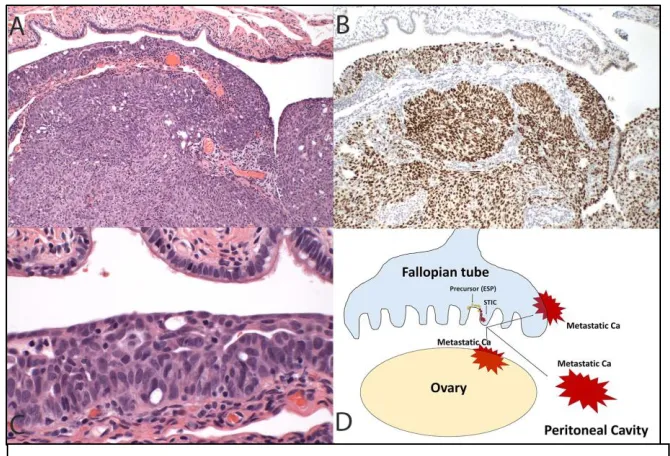
fallopian tube and over time extends to the ovary or local peritoneal surfaces, after which the tumor becomes more widespread (Figure 1). Alternatively, these tumors might invade and then spread directly to lymph nodes, which we have seen in tubal carcinomas. Still another possibility is what is seen in
Figure 1. In this more conventional model of high-grade serous cancer development, the tumor initiates in the fallopian tube (A) and
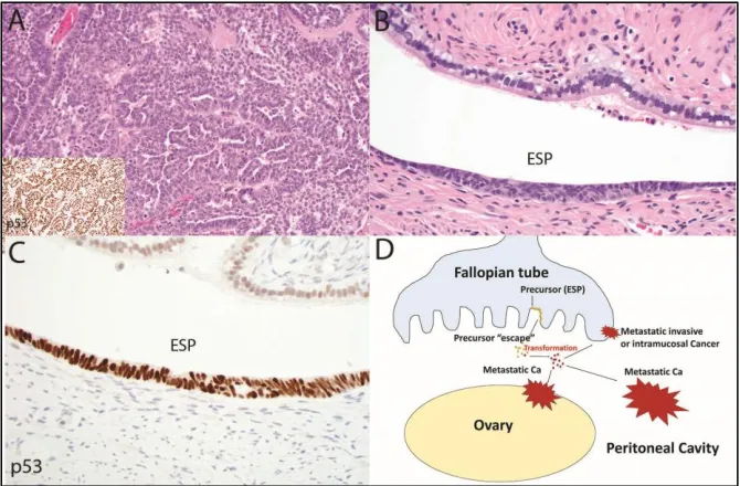
women with BRCA mutations who present with isolated serous tubal intraepithelial carcinomas. Approximately one in 20 of these patients will eventually manifest with a metastatic HGSC. This speaks to the fact that many STICs do not possess sufficient biologic potential to culminate in metastatic HGSC. The scenario which involves precursor escape and eventual intra-abdominal malignancy is very similar then to the latter pathway, the only difference being that the initiating lesion of record will not be recognized as a STIC (Figure 2). This may appear counterintuitive; however, the potential for
nonmalignant but genetically altered Mullerian epithelium to exist suspended in the peritoneal fluid or to travel from one site to
another is plausible, particularly when one considers the
pathogenesis of endometriosis.35
Unanswered questions
The promise of
"precursor escape" or a similar mechanism is the possibility that it will settle the question of HGSC origin. If precursor escape is an important mechanism of serous
carcinogenesis, other existing models must be more critically reevaluated. Neither the ovarian surface epithelium/cortical inclusion cyst nor the secondary Mullerian system model is strongly supported by a
recognizable precursor with a TP53 mutation. Moreover, it can be argued that if serous tubal
intraepithelial carcinomas and early serous proliferations in the tube occasionally possess the capability to persist and reemerged as metastatic HGSC, it is entirely conceivable that less conspicuous but genetically similar lesions could contribute to this disease. This possibility must be excluded by rigorous pathologic and molecular analysis of fallopian tubes of women with HGSC to establish the extent of lineage relationship between these early proliferations and advanced malignancy. If proven, this association will certainly inform future strategies for serous cancer prevention, both in the general population and women at high risk for this disease. In the process of this research a more complete understanding of the biologic events taking place during the occult phase of serous cancer development could come to light.
Bibliography
1 Cannistra SA. Cancer of the ovary. N Engl J Med. 2004;351:2519-29. Erratum in: N Engl J Med. 2005 Jan
6;352(1):104
2 Scully RE, Yo7ung RH, Clement PB. Atlas of Tumor Pathology; Tumors of the ovary, maldeveloped gonads,
fallopian tube and broad ligament. American Registry of Pathology, AFIP, 1998, Bethesda MD, p51
3 Friedman LS, Ostermeyer EA, Szabo CI, et al. Confirmation of BRCA1 by analysis of germline mutations linked to
breast and ovarian cancer in ten families. Nat Genet. 1994;8:399-404
4 Bell DA, Scully RE. Early de novo ovarian carcinoma. A study of fourteen cases. Cancer. 1994;73:1859-64
5 Henderson JT, Webber EM, Sawaya GF. Screening for Ovarian Cancer: An Updated Evidence Review for the U.S.
Preventive Services Task Force [Internet]. Rockville (MD): Agency for Healthcare Research and Quality (US); 2018
Feb. Report No.: 17-05231-EF-1. U.S. Preventive Services Task Force Evidence Syntheses, formerly Systematic
Evidence Reviews.
6 Zweemer RP, van Diest PJ, Verheijen RH, et al. Molecular evidence linking primary cancer of the fallopian tube to
7 Piek JM, van Diest PJ, Zweemer RP, et al. Dysplastic changes in prophylactically removed Fallopian tubes of
women predisposed to developing ovarian cancer. J Pathol 2001;195:451-6
8 Colgan TJ. Challenges in the early diagnosis and staging of Fallopian-tube carcinomas associated with BRCA
mutations. Int J Gynecol Pathol. 2003;22:109-20. Review
9 Medeiros F, Muto MG, Lee Y, et al. The tubal fimbria is a preferred site for early adenocarcinoma in women with
familial ovarian cancer syndrome. Am J Surg Pathol 2006; 30: 230–236
10 Howitt BE, Hanamornroongruang S, Lin DI, et al. Evidence for a dualistic model of high-grade serous carcinoma:
BRCA mutation status, histology, and tubal intraepithelial carcinoma. Am J Surg Pathol 2015; 39: 287–93.
11 Jarboe EA, Pizer ES, Miron A, et al. Evidence for a latent precursor (p53 signature) that may precede serous
endometrial intraepithelial carcinoma. Mod Pathol 2009;22:345-50.
12Callahan MJ, Crum CP, Medeiros F, et al. Primary fallopian tube malignancies in BRCA-positive women
undergoing surgery for ovarian cancer risk reduction. J Clin Oncol. 2007;25:3985-90
13 Vang R, Shih IeM, Kurman RJ. Fallopian tube precursors of ovarian low- and high-grade serous neoplasms.
Histopathology. 2013;62:44-58
14 Kindelberger DW, Lee Y, Miron A, et al. Intraepithelial carcinoma of the fimbria and pelvic serous carcinoma:
Evidence for a causal relationship. Am J Surg Pathol. 2007;31:161-9
15 Singh N, Gilks CB, Hirschowitz L, et al. Primary site assignment in tubo-ovarian high-grade serous carcinoma:
Consensus statement on unifying practice worldwide. Gynecol Oncol 2016; 141: 195–198
16 Gockley AA, Elias KM. Fallopian tube tumorigenesis and clinical implications for ovarian cancer risk-reduction.
Cancer Treat Rev. 2018;69:66-71
17 Nebgen DR, Hurteau J, Holman LL, et al. Bilateral salpingectomy with delayed oophorectomy for ovarian cancer
risk reduction: A pilot study in women with BRCA1/2 mutations. Gynecol Oncol. 2018;150:79-84.
18 Chen F, Gaitskell K, Garcia MJ, et al. Serous tubal intraepithelial carcinomas associated with high-grade serous
ovarian carcinomas: a systematic review. Brit J Obstet Gynecol 2017; 124: 872–878
19 Kim J, Coffey DM, Ma L, et al. The ovary is an alternative site of origin for high-grade serous ovarian cancer in
20 Auersperg N. Ovarian surface epithelium as a source of ovarian cancers: unwarranted speculation or
evidence-based hypothesis? Gynecol Oncol. 2013;130:246-51.
21 Dubeau L. The cell of origin of ovarian epithelial tumours. Lancet Oncol. 2008;9:1191-7.
22 McDaniel AS, Stall JN, Hovelson DH et al. Next-generation sequencing of tubal intraepithelial carcinomas. JAMA
Oncol 2015; 1: 1128–1132.
23 Folkins AK, Jarboe EA, Saleemuddin A, et al. A candidate precursor to pelvic serous cancer (p53 signature) and its
prevalence in ovaries and fallopian tubes from women with BRCA mutations. Gynecol Oncol. 2008;109:168-73
24 Barakat RR1, Federici MG, Saigo PE, et al. Absence of premalignant histologic, molecular, or cell biologic
alterations in prophylactic oophorectomy specimens from BRCA1 heterozygotes. Cancer. 2000;89:383-90.
25 Howitt BE, Hanamornroongruang S, Lin DI, et al. Evidence for a dualistic model of high-grade serous carcinoma:
BRCA mutation status, histology, and tubal intraepithelial carcinoma. Am J Surg Pathol. 2015;39:287-93.
26 Kotsopoulos J, Gronwald J, Karlan B, et al.; Hereditary Ovarian Cancer Clinical Study Group. Age-specific ovarian
cancer risks among women with a BRCA1 or BRCA2 mutation. Gynecol Oncol. 2018;150:85-91
27 Brown PO, Palmer C. The preclinical natural history of serous ovarian cancer: defining the target for early
detection. PLoS Med 2009; 6: e1000114
28 Ducie J, Dao F, Considine M, et al. Molecular analysis of high-grade serous ovarian carcinoma with and without
associated serous tubal intra-epithelial carcinoma. Nat Commun. 2017;8:990
29 Mehra KK, Chang MC, Folkins AK, et al. The impact of tissue block sampling on the detection of p53 signatures in
fallopian tubes from women with BRCA 1 or 2 mutations (BRCA+) and controls. Mod Pathol 2011; 24: 152–156.
30 Lee Y, Miron A, Drapkin R, et al. A candidate precursor to serous carcinoma that originates in the distal fallopian
tube. J Pathol. 2007;211:26-35
31 Labidi-Galy SI, Papp E, Hallberg D, et al. High grade serous ovarian carcinomas originate in the fallopian tube.
Nat Commun. 2017 Oct23;8:1093
32 Vang R, Visvanathan K, Gross A et al. Validation of an algorithm for the diagnosis of serous tubal intraepithelial
carcinoma. Int J Gynecol Pathol 2012; 31: 243–253.
33 Soong TR, Howitt BE, Miron A, et al. Evidence for lineage continuity between early serous proliferations (ESPs) in
34 Zakhour M, Danovitch Y, Lester J, et al. Occult and subsequent cancer incidence following risk-reducing surgery
in BRCA mutation carriers. Gynecol Oncol. 2016;143:231-235
35 Anglesio MS, Wang YK, Maassen M, et al. Synchronous Endometrial and Ovarian Carcinomas: Evidence of