Open Access
Research
Increase in number of circulating disseminated epithelial cells after
surgery for non-small cell lung cancer monitored by MAINTRAC
®
is
a predictor for relapse: A preliminary report
Axel Rolle
1, Rainer Günzel
1, Ulrich Pachmann
2, Babette Willen
2,
Klaus Höffken
3and Katharina Pachmann*
2,3Address: 1Fachkrankenhaus Coswig/Dresden, der Friedrich-Schiller-Universität Jena, Germany, 2TZB Transfusionsmedizinisches Zentrum
Bayreuth, der Friedrich-Schiller-Universität Jena, Germany and 3Klinik für Innere Medizin II der Friedrich-Schiller-Universität Jena, Germany
Email: Axel Rolle - [email protected]; Rainer Günzel - [email protected]; Ulrich Pachmann - [email protected]; Babette Willen - [email protected]@t-online.de; Klaus Höffken - [email protected];
Katharina Pachmann* - [email protected] * Corresponding author
Abstract
Background: Lung cancer still remains one of the most commonly occurring solid tumors and even in stage Ia, surgery fails in 30% of patients who develop distant metastases. It is hypothesized that these must have developed from occult circulating tumor cells present at the time of surgery, or before. The aim of the present study was to detect such cells in the peripheral blood and to monitor these cells following surgery.
Methods: 30 patients treated for lung cancer with surgery were monitored for circulating epithelial cells (CEC) by taking peripheral blood samples before, 2 weeks and 5 months after surgery and/or radiotherapy (RT) chemotherapy (CT) or combined RT/CT using magnetic bead enrichment and laser scanning cytometry (MAINTRAC®) for quantification of these cells.
Results: In 86% of the patients CEC were detected before surgery and in 100% at 2 weeks and 5 months after surgery. In the control group, which consisted of 100 normal donors without cancer, 97 % were negative for CEC. A significantly higher number of CEC was found preoperatively in patients with squamous cell carcinoma than in those with adenocarcinoma. In correlation to the extent of parenchymal manipulation 2 weeks after surgery, an increase in numbers of CEC was observed with limited resections (18/21) whereas pneumonectomy led to a decrease (5/8) of CEC, 2 weeks after surgery. The third analysis done 5 months after surgery identified 3 groups of patients. In the group of 5 patients who received neo-or adjuvant chemo/radiotherapy there was evidence that monitneo-oring of CEC can evaluate the effects of therapy. Another group of 7 patients who underwent surgery only showed a decrease of CEC and no signs of relapse. A third group of 11 patients who had surgery only, showed an increase of CEC (4 with an initial decrease after surgery and 7 with continuous increase). In the group with a continuous increase during the following 24 months, 2 early relapses in patients with stage Ia adenocarcinoma were observed. The increase of CEC preceded clinical detection by six months.
Conclusion: We consider, therefore, that patients with adenocarcinoma and a continuous increase of CEC after complete resection for lung cancer are at an increased risk of early relapse.
Published: 31 March 2005
World Journal of Surgical Oncology 2005, 3:18 doi:10.1186/1477-7819-3-18
Received: 29 November 2004 Accepted: 31 March 2005
This article is available from: http://www.wjso.com/content/3/1/18
© 2005 Rolle et al; licensee BioMed Central Ltd.
Background
Lung cancer still remains the most frequent solid tumour in men and represents the leading cause of death in women since the 1980s in the USA [1]. Due to a steady increase in cigarette smoking, even in Europe and Ger-many, lung cancer has become three times more common in women during the last 20 years [2].
Despite good progress in diagnosis, operative and multi-modality treatment of patients with lung cancer the prog-nosis has remained persistently bad during the last 50 years, with 5 years survival rates of 9% for all patients [3]. The therapeutic dilemma becomes evident if results from stage Ia lung cancers are examined. If patients undergo a complete resection, the therapy of choice, then the 5 year survival is 70%. But treatment has failed for the 30% of patients who develop distant metastases in the two years following surgery [4]. The metastases in these patients must have developed from occult circulating tumor cells present at the time of their original surgery, or before, leading to an underestimation of the true tumor stage. Pantel et al [5] detected epithelial cells in 20–25 % of biopsies taken intra-operatively from bone marrow after resection of lung cancers. Nevertheless, even today the role of cytokeratin positive cells in bone marrow remains unclear. They may have come from the tumor at an early stage, or seeded into the circulation during surgery. lt is also not clear whether these cells have proliferative activ-ity or are so-called dormant cells [6].
Using a method for rapid determination of circulating epithelial cells (CEC) in peripheral blood [7], which allows monitoring of these cells under therapy, we dem-onstrate that a single examination is not sufficient to determine outcome. However, it is necessary to monitor the behavior of these cells over time to be able to predict tumor recurrence.
Methods and methods
Anti-coagulated peripheral blood samples were drawn after informed consent from a control group of 100 nor-mal subjects aged between 17 and 73 years and from 30 successive patients before, and two weeks after, surgery for lung cancer. A third sample was obtained 3 to 5 months postoperatively when the patients were seen for follow-up. In all patients routine controls were continued up to 27 months or death with a median follow-up of 22 months.
Seven patients were excluded. In one patient lung cancer was not confirmed after histological examination and in another patient lung cancer was combined with advanced sarcoidosis. In 5 patients one or two samples were missing therefore these cases were omitted as well, despite there being no further discrepancy. Of the remaining 23
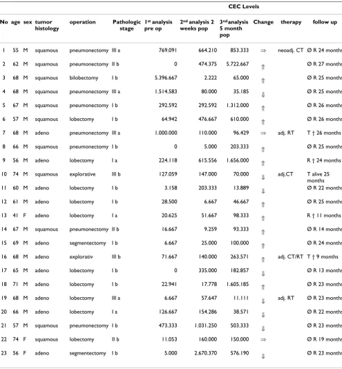
patients, for which all data and follow-up were complete, 20 were male (mean age 64 years, range 55 – 74 years) and 3 were female (mean age 53 years, range 40 – 74 years) with a balanced proportion of early to advanced stages of lung cancer (12:11). Staging was performed by chest x-ray, endobrochial bronchoscopy, computerized tomography (CT), bone scan, operative lymph node dis-section and size of tumor-free margin. Further informa-tion about surgery, histological type of tumor stage and the number of CEC is depicted in table 1.
Cell preparation
Red blood cells from 10 ml samples of blood were lysed, the white blood cells spun down and epithelial cells were enriched using micro-beads from Miltenyi (Bergisch Gladbach, Germany) [7] or the method by Labsoft Diag-nostics (Halle, Germany) [8]. Both methods yielded com-parable results. The same method was always used in follow-up samples from the same patient. Cells were stained with FITC conjugated mouse anti-human epithe-lial antibody (HEA, Milteny, Bergisch Gladbach, Ger-many), reacted either with HEA-micro-beads Miltenyi (Bergisch Gladbach, Germany) or Cell Select-micro-beads (Labsoft Diagnostics Halle, Germany) and then either separated over the columns provided by the Miltenyi company according to their description or the vials con-taining the cells were applied to magnets provided by the Labsoft company. Negative cells not bound in the col-umns were washed out, the colcol-umns removed from the magnet and the retained cells eluted. In the Labsoft method cells not attached to the vial wall were sucked off, the vials removed from the magnet and the cells retained in the vials were then flushed off the wall and studied.
Analysis
For measurements the cells were applied to adhesion slides (Menzel, Gläser, Germany) and measurements started when the cells had settled using a Laser Scanning Cytometer (LSC® Compucyte Corporation, Cambridge, MA, USA). The cells could easily and unequivocally be contoured using forward scatter as a threshold parameter. Background fluorescence was determined dynamically to calculate both peak and integral fluorescence on a per-cell basis making the fluorescent calculation equivalent for all cells. The green fluorescence was displayed as scattergrams and histograms.
Results
Circulating epithelial cells (CEC) were detectable in blood samples from 25/29 patients (86%) before surgery, whereas 97% of normal controls were negative for these
cells. The number of CEC was calculated in relation to the analyzed blood volume and not to white blood cells, which vary considerably during the course of disease. For 26 patients histological evaluation was available and
Table 1: Data from 23 patients operated for lung cancer
CEC Levels
No age sex tumor histology
operation Pathologic stage
1st analysis
pre op
2nd analysis 2
weeks pop
3nd analysis
5 month pop
Change therapy follow up
1 55 M squamous pneumonectomy III a 769.091 664.210 853.333 ⇒ neoadj. CT Ø R 24 months
2 62 M squamous pneumonectomy II b 0 474.375 5.722.667 Ø R 27 months
3 68 M squamous bilobectomy I b 5.396.667 2.222 65.000 Ø R 25 months
4 68 M squamous pneumonectomy III a 1.514.583 80.000 35.185 Ø R 25 months
5 67 M squamous pneumonectomy I b 292.592 292.592 1.312.000 Ø R 26 months
6 57 M squamous lobectomy I b 64.942 476.667 610.000 Ø R 26 months
7 68 M adeno pneumonectomy III a 1.000.000 110.000 96.429 ⇒ adj. RT T † 26 months
8 66 M squamous pneumonectomy I b 0 5.000 203.333 Ø R 25 months
9 56 M adeno lobectomy I a 224.118 615.556 1.656.000 R † 24 months
10 74 M squamous explorative III b 127.059 147.000 70.000 adj.CT T alive 25
months
11 60 M adeno lobectomy I b 3.158 203.333 13.889 Ø R 22 months
12 61 M adeno lobectomy I b 28.500 6.667 46.667 Ø R 25 months
13 41 F adeno lobectomy I a 20.625 51.667 98.333 R † 11 months
14 67 M squamous pneumonectomy II b 16.667 9.259 93.333 Ø R 14 months
15 69 M adeno segmentectomy I b 6.667 25.000 100.000 Ø R 24 months
16 68 M adeno explorativ III b 71.667 140.000 263.571 adj. CT/RT T † 9 months
17 65 M adeno lobectomy I b 0 335.000 182.857 Ø R 13 months
18 71 M adeno lobectomy I b 22.941 17.778 1.605.185 Ø R 23 months
19 68 M adeno lobectomy III a 6.667 57.647 11.111 adj. RT Ø R 23 months
20 66 M adeno lobectomy I a 126.667 154.286 38.571 Ø R 22 months
21 57 M squamous pneumonectomy I b 473.333 1.031.250 503.333 Ø R 23 months
22 74 F squamous lobectomy II b 11.053 160.000 150.000 ⇒ Ø R 19 months
23 56 F adeno segmentectomy I b 5.000 2.670.370 576.190 Ø R 23 months
CT = Chemotherapy, RT = Radiotherapy, R = Recurrence, ∅ R = no Recurrence, T = Tumor, T † = died with tumor, adj. = adjuvant, neoadj. = neoadjuvant, Pop = Postoperatively, Adeno = Adenocarcinoma, Squamous = Squamous carcinoma
⇑
⇑
⇓
⇑
⇑
⇑
⇑
⇓
⇓
⇑
⇑
⇑
⇑
⇑
⇓
⇑
⇓
⇓
⇓
revealed 15 cases of adenocarcinoma and 11 cases of squamous cell carcinoma. The comparison of preopera-tive numbers of CEC revealed higher values in patients with squamous cell than in those with adenocarcinoma and these values differed significantly (p = 0.046) (Figure 2). Neither tumor size nor positivity of lymph nodes cor-related with the number of circulating tumor cells before surgery. With regard to the extent of resection 1
pleurec-tomy, 8 pneumonectomies, 1 bilobecpleurec-tomy, 12 lobecto-mies, 4 segmental resections and 3 explorative thoracotomies were performed. Lobectomy was more fre-quent than pneumonectomy in patients with adenocarci-noma (69% vs. 36%) and pneumonectomy more frequent than lobectomy in those with squamous cell car-cinoma (54% vs. 6%) (Figure 3).
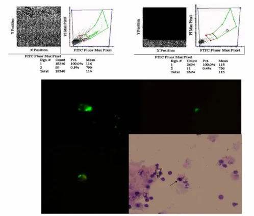
Example of an analysis of a slide with epithelial antigen positive and negative cells: a) fluorescence intensity displayed as a dot plot with all cells in region 1 and the positive cells gated in region 2 (green region)
Figure 1
Fourteen days postoperatively, CEC were detected in all patients including the 3 where no cells had been detecta-ble before surgery. In cases with limited resection (3 exploratory thoracotomies, 4 segmental resections, 12 lobectomies, 1 bilobectomy) parenchymal manipulation was more extensive. This resulted more frequently in an increase (85.7%) than in a decrease (14.3%) in CEC, whereas in cases of complete resection of the lung (pneumonectomy) numbers of CEC decreased (62.5%) more frequently than increased (37.5%) (Figure 4). We, also, investigated CEC in one patient who had surgery for pneumothorax and observed a comparable increase in CEC as in the tumor patients but with a rapid re-decrease already after another two weeks (Figure 5a).
A third analysis of CEC was performed between 3 and 5 months after surgery. In 23 (76%) of 30 consecutive patients considered to have a resectable non small cell lung cancer (NSCLC) complete data were available with a median follow-up of 22 months (range 9 – 27 months) (Table 1).
Five out of 23 patients underwent chemotherapy (CT) or chemo/radiotherapy (CT/RT). These patients were consid-ered separately from the 18 patients who had complete curative resection of their tumors without adjuvant treat-ment. Two patients underwent exploratory thoracotomy: one with squamous cell carcinoma (patient 10) was treated with adjuvant CT with a subsequent decrease in CEC and is still alive 25 months after surgery. The other patient with adenocarcinoma (patient 16) showed an
increase in CEC in spite of combined RT/CT and died 9 months later with tumor progression.
Three of the 21 patients with tumor resection received additional treatment: 1 lobectomy patient with adenocar-cinoma had N2 lymph node status (patient 19). This patient with one level N2 status received adjuvant RT. He subsequently showed a decrease in CEC and is alive, 23 months after surgery without signs of relapse. One patient with squamous cell carcinoma (patient 1) received neoad-juvant therapy and subsequently was treated with pneumonectomy. He showed a slight increase in CEC but is still alive without signs of relapse. Another patient with adenocarcinoma (patient 7) underwent RT but with no significant decrease in CEC and died 26 months later with Number of preoperative circulating tumor cells in
compari-son to histological classification
Figure 2
Number of preoperative circulating tumor cells in compari-son to histological classification.
Frequency of surgical interventions in comparison to histo-logical classification
Figure 3
Frequency of surgical interventions in comparison to histo-logical classification.
Changes in cell numbers (increase or decrease) following surgical intervention
Figure 4
tumor progression. Among these patients receiving additional radio- and/or chemotherapy relapses occurred only in those with adenocarcinoma and increasing CEC numbers in spite of treatment.
Of the remaining 18 patients who had complete curative resection of their tumors, 7 showed a decrease, and 11 showed an increase in CEC during the 3–5 months fol-low-up after surgery.
Of the former 7 patients 6 had had cell numbers increas-ing after surgery and subsequently decreasincreas-ing durincreas-ing the
following months (patients 11, 17, 20, 21, 22, 23), 4 with adeno- and 2 with squamous cell carcinoma and all but one (patient 21) had lobectomy (Figure 5a). All are in complete continuous remission. 1 patient (patient 4) with squamous cell carcinoma had pneumectomy and contin-uously decreasing cell numbers (Figure 5c) and he, too, is in complete continuous remission.
Among the 11 patients with increasing CEC there were 4 with an initial decrease after surgery followed by a subse-quent increase (Figure 5c). None has shown early relapse so far. Seven had a continuous increase from pre-surgery Trends of CEC numbers in individual patients after complete resection without further treatment
Figure 5
to 5 months after treatment (Figure 5b), 4 with squamous cell carcinoma and 3 with adenocarcinoma. Two relapses were observed amongst these patients during the subse-quent 2 years of follow-up. Both, a male (56 years of age) and a female (41 years of age), had adenocarcinoma stage Ia. They had continuously increasing CEC and the increase occurred six months before repeated clinical examinations could verify disease relapse. Both received chemotherapy after diagnosis of relapse but died despite 9 and 24 months after surgery with systemic spread of their tumor.
In summary, amongst the patients with adjuvant treat-ment after resection, as well as in curatively resected patients, patients with adenocarcinoma and a continuous increase in CEC during the course of their disease have a significantly higher risk of early relapse than those with decreasing cell numbers and than those with squamous cell carcinoma, even if the latter show increasing numbers of CEC.
In the relapsed patients not single values of circulating tumor cells at different time points but only the increase in cell numbers was a predictor of relapse.
Discussion
Analyzing circulating epithelial cells in peripheral blood from patients with non small cell lung cancer, we were able to detect live CEC in 86% of patients before surgery. This represents a significantly higher number and fre-quency than reported from other groups analyzing micrometastases taken from bone marrow [9,10] but cell numbers are in the same range as those reported by Cristofanilli and coworkers [11]. This discrepancy may be explained by several modifications of pre-analytical han-dling: 1) In contrast to the former authors [9,10] we used red blood cell lysis [7] for white blood cell preparation. During ficoll-separation used for preparation of cells from bone marrow aspirates we observed a loss of up to 90% epithelial antigen positive cells and there is no reason why circulating epithelial cells should have the same density properties as mononuclear cells (Pachmann submitted for publication]. 2) Micrometastatic cells can be hidden in the fat lumps of the bone marrow aspirates and thus be lost for analysis when preparing mononuclear cell suspen-sions. 3) The portion of blood admixed to marrow aspi-rates can vary between 10 and 90% resulting in a high variability of the material used for analysis. Although the method applied by us is also suitable for bone marrow samples, we preferred analysis of peripheral blood samples which are easy to obtain and can be drawn repeatedly.
The number of CEC detected by us in lung cancer patients is somewhat lower than the number reported recently by
Cristofanilli et al [11] in metastatic breast cancer patients. In positive patients we found between 2000 and 6 × 10⁶ epithelial cells calculating the number of cells in the circu-lation assuming that a patient has about 5 liters of blood. The values of 5 to 22000 cells per 7.5 ml blood reported by Cristofanilli et al [11] would translate into 3000 to 13 × 106 cells in the circulation. The frequency of preopera-tively CEC positive patients was comparable to that reported by Racila et al [12] and Krag et al [13] who also used a similar method for analyzing blood from breast cancer patients.
We preferred detection of fluorescent antibody bound to the surface of live epithelial antigen positive cells per-formed by an automated Laser Scanning Cytometer LSC® to PCR based methods. PCR methods are currently regarded as the most sensitive procedures, but there is a reported high frequency of false positives [14-16]. In addi-tion, our procedure allowed subsequent separation and even culturing of the isolated cells. Culturing will defi-nitely allow determination whether the cells are dormant or have the potency of differentiating into tumor cells.
It should be mentioned that we observed significantly higher numbers of CEC preoperatively in patients with squamous cell carcinoma than in those with adenocarci-noma. This could be due to the former being more cen-trally located (next to the great vessels) which might lead to a higher dissemination than occurs in more peripher-ally located tumors.
Two weeks after surgery all patients were positive for CEC, but there was no correlation to stage, tumor size or lymph node involvement. Unexpectedly we found a strong corre-lation between postoperative increase and limited resec-tions (segmental resecresec-tions and lobectomies) and a decrease of CEC after pneumonectomies. We can only explain this observation by the extended parenchymal manipulation involving the separation of parenchymal bridges between lobes and late ligation of pulmonary ves-sels during lobectomies or segmentectomies. In contrast, pneumonectomies normally start with ligation of the ves-sels and subsequent resection of the lung. Perhaps our data confirm once more the importance of careful "No touch" preparation during oncological resections [17].
dis-seminated normal cells will die in a short time and only the portion of cells that are malignant will have a chance of survival for unknown periods of time. [18]. Monitoring the CEC after five months would, therefore, be expected to detect only surviving malignant epithelial cells. Once again we found CEC in all patients.
Analysis of the increase or reduction of CEC in the 5 patients receiving neoadjuvant and/or adjuvant CT and RT due to their nodal status indicated that this parameter can possibly become a tool for monitoring the effective-ness of therapeutic interventions, an effect that we are actually investigating now in patients receiving CT for lung cancer.
Of the 18 patients treated with complete curative resec-tion all 7 patients of the group with decreasing numbers of CEC were without signs of relapse up to 27 months after surgery. In the group of 11 patients with increasing numbers of CEC, 6 had squamous cell carcinomas and none has relapsed during the observation time of 2 years in accordance with published data [20]. In contrast, in the group of adenocarcinomas 2/5 early relapses were observed, both in patients with stage Ia with the increase in CEC preceded overt clinical relapse by 6 months.
Conclusion
Thus, postoperative monitoring of CEC may become a reliable prognostic predictor of relapse. The observation that only 20% of the patients in the group at risk develop early relapse confirmed by other studies [21] and that patients with squamous cell carcinomas can have high and over time even increasing numbers of CEC necessitate additional analyses on the CEC to obtain more informa-tion about their metastatic potential [22]. Such analyses on enriched and cultured isolated CEC are now under way in our laboratory.
Competing interests
The author(s) declare that they have no competing interests.
Authors' contributions
AR: carried out the studies, and drafted the manuscript
RG: conceived of the study, and participated in its design and coordination
UP: participated in the design of the study and performed the statistical analysis
BW: carried out the immunoassays
KH: preparation of the manuscript
KP: participated in the design of the study and performed the statistical analysis
All authors read and approved the manuscript
Research grant
Not declaredAcknowledgements
NoneReferences
1. Wingo PA, Cardinez CJ, Landis SH, Greenlee RT, Ries LA, Anderson RN, Thun MJ: Long term trends in cancer mortality in the United States 1996–1998. Cancer 2003, 97:3133-3275.
2. Bray F, Tykczynski JE, Parkin DM: Going up or coming down? The changing phases of the lung cancer epidemic from 1966– 1999 in the 15 European Union countries.Eur J Cancer 2004,
40:96-125.
3. Xie L, Ugnat AM, Morriss J, Scmenciw R, Mao Y: Histology related variation in the treatment and survival of patients with lung carcinoma in Canada. Lung Cancer 2003, 42:127-139.
4. Takita H, Pitoniak RF: Induction therapy for locoregional lung cancer using paclitaxel combination. A preliminary report. J Exp Clin Cancer Res 2000, 19:291-293.
5. Pantel K, von Knebel Doeberitz M, Izbicki JR, Riethmüller G: Dis-seminierte Tumourzellen: Diagnostik, prognostische Rele-vanz, Phänotypisierung und therapeutische Strategien.
Chirurg 1997, 68:1241-1250.
6. Bessa X, Elizalde JI, Boix L, Pinol V, Lacy AM, Salo J, Pique JM, Castells A: Lack of prognostic influence of circulating tumor cells in peripheral blood of patients with colorectal cancer. Gastroen-terology 2001, 120:1084-1092.
7. Pachmann K, Heiß P, Demel U, Tilz G: Detection and quantifica-tion of small numbers of circulating tumor cells in peripheral blood using Laser Scanning Cytometry (LSC®). Clin Chem Lab
Med 2001, 39:811-817.
8. Pachmann K, Lychatz S, Oblonczek G, Willen B: Tumourzell-Ana-lyse mit kombinierter Magnetanreicherung, Laser-Scanning Zytometrie und visueller Kontrolle. Laborwelt 2001, Nr. N:23-26.
9. Schlimok G, Funke I, Pantel K, Strobel F, Lindemann F, Witte J, Rieth-müller G: Micrometastatic tumour cells in bone marrow' of patients with gastric cancer: methodological aspects of detection and prognostic significance. Eur J Cancer 1991,
27:1461-1465.
10. Cote RJ, Beattie EJ, Chaiwun B, Sevi SR, Harry J, Chen SC, Sherrod AF, Groshen S, Taylor CR: Detection of occult bone marrow micrometastases in patients with operable lung carcinoma.
Ann Surg 1995, 222:423-425.
11. Cristofanilli M, Budd GT, Ellis MJ, Stopeck A, Matera J, Miller MC, Reuben JM, Doyle GV, Allard WJ, Terstappen LW, Hayes DF: Circu-lating tumor cells, disease progression, and survival in meta-static breast cancer. New Engl J Med 2004, 351:781-791. 12. Racila E, Euhus D, Weiss AJ, Rao C, Mc Connell J, Terstappen LW,
Uhr JW: Detection and characterization of carcinoma cells in the blood. Proc Natl Acad Sci USA 1998, 95:4589-4594.
13. Krag DN, Ashikaga T, Moss TS, Kusminsky RE, Feldman S, Carp NZ, Moffat FL Jr, Beitsch PD, Frazier TG, Gaskin TA, Shook JW, Harlow SP, Weaver DL: Breast cancer cells in the blood; a pilot study.
The Breast J 1999, 5:354-358.
14. Jung R, Petersen K, Krugar W, Wolf M, Wagener C, Zander A, Neu-maier M: Detection of micrometastasis by cytokeratin 20 RT-PCR is limited due to stable background transcription in granulocytes. Br J Cancer 1999, 81:870-873.
15. Suchy B, Austrup F, Driesel G, Eder C, Kusiak I, Uciechowski P, Grill HJ, Giesing M: Detection of mammaglobin expressing cells in blood of breast cancer patients. Cancer Lett 2000, 158:171-178. 16. Wong IH, Chan AT, Johnson PJ: Quantitative analysis of
Publish with BioMed Central and every scientist can read your work free of charge "BioMed Central will be the most significant development for disseminating the results of biomedical researc h in our lifetime."
Sir Paul Nurse, Cancer Research UK
Your research papers will be:
available free of charge to the entire biomedical community
peer reviewed and published immediately upon acceptance
cited in PubMed and archived on PubMed Central
yours — you keep the copyright
Submit your manuscript here:
http://www.biomedcentral.com/info/publishing_adv.asp
BioMedcentral 17. Martin J, Ginsberg RJ, Venkatrman ES, Bains MS, Downey RJ, Korst RJ,
Kris MG, Rusch VW: Long-term results of combined-modality therapy in resectable non-small cell lung cancer. J Clin Oncol 2002, 120:1989-1995.
18. Schleiermacher G, Delattre O: Detection of micrometastases and circulating tumour cells using molecular biology tech-niques in solid tumors. Bull Cancer 2001, 88:561-570.
19. Mehes G, Luegmayr A, Ambros IM, Ladenstein R, Ambros PF: Com-bined automatic immunological and molecular cytogenetic analysis allows exact identification and quantification of tumor cells in the bone marrow. Clin Canc Res 2001,
7:1969-1975.
20. Ghosh S, Ahmed K, Hopkinson DN, Vaughan R: Pulmonary aden-ocarcinoma is associated with poor long-term survival after surgical resection. Effect of allogeneic blood transfusion. Can-cer 2004, 101:2058-2066.
21. Boldrini L, Cacinai A, Sarnaritani E, Pistolesi F, Mussi A, Lucchi M, Angeletti CA, Basolo F, Fontanini G: Tumor necrosis factor alpha and transforming growth factor beta are significantly associ-ated with better prognosis in non small cell lung carcinoma: putative relation with BCL-2 mediated neovascularization.
Br J Cancer 2000, 83:480-486.