324 | P a g e
AUTOMATED DETECTION AND CLASSIFICATION
OF LEUKEMIA USING IMAGE PROCESSING AND
MACHINE LEARNING
Preeti Jagadev
Goa College of Engineering
ABSTRACT
Leukaemia stands for blood cancer that begins in the bone marrow and results in the generation of abnormal
cells. Leukaemia is mainly classified as acute lymphoblastic leukaemia (ALL), acute myeloid leukaemia (AML),
chronic lymphocytic leukaemia (CLL) and chronic myeloid leukaemia (CML).This thesis makes an effort to
devise a methodology for the detection and classification of Leukaemia. The images have been segmented using
HSV colour based segmentation algorithm. The morphological components of normal and Leukemic
lymphocytes differ significantly; hence various features are extracted from the segmented lymphocyte images,
for detection purpose. The leukaemia is classified using SVM classifier.
Keywords: HSV colour based segmentation algorithm,SVM Classifier
.
I. INTRODUCTION
Figure.1- Components of blood
325 | P a g e RGB (Red, Green, and Blue) colour space [14]. The three Components of RGB colour model is highly correlated, so the chromatic information is not suitable for direct processing. Due to colour space, it is convenient to convert from RGB to HSV (Hue, Saturation and Value) colour space [11]. RGB images is difficult for segmentation, hence is converted into the HSV colour space to make segmentation easy[14].This reduces correlation between the colour channels (compared to RGB) and enables dealing with three H, S and V channels separately [24]. The feature extraction technique which is carried out after the segmentation process plays a vital role in differentiating the normal cells from the leukemic ones. The features are extracted from the nucleus of the wbc[29].Subrajeet Mohapatra suggested that features such as fractal dimension, shape features including contour signature and texture could be extracted for detection of leukaemia [2, 7]. Once leukaemia is detected, it needs to be classified into its types (Acute or Chronic). Subrajeet Mohapatra suggested the use of SVM classifier and FLANN-Functional Link Artificial Neural Network for classification purpose [2, 7].The Support Vector Machine (SVM) classifier has been most commonly used for classification [27]. If the patterns are veryclose in the feature space,support vector machine (SVM) is a suitable choice for classification. It is a powerful tool for data classification based on hyper plane [24].N. Z. Supardi proposed to use k-nearest neighbour to classify blasts in acute leukaemia into two types which are AML and ALL [9].
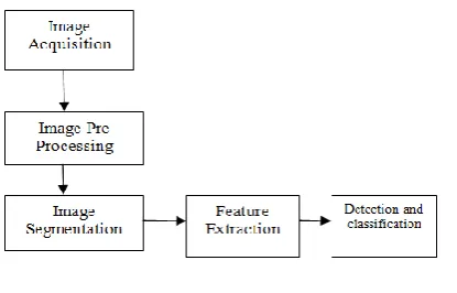
II. PROCESS OVERVIEW
Figure 2 gives a detailed description of the sequence of steps that have been followed for efficient detection and classification of leukaemia.
Figure. 2-Process flow Image
Acquisition-Images of the blood smears of leukemic patients and images of the blood smears of non leukemic patients; have been obtained from online databases.
Image Pre-processing-
Image pre-processing is a technique by means of which the signal to noise ratio and image quality can be improved, that will be helpful for further processing processes. The images that were obtained were in CMYK form, hence were pre processed and converted to RGB form.
Image segmentation algorithm-
326 | P a g e HSV colour space contains most of the WBC information while the S component contains the structure information of the WBC nucleus.
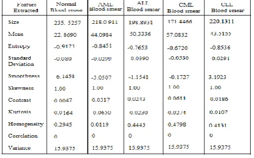
Feature extraction-
While analysing data, the major problem arises due to the number of variables involved that require a large amount of memory and computation. This problem is overcome by feature extraction. The features that were extracted are shape based features and statistical features.
Classification-The classification model chosen for this phase is the Support Vector Machine, which is a machine learning technique. Since the patterns are very close in the feature space, support vector machine is a suitable choice for classification. It is a powerful tool for data classification based on hyper plane classifier.
III. EXPERIMENTAL RESULTS
The dataset under consideration consists of 400 images of blood smear. Figure 3 shows the segmentation output obtained after implementing the HSV colour based segmentation.
Figure 3 Segmentation of blood smear image using HSV colour based segmentation
The image segmentation and feature extraction is followed by the detection and classification process. The SVM classifier is used to detect whether the person has leukaemia or not. If the person is detected with leukaemia, then the type of leukaemia is detected i.e. whether it is ALL, AML, CML or CLL.
327 | P a g e
IV. CONCLUSION AND FUTURE WORK
The paper mainly focuses on the detection of Leukaemia and provides a broader range of Leukaemia classification into its four main types. HSV colour based segmentation was used for image segmentation. A large number of features were extracted to make the detection process more accurate. This work can further be extended by detecting the subtypes of leukaemia types, e.g. AML M3 is a subtype of AML. More segmentation algorithms can be explored, so as to obtain better results as compared to the previous ones.
V. ACKNOWLEDGEMENT
This project has endured a long journey from concept to structured framework and then to its final implementation. First of all I would like to thank my internal guide, Dr. H. G. Virani for allowing me to carry out this project under his supervision. I am grateful for all the advices and encouragement given by him. I would also like to express my gratitude to Dr. V. N. Shet, Principal, Goa College of Engineering for allowing me to use the college facilities. I would like to thank Professor Yeshudas.A.Muttu and Professor Mohini.N.Naik for all the advices and help. I am thankful to Dr Merline Augustine for providing excellent guidance related to the medical aspects of my thesis. Last, but not the least I want to thank God and my family for their unconditional support and encouragement provided throughout the journey of the project.
REFERENCES
[1] Himali P. Vaghela, Hardik Modi, Manoj Pandya and M.B. Potdar ,Leukaemia Detection using Digital Image Processing Techniques", November 2015 - International Journal of Applied Information Systems (IJAIS)
[2] Subrajeet Mohapatra and Dipti Patra, Automated Leukaemia Detection using Hausdroff Dimension in Blood Microscopic Images ", 2010 IEEE
[3] Emad A. Mohammed, MostafaM.A.Mohamed, Christopher Naugler and Behrouz.H.Far, Chronic lymphocytic leukaemia cell segmentation from microscopic blood images using watershed algorithm and optimal thresholding", 2013 26th IEEE Canadian Conference of Electrical and Computer Engineering (CCECE) 2010
[4] Sos Agaian, and Anthony T. Chronopoulos .Automated Screening System for Acute Myelogenous Leukaemia Detection in Blood Microscopic Images, IEEE SYSTEMS JOURNAL, VOL. 8, NO. 3, SEPTEMBER 2014
[5] Jyoti Rawat.A.Singh, H.S. Bhadauria.kumar, Comparative analysis of segmentation algorithms for leukocyte extraction in the acute lymphoblastic images", 2014 International Conference on Parallel, Distributed and Grid Computing
[6] Adnan Khashman and Hayder Hassan Abbas, Acute Lymphoblastic Leukaemia Identification Using Blood Smear Images and a Neural Classifier", 2014 International Conference on Parallel, Springer-Verlag Berlin Heidelberg 2013
328 | P a g e [8] Arjun Nelikanti Segmentation and Analysis of Cancer Cells in Blood Samples, Indian Journal of
Computer Science and Engineering (IJCSE).
[9] R. Hassan, Classification of Blasts in Acute Leukaemia Blood Samples Using K-Nearest Neighbour", 2012 IEEE 8th International Colloquium on Signal Processing and its Applications
[10] A.S.Abdul Nasir, M.Y.Mashor, Unsupervised Colour Segmentation of White Blood Cell for Acute Leukaemia Images", 2011 IEEE
[11] A.S.Abdul Nasir, M.Y.Mashor, Nucleus Segmentation Technique for Acute Leukaemia", 2011 IEEE 7th International Colloquium on Signal Processing and its Applications
[12] A.S.Abdul Nasir, M.Y.Mashor, Colour Image Enhancement Techniques for Acute Leukaemia Blood Cell Morphological Features", 2010 IEEE
[13] Hayan T. Madhloom, Sameem Abdul Kareem, A Robust Feature Extraction and Selection Method for the Recognition of Lymphocytes versus Acute Lymphoblastic Leukemia"2012International Conference on Advanced Computer Science Applications and Technologies.
[14] Tejashri G. Patil, V. B. Raskar, Blood Microscopic Image Segmentation Acute Leukaemia Detection “International Journal of Emerging Research in Management Technology (Volume-4, Issue-9)
[15] A.S.Abdul Nasir, M.Y.Mashor, Comparison of Acute Leukaemia Image Segmentation using HSI and RGB Colour Space"2010 IEEE
[16] Fabio Scotti, Automatic Morphological Analysis for Acute Leukaemia Identification in Peripheral Blood Microscope Images" CIMSA 2005 IEEE International Conference on Computational Intelligence for Measurement Systems and Applications
[17] Vanika Singhal and Preety Singh, Texture Features for the Detection of Acute Lymphoblastic Leukaemia" Proceedings of International Conference on ICT for Sustainable Development Springer [18] Ahmed Faraq, Computer Based Acute Leukaemia Classification" 2004 IEEE
[19] Kuntal Barua1, Prasun Chakrabarti, Detection and Classification for Blood Cancer a Survey “International Journal of Computer Trends and Technology (IJCTT) Volume Number 2 - June 2016 [20] Subrajeet Mohapatra and Dipti Patra Lymphocyte Image Segmentation Using Functional Link Neural
Architecture for Acute Leukemia Detection" The Korean Society of Medical Biological Engineering and Springer 2012
[21] Mr. Rajeev R Menon, Mr. Ranjith S, Automated Detection of Acute Myelogenous Leukaemia Using Neural Classifier” International Journal of Engineering and Technical Research (IJETR) March 2016 [22] Chastine Fatichah, Martin L. Tangel, Fei Yan, Janet P. Betancourt, M. Rahmat Widyanto, Fangyan Dong
and Kaoru Hirota,Fuzzy Feature Representation for White Blood Cell Differential Counting in Acute Leukaemia Diagnosis” Springer 2015
[23] Shubhangi Khobragade, Dheeraj D Mor, Dr. C.Y.Patil, Detection of Leukaemia in Microscopic White Blood Cell Images" 2015 IEEE
329 | P a g e [25] lK. Raghul, A. Shriram Raj and P.U. Ilavarasi, Acute Lymphocytic Leukaemia Detection by Image
Processing Using Matlab" Middle-East Journal of Scientific Research, 2016 proceedings
[26] Rajivegandhi, Animesh Mrinal, N. Sanjana, Sumeet Shekhar,Acute Mylogenous Leukaemia Detection Using Blood Microscopic Images “International Journal for Research in Applied Science Engineering Technology (IJRASET)Volume 3 Issue IV, April 2015
[27] Yogesh Ambadas Gajul, Rupali Shelke, Computerized Detection System for Acute Myelogenous Leukaemia in Blood Microscopic Images" International Journal of Innovative Research in Science, Engineering and Technology June 2016
[28] Renuka devi1, C.V.Gnana, Classification of Acute Myelogenous Leukaemia in Blood Microscopic Images Using Supervised Classifier" 2015 IJESC