R E S E A R C H A R T I C L E
Open Access
Mechanical testing of a device for subcutaneous
internal anterior pelvic ring fixation versus
external pelvic ring fixation
Georg Osterhoff
1*, Simon Tiziani
1, Stephen J Ferguson
2, Gregor Spreiter
2, Max J Scheyerer
1, Gian-Leza Spinas
1,
Guido A Wanner
1, Hans-Peter Simmen
1and Clément ML Werner
1Abstract
Background:Although useful in the emergency treatment of pelvic ring injuries, external fixation is associated with pin tract infections, the patient’s limited mobility and a restricted surgical accessibility to the lower abdomen. In this study, the mechanical stability of a subcutaneous internal anterior fixation (SIAF) system is investigated.
Methods:A standard external fixation and a SIAF system were tested on pairs of Polyoxymethylene testing cylinders using a universal testing machine. Each specimen was subjected to a total of 2000 consecutive cyclic loadings at 1 Hz with sinusoidal lateral compression/distraction (+/−50 N) and torque (+/−0.5 Nm) loading alternating every 200 cycles. Translational and rotational stiffness were determined at 100, 300, 500, 700 and 900 cycles.
Results:There was no significant difference in translational stiffness between the SIAF and the standard external fixation when compared at 500 (p = .089), 700 (p = .081), and 900 (p = .266) cycles. Rotational stiffness observed for the SIAF was about 50 percent higher than the standard external fixation at 300 (p = .005), 500 (p = .020), and 900 (p = .005) cycles. No loosening or failure of the rod-pin/rod-screw interfaces was seen.
Conclusions:In comparison with the standard external fixation system, the tested device for subcutaneous internal anterior fixation (SIAF)in vitrohas similar translational and superior rotational stiffness.
Keywords:Pelvic fracture, Pelvic ring injury, Internal fixation, External fixation, Subcutaneous internal anterior fixation
Background
External fixation has been an established technique in the emergency treatment of pelvic ring injuries [1,2]. With external fixation, reduction of the inner pelvic diameter can be achieved within minutes, thereby limit-ing venous haemorrhage into the lesser pelvis [3]. Be-sides this direct effect on the extent of blood loss in the acute situation, external fixation reduces motion be-tween the pelvic fragments and thus helps to maintain haemostasis during the first days after the trauma [4,5].
It has been shown that supra-acetabular positioning of the pins leads to improved anchorage of the pins and a higher construct stability when compared to a pin place-ment into the iliac crest [6].
Particularly for pelvic injuries with posterior instability, appropriate stability can only be achieved by combined anterior and posterior fixation or a sufficient posterior fixation alone [7,8].
Thus, definitive internal pelvic stabilization is usually applied as soon as the patient’s condition allows for more extensive surgery [9].
Besides usually serving only as a temporary stabilization, the use of external fixation of pelvic fractures is associ-ated with further disadvantages. Pin tract infections are common and can occur in up to 50% of cases [10]. Roll-ing the patient side-to-side, sittRoll-ing and lyRoll-ing in prone position is limited. Especially in patients with concomi-tant intra-abdominal injuries, where multiple revisions might be necessary, an anterior frame impairs the access to the lower abdomen. In addition, achieving stability by external fixation in severely obese patients with a large distance between the pelvis and the connecting rods
* Correspondence:[email protected]
1
Division of Trauma Surgery, University of Zurich, Raemistrasse 100, Zurich 8091, Switzerland
Full list of author information is available at the end of the article
is almost impossible, due to abundant abdominal soft tissue.
Therefore, a subcutaneous internal anterior screw-rod fixation has been suggested that can be applied within a comparable time to an external fixateur, while avoiding the disadvantages of the external implant [11]. This new technique has already been tested in several case series, where indeed very low rates of wound infections were observed when compared to those of external fixation. Yet, it was associated new complications like irritation of the lateral femoral cutaneous nerve or heterotopic os-sifications [11-13].
With the development of minimal-invasive spinal in-strumentation systems, there are new possibilities to per-form this technique of subcutaneous internal anterior fixation (SIAF) in a less invasive fashion.
Little is known about the mechanical properties of such instrumentation. Failure to achieve stable fixation can result in the detachment of blood clots at the fracture site and therefore put the patient at risk for re-current bleeding. As well, loosening of the rod-screw interface would require revision surgery.
The purpose of this study was to investigate the in vitromechanical stability of a device for subcutaneous internal anterior fixation and compare it to a standard external fixation.
Methods Testing samples
The experimental setup was designed to progressively measure the fatigue life or damage accumulation of the rod-pin/rod-screw interfaces. Two fixation techniques were tested on pairs of Polyoxymethylene testing cylinders
with a diameter of 70 mm (Delrin, DuPont, Wilmington, DE, USA). These cylinders provided sound purchase of the pelvic pins and screws thereby simulating optimum anchorage in the pelvic bone.
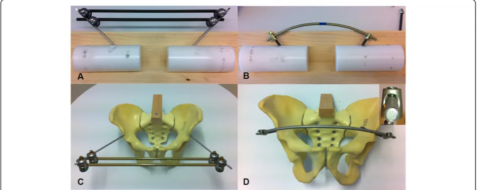
In the group EXTERNAL (n = 3), a standard external fixateur (Hoffmann II, Stryker, Kalamazoo, MI, USA) was mounted (Figure 1A) using two apex self-drilling pins (diameter 5 mm) each anchored in one of on two separate testing cylinders. Angles and distances between the two pins were chosen in a way to reproduce the con-ditions as measured for the supra-acetabular channel on a pelvic model (Pelvis, Synbone, Malans, Switzerland) (Figure 1C). Two connecting carbon fiber rods (diameter 8 mm) were attached using pin-to-rod couplings accord-ing to the manufacturer’s instructions. Like in clinical routine, we aimed for torque forces exceeding that of a torque-limiter. Thus, all couplings were tightened with maximum effort by the same person (S. T.).
In the group SIAF (n = 3), two 8-mm-screws (Iliac Multiaxial Screw, Legacy, Medtronic, Minneapolis, MN, USA) were placed in two testing cylinders in exactly the same way as described for the supra-acetabular pins in the EXTERNAL group Figure 1B, D). The screws were connected by a contoured titanium rod (diameter 8.5; Longitude, Medtronic, Minneapolis, MN, USA). Cou-plings in the polyaxial screw heads were tightened by the same person (S. T.) with a torque screwdriver ac-cording to the manufacturer’s instructions (Legacy, Med-tronic, Minneapolis, MN, USA).
Mechanical testing
[image:2.595.56.540.501.694.2]In patients, two basic loading scenarios are likely after provisional stabilization of pelvic ring fractures. First,
rolling the patient side-to-side is common (e.g. when changing bed-linen) which would result in lateral com-pression forces on the pelvic fixation construct in the transversal plane. Second, bending of the hip or transfer to a chair would result in torque forces in the sagittal plane.
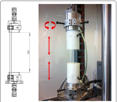
Thus both fixation constructs were tested by alternating cycles of lateral compression/distraction (i.e. following the cylinder axis) and torque forces (i.e. counter-rotation of the cylinders about the central axis) using a universal testing machine (ElectroPulse E10000, Instron, High Wycombe, UK). The cylinder axes of the samples were aligned with the axis of the actuator using universal joints to eliminate confounding bending moments (Figure 2). After observing a displacement of clinical relevance (> 25 mm) at +/−50 N, this load was chosen for the experi-mental setup.
Each specimen was subjected to total of 2000 consecu-tive cyclic loadings at 1 Hz with sinusoidal lateral com-pression/distraction (+/−50 N) and torque (+/−0.5 Nm). One thousand cycles of rotation and 1000 cycles of com-pression/distraction with the mode of loading alternating every 200 cycles were applied.
During testing, loading forces and displacement were registered by a +/−1000 N (compression/distraction forces) and a 25 Nm (torque forces) load cell and trans-lational and rotational stiffness were calculated. Failure resulting in immediate stop of testing included: displace-ment beyond the machine’s range of motion (+/−30 mm
in the transversal plane, +/− 135 degrees rotation) or interface failure.
The following parameters were evaluated:
Translational stiffness [N/mm]
A measure of the extent to which the fixation system resisted deformation in response to a force simulating lateral compression of the pelvis. This value was deter-mined at 100, 300, 500, 700 and 900 cycles.
Rotational stiffness [Nm/degree]
A measure of the extent to which the fixation system resisted deformation in response to a moment simulat-ing torque of the pelvis. This value was determined at 100, 300, 500, 700 and 900 cycles.
Statistical analysis
After preliminary testing, a sample size calculation was performed using PS Power and Sample Size Calculations 3.0 (alpha error: 0.05) [14].
With an expected difference in means of 0.5 N/mm and a standard deviation of 0.15 N/mm for translational stiffness and an expected difference in means of 0.08 Nm/degree and a standard deviation of 0.025 Nm/ degree for rotational stiffness the calculated number of samples to be able to reject the null hypothesis that the population means of the experimental and control groups are equal with a probability (power) of 0.8 was 3 per group.
After testing for normal distribution by a Shapiro-Wilk test, comparison of translational and rotational stiffness was done using a paired T-Test with SPSS for Windows V14.0 (SPSS, Chicago, Illinois, USA). Differ-ences were considered significant for values ofp< 0.05.
Results
Translational stiffness
There was a slight but not significant difference, when tested in response to a force simulating lateral compres-sion of the pelvis, between the stiffness observed for the internal anterior fixation (SIAF) and the standard external fixation (EXTERNAL) when compared at 500 (p = .089), 700 (p = .081), and 900 (p = .266) cycles. At 100 (p = .038) and 300 (p = .021) cycles, however, the translational stiff-ness was even slightly higher in the SIAF group (Figure 3).
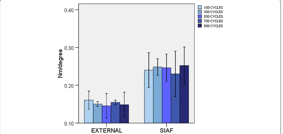
Rotational stiffness
[image:3.595.56.292.459.666.2]When tested in response to a force simulating torque of the pelvis, the stiffness observed for the subcutaneous internal anterior fixation (SIAF) was about 50 percent higher than the standard external fixation (EXTERNAL) at all times of measurement. This was statistically signifi-cant when compared at 300 (p = .005), 500 (p = .020), and 900 (p = .005) cycles. At 100 (p = .050) and 700 Figure 2Experimental setup.On a universal testing machine,
(p = .050) cycles, the level of significance was not achieved (Figure 4).
Temporary deformation was solely caused by elastic bending of the rods, screws and pins, the rod-pin/rod-screw interfaces did not show any loosening or failure with the applied loads. Persistent plastic deformation was not seen.
Discussion
[image:4.595.57.542.476.706.2]Supra-acetabular external fixation of pelvic ring injuries is an efficient tool to minimize blood loss in the acute situation and to maintain haemostasis during the first days after the trauma [1-6]. However, pin tract infec-tions, the patients’limited mobility and a restricted sur-gical accessibility to the lower abdomen are noticeable Figure 3Translational stiffness.EXTERNAL: standard external fixation. SIAF: subcutaneous internal anterior fixation. Error bars represent 2 standard deviations.
disadvantages of this technique. In 2009, Kuttner et al. introduced the idea of a subcutaneous internal anterior fixation of the pelvis [11].
This study tested the in vitro mechanical stability of a subcutaneous internal anterior fixation system (SIAF) in comparison to a standard external fixation (EXTERNAL). We used an experimental setup with a focus on the rod-pin/rod-screw interface, which was thought to be the weakest link within both fixation techniques and simu-lated forces resulting from side-to-side rolling of a patient (translational stiffness) and forces resulting from bending of the hip (rotational stiffness). The SIAF system showed a similar to slightly higher translational stiffness than the EXTERNAL system and a significantly higher rotational stiffness at up to 900 loading cycles. It is important to note that this combined implant-construction stiffness is a function of rod- and pin/screw stiffness, pin/screw-rod interface motility and the swinging distance of the implant over the bone surrogate. It is our conviction that this dif-ference is largely caused by the large swinging distance of the pins over the bone in external fixation. This problem is partly eliminated by the positioning of the rod in SIAF.
Several case series using a subcutaneous internal anter-ior fixation showed good short-term results with only few complications related to implant failure [11-13]. Our in vitrodata is consistent with these clinical experiences. In addition, there is one recent mechanical study compar-ing a similar subcutaneous anterior fixation technique with anterior external and plate fixation suggesting super-ior stiffness of the SIAF-like system [15]. The latter study, however, uses only one single connecting rod with the ex-ternal fixation, which is known to have inferior mechan-ical stability [16] and therefore usually does not represent the clinical routine. In contrast to our study, a cyclic load-ing setup was not used by Vigdorchik et al. [15]. In most in vivocases, however, postoperative fragment dislocation is not a result of a single trauma or force, but rather oc-curs due to repetitive motion (e.g. by nursing bedside as-sistance or physiotherapy) as is suggested by the typical appearance of symmetric aseptic loosening of the pins in many cases [10]. Therefore, cyclic loading seems to better approximate clinical loading scenarios.
To date, there exists no standard in mechanical testing of pelvic ring fixations. Generally, a cadaver or synthetic pelvis is tested with large variations in force vectors’ dir-ection, quantity and point of application [4,15,17-21]. Most experimental setups ignore forces resulting by ac-celeration when walking and ignore force vector realign-ment by abductor and adductor muscles [17,22,23].
There is also no gold standard for external fixation configurations in the treatment of pelvic ring injuries, even single-rod fixation has been described [15]. A double-rod configuration as used in our study, however, has been considered to provide sufficient stability [16].
The stability of pelvic fixation systems is defined by their stiffness and by possible failure of the rod-pin/rod-screw interfaces. As data forin vivoloading of the pelvic ring are limited, the test was designed to progressively measure the fatigue life or damage accumulation of these rod-pin/rod-screw interfaces [24]. A simplified setup was chosen with a focus on the stiffness of the im-plants themselves as well as on the specified interfaces. By choosing this test protocol, confounding factors were avoided, such as the quality of screw/pin anchorage, which depends on bone quality and surgical application. With the results of this study, no conclusions can be drawn on the implant’s behaviour at the implant-bone-interface. It is possible that the increased stiffness of the SIAF system prevents loosening of bone anchorage, or also that it even promotes loosening by increased stresses at the implant-bone-interface [25]. The very low implant loosening rates in the few known clinical series using the SIAF [11-13] support the idea of improved an-chorage–due to a higher stiffness and stability and due to the greater diameter of the SIAF screws.
All the latter clinical studies applied the SIAF in com-bination with posterior fixation. Future studies may fur-ther analyse the implant-bone-interface by the use of cadaver testings or finite element models [26].
Ourin vitrodata suggest the possibility of even an iso-lated use of the SIAF for fractures where external fix-ation alone would be considered adequate.
This would provide a quick intervention with early and simplified mobilization of the patient. It is known that, in the treatment of pelvic ring injuries, early mobilization significantly reduces posttraumatic compli-cations and morbidity [5]. In fractures with intact pos-terior sacroiliac and sacro-tuberous ligaments (like LC II and APC II), posterior stabilization has to be postponed in some cases. By providing higher stability and lower infection and loosening rates [11-13], isolated prelimin-ary anterior fixation with SIAF might be an alternative to external fixation. However, the promising results of this study have yet to be provenin vivo.
In this context, it is important to mention that SIAF seems to be associated with irritation of the lateral fem-oral cutaneous nerve in up to 27% of cases and with het-erotopic ossifications in up to 32% [11-13]. In addition, components of SIAF are close to important anatomic structures, like the femoral vascular bundle and the urinary bladder [27].
Hence, careful adherence to the surgical anatomy is crucial.
Conclusions
superior rotational stiffness. This technique might pro-vide a useful tool in the treatment of pelvic ring injuries, especially in case of patients with concomitant abdom-inal injuries or risk factors for pin tract infection.
Competing interests
The authors declare that they have no competing interests.
Authors’contributions
GO, ST, SF, and CW participated in the study’s design and planning. GO, ST, SF, GS, G-LS, and CW carried out the testing. GO, SF, and MS performed the data analysis. GO, SF, GW, H-PS, and CW drafted/revised the final manuscript. All authors read and approved the final manuscript.
Acknowledgements
We would like to thank Stryker (Kalamazoo, MI, USA) and Medtronic (Minneapolis, MN, USA) for donating the implants that were used for this study.
Author details
1Division of Trauma Surgery, University of Zurich, Raemistrasse 100, Zurich
8091, Switzerland.2Institute for Biomechanics, ETH Zurich, HCI-E355.2
Wolfgang-Pauli-Strasse 10, Zurich 8093, Switzerland.
Received: 29 April 2013 Accepted: 28 March 2014 Published: 31 March 2014
References
1. Burgess AR, Eastridge BJ, Young JW, Ellison TS, Ellison PSJ, Poka A, Bathon GH, Brumback RJ:Pelvic ring disruptions: effective classification system and treatment protocols.J Trauma1990,30(7):848–856.
2. Miller PR, Moore PS, Mansell E, Meredith JW, Chang MC:External fixation or arteriogram in bleeding pelvic fracture: initial therapy guided by markers of arterial hemorrhage.J Trauma2003,54(3):437–443. 3. Grimm MR, Vrahas MS, Thomas KA:Pressure-volume characteristics of the
intact and disrupted pelvic retroperitoneum.J Trauma1998,
44(3):454–459.
4. Gardner MJ, Kendoff D, Ostermeier S, Citak M, Hufner T, Krettek C, Nork SE:
Sacroiliac joint compression using an anterior pelvic compressor: a mechanical study in synthetic bone.J Orthop Trauma2007,21(7):435–441. 5. Riemer BL, Butterfield SL, Diamond DL, Young JC, Raves JJ, Cottington E,
Kislan K:Acute mortality associated with injuries to the pelvic ring: the role of early patient mobilization and external fixation.J Trauma1993,
35(5):671–675. discussion 676–677.
6. Kim WY, Hearn TC, Seleem O, Mahalingam E, Stephen D, Tile M:Effect of pin location on stability of pelvic external fixation.Clin Orthop Relat Res
1999,361:237–244.
7. Pohlemann T, Krettek C, Hoffmann R, Culemann U, Gansslen A:
Biomechanical comparison of various emergency stabilization measures of the pelvic ring.Unfallchirurg1994,97(10):503–510.
8. Osterhoff G, Ossendorf C, Wanner GA, Simmen HP, Werner CM:Posterior screw fixation in rotationally unstable pelvic ring injuries.Injury2011,
42(10):992–996.
9. Wanner GA, Keel M, Steckholzer U, Beier W, Stocker R, Ertel W:Relationship between procalcitonin plasma levels and severity of injury, sepsis, organ failure, and mortality in injured patients.Crit Care Med2000,
28(4):950–957.
10. Mason WT, Khan SN, James CL, Chesser TJ, Ward AJ:Complications of temporary and definitive external fixation of pelvic ring injuries.Injury
2005,36(5):599–604.
11. Kuttner M, Klaiber A, Lorenz T, Fuchtmeier B, Neugebauer R:The pelvic subcutaneous cross-over internal fixator.Unfallchirurg2009,
112(7):661–669.
12. Vaidya R, Kubiak EN, Bergin PF, Dombroski DG, Critchlow RJ, Sethi A, Starr AJ:Complications of anterior subcutaneous internal fixation for unstable pelvis fractures: a multicenter study.Clin Orthop Relat Res2012,
470(8):2124–2131.
13. Gardner MJ, Mehta S, Mirza A, Ricci WM:Anterior pelvic reduction and fixation using a subcutaneous internal fixator.J Orthop Trauma2011,
26(5):314–321.
14. Dupont WD, Plummer WD:Power and Sample Size Calculations for studies Involving Linear Regression.Controlled Clinical Trials1998,
19:589–601.
15. Vigdorchik JM, Esquivel AO, Jin X, Yang KH, Onwudiwe NA, Vaidya R:
Biomechanical stability of a supra-acetabular pedicle screw Internal Fixation device (INFIX) vs External Fixation and plates for vertically unstable pelvic fractures.J Orthop Surg Res2012,7(1):31.
16. Moroz TK, Finlay JB, Rorabeck CH, Bourne RB:External skeletal fixation: choosing a system based on biomechanical stability.J Orthop Trauma
1988,2(4):284–296.
17. Papathanasopoulos A, Tzioupis C, Giannoudis VP, Roberts C, Giannoudis PV:
Biomechanical aspects of pelvic ring reconstruction techniques: Evidence today.Injury2010,41(12):1220–1227.
18. Ponsen KJ, Hoek van Dijke GA, Joosse P, Snijders CJ:External fixators for pelvic fractures: comparison of the stiffness of current systems. Acta Orthop Scand2003,74(2):165–171.
19. Rieger H, Winckler S, Wetterkamp D, Overbeck J:Clinical and biomechanical aspects of external fixation of the pelvis.Clin Biomech (Bristol, Avon)1996,11(6):322–327.
20. van den Bosch EW, van Zwienen CM, van Dijke GA H, Snijders CJ, van Vugt AB:Sacroiliac screw fixation for tile B fractures.J Trauma2003,
55(5):962–965.
21. Wang M, Dumas GA:Mechanical behavior of the female sacroiliac joint and influence of the anterior and posterior sacroiliac ligaments under sagittal loads.Clin Biomech (Bristol, Avon)1998,13(4–5):293–299. 22. Bergmann G, Graichen F, Rohlmann A, Bender A, Heinlein B, Duda GN,
Heller MO, Morlock MM:Realistic loads for testing hip implants.Biomed Mater Eng2010,20(2):65–75.
23. Goel VK, Valliappan S, Svensson NL:Stresses in the normal pelvis.Comput Biol Med1978,8(2):91–104.
24. Ferguson SJ, Winkler F, Nolte LP:Anterior fixation in the osteoporotic spine: cut-out and pullout characteristics of implants.Eur Spine J2002,
11(6):527–534.
25. Lill H, Hepp P, Korner J, Kassi JP, Verheyden AP, Josten C, Duda GN:
Proximal humeral fractures: how stiff should an implant be? A comparative mechanical study with new implants in human specimens. Arch Orthop Trauma Surg2003,123(2–3):74–81.
26. Bohme J, Shim V, Hoch A, Mutze M, Muller C, Josten C:Clinical implementation of finite element models in pelvic ring surgery for prediction of implant behavior: a case report.Clin Biomech (Bristol, Avon)
2012,27(9):872–878.
27. Merriman DJ, Ricci WM, McAndrew CM, Gardner MJ:Is application of an internal anterior pelvic fixator anatomically feasible?Clin Orthop Relat Res
2012,470(8):2111–2115.
doi:10.1186/1471-2474-15-111
Cite this article as:Osterhoffet al.:Mechanical testing of a device for subcutaneous internal anterior pelvic ring fixation versus external pelvic ring fixation.BMC Musculoskeletal Disorders201415:111.
Submit your next manuscript to BioMed Central and take full advantage of:
• Convenient online submission
• Thorough peer review
• No space constraints or color figure charges
• Immediate publication on acceptance
• Inclusion in PubMed, CAS, Scopus and Google Scholar
• Research which is freely available for redistribution