International Journal of Medical Science and Current Research (IJMSCR)
Available online at: www.ijmscr.com
Volume1, Issue 2,Page No: 159-164
July-August 2018
159
Biochemical Evaluation Of Renal Function And Liver Function In Patients Of
Different Types Of Renal Stones
1
Dr Ajay Meshram - Professor& HOD, 2 Dr Yogita Dubey ,PG Scholar
Department of biochemistry JNMC Sawangi (Meghe) Wardha 3 Dr Komal Meshram - Assistant Professor, Department of physiology,JNMC Sawangi (Meghe) Wardha
Corresponding Author: Dr Yogita Dubey ,
PG Scholar Department of biochemistry JNMC Sawangi (Meghe) Wardha
Type of Publication: Original Research Paper Conflicts of Interest: Nil
ABSTRACT
Introduction - Kidney stone disease is a common chronic disorder with male – female ratio of 2:1. It is multi-factorial disorder resulting from combined influence of epidemiological, biochemical and genetic risk factors. These are hardened mineral deposits in kidney and impose much impact on many metabolic processes like liver function and kidney function etc.
Aim & objective – Evaluation of renal function and liver function biochemically in patients of renal stone and compare with controls to observe the impact of urinary stones to minimize the complications.
Method and material - This, case control study included . 30 cases of Renal stone both male(21) and female(9) and 30 controls. Diagnosis of cases were based on history, clinical examination and KUB x-ray to differentiate radiopaque from radiolucent stones All the tests are done in Randox chemical auto analyzer by using kit method.
Result - Impaired renal function is seen in both type of stone formers. Serum calcium is significantly increased in radio-opaque stones, while serum uric acid level is increased in radiolucent stone formers. In Liver function tests, significant elevation in serum AST, ALT, ALP, while TP is lowered significantly in renal stone patients as compared with control group.
Conclusion - Nephrolithiasis is not only a stone disease, but also as a kidney and liver function impairing disease. Stone components were associated with an impaired renal function. Hence, stone analysis is important and should be carefully evaluated to preserve the renal function.
Keywords: renal stones, renal function tests, liver function tests
INTRODUCTION
Kidney stone disease is a common chronic disorder associated with painful stone episodes, lost work days and significant health care costs [1], affecting up to 10–12 % of men and 5–6 % of women [2]. In recent years, kidney stones have been associated with increased risk of chronic kidney disease (CKD) [3], and even end-stage renal disease (ESRD) [4, 5].
Urinary calculi have worldwide distribution but are particularly common in some geographic locals such as United States, South Africa, India and other South East Asian Countries. It is estimated that approximately 2% of the population experiences renal stone disease at sometime in their life with male
e
160
e
160
e160
e
160
e160
e
160
e160
e
160
e160
e
160
e
160
e
160
e
160
e160
e
160
e160
e
160
e160
e
160
e160
e
160
.Previous studies have considered kidney stones as a potential risk factor for chronic kidney disease (CKD), associated with significant morbidity, mortality, and heath care cost [12,13,14]. To date, the mechanism accounting for this association is not completely understood. There are several components of urinary stones, such as calcium oxalate, calcium phosphate, uric acid, and struvite
[15]. The relationships between stone components and the change of renal function are unclear .
Because the liver has many crucial roles in the maintenance of a healthy body, any level of liver dysfunction can be problematic. The most commonly used serum liver function tests include serum ALT and AST activities that reflect the patocellular injury and serum ALP that reflect impaired bile excretion and bile flow and serum albumin that represent the synthetic function of the liver[16]. This study is aimed the assessment of the changes in liver function and kidney function in patients of different types of kidney stone .
Method and materials :
This cross sectional, case control study was conducted in Department of Biochemistry, JNMC, Sawangi, Wardha after obtaining clearance from the Institutional Human Ethical committee. Patients were taken from indoor admitted in Achrya Vinobha Bhave Rural Hospital from October 2017 to March 2018. Informed consent was obtained from each participant.
Study group comprises of 60 subjects. 30 cases of Renal stone of age group of 15 – 75 yrs both male(21) and female(9) and 30 controls of normal healthy persons same age and sex, from general population. We reviewed the medical records of patients. The diagnosis of the cases were done in the hospital, based on history, clinical examination followed by KUB x-ray, ultrasonography, and urinalysis
Stone composition - Results of stone analysis were
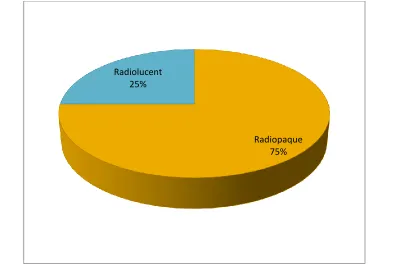
obtained from medical records and all available medical imaging reports in order to differentiate radiopaque from radiolucent stones. Stones visualized radiopaque were considered as calcium containing stones and radiolucent stones were considered as uric acid stones. There were 24 cases of radiopaque and 6 cases of radiolucent stones.
Exclusion criteria - The exclusion criteria were
chronic renal failure, chronic ureteral obstruction in kidneys , congenital renal anomalies, and polycystic kidney disease.
Collection of blood samples - Five mL of venous
blood samples was withdrawn in plain bulb. After centrifugation, we got clear serum. The serum samples were isolated with proper labeling, and used for biochemical measurements
Biochemical measurement - For liver function test,
we measured serum AST, ALT,ALP, total billirubin, and total protein. For renal function test we measured serum urea, creatinine. We also measured serum calcium and uric acid . All the tests are done in Randox auto analyzer by using kit method.
Statistical analysis - Statistical analysis was done by
using descriptive and inferential statistics using one way ANOVA , Multiple Comparison: Tukey Test and z-test for difference between two means and software used in the analysis were SPSS 22.0 version and EPI-INFO 6.0 version and p<0.05 is considered as level of significance.
Result
Pag
e
161
Pag
e
161
Pag
e
161
Pag
e
161
Pag
e
161
Pag
e
161
Pag
e
161
Pag
e
161
Pag
e
161
Pag
e
161
Pag
e
161
Pag
e
161
Pag
e
161
Pag
e
161
Pag
e
161
Pag
e
161
Pag
e
161
Pag
e
161
Pag
e
161
Pag
e
161
Pag
e
161
Renal function tests - Results showed (Table – 1) impaired renal function in both type of stone formers.
Increased mean serum urea (radio-opaque- 55.04 )(radiolucent- 52.00) and increased mean serum creatinine (radio-opaque- 2.63)(radiolucent-2.39 )is seen when compared with control(mean serum urea-22.23, mean serum creatinine-0.92). Serum calcium is significantly increased in radio-opaque stones, while serum uric acid level is increased in radiolucent stone formers.
Table – 1 Comparison of kidney function tests in cases and conrols
Radiopaque 75% Radiolucent
e
162
e
162
e162
e
162
e162
e
162
e162
e
162
e162
e
162
e
162
e
162
e
162
e162
e
162
e162
e
162
e162
e
162
e16
2
e
162
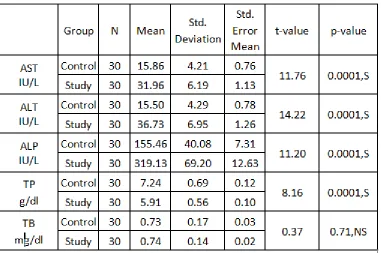
Liver function tests - The result of the current study showed significant elevation ( P< 0.05) in serum AST,
ALT, ALP, concentrations ( mean 31.96, 36.73 & 319.13 respectively)in renal stone former patients when compared with concentrations in the control group, while TP (mean 5.91)is lowered significantly (P< 0.05) in renal stone former patients as compared with control group. Farther more results showed non significant differences ( P< 0.05) in the serum bilirubin concentrations between renal stone former patients and controls groups as shown in (Table – 2).
Table – 2 Comparison of Liver function test in cases and contro
Discussion
Stone formation in the urinary tract has been recognized for thousands of years, but during the last few decades the pattern and incidence of the disease have changed markedly. Urinary stone affect 10-12% of the population in industrialized countries. The average lifetime risk of stone formation has been reported in the range of 5-10%. .The incidence of Urinary stones has been increasing recently. With a prevalence of >10% and an expected recurrence rate of nearly 50%, stone disease has an important effect on the health care system.
In our study the male female ratio was 2.3:1.Our study revealed that nephrolithiasis is more common in men (12%) than in women (6%) and is more prevalent between the ages of 20 to 45 in both sexes.
Urolithiasis may result in loss of function by two general mechanisms, the first mechanism include episodic events, such as urethral obstruction during stone passage, or because of procedures needed for stone removal, and their attendant complications, and the second mechanism include continuous events, as
a result of a disordered physiology that underlies stone formation[17].
Kidney function test - In the present study, we
found a significantly lower level of kidney function in patients with kidney stone disease than in age- and gender-matched control subjects and it is more deteriorated in radiopaque subset.
In a study by Yii-Her Chou et al [18] they found better renal function in calcium containing stones compared with those with non-calcium containing stones (struvite and uric acid, p < 0.01)
In another study by Vaka K. Sigurjonsdottir et al [19] they found the prevalence of CKD was significantly higher among the patients with radiolucent stones compared with calcium stone formers (p < 0.001).
Pag e
163
Pag e163
Pag e163
Pag e163
Pag e163
Pag e163
Pag e163
Pag e163
Pag e163
Pag e163
Pag e163
Pag e163
Pag e163
Pag e163
Pag e163
Pag e163
Pag e163
Pag e163
Pag e163
Pag e163
Pag e163
flow reduction, can lead to significant ischemia and permanent renal parenchymal damage [21]. Renal parenchymal crystal deposition and the associated inflammation and fibrosis have been well described in patients with primary hyperoxaluria and APRT deficiency[22] and similar histological changes have also been observed in patients with uric acid and struvite stones [23].
Furthermore, Evan et al. [24] shown that obstruction of the ducts of Bellini, commonly seen in stone formers, is strongly associated with tubular atrophy, interstitial fibrosis and glomerulosclerosis [25]. While crystal nephropathies can cause inflammation and fibrosis leading to severe kidney damage, the underlying pathobiological mechanisms of CKD in ordinary human kidney stone disease remain elusive. In animal models, however, the best characterized pathway of calcium oxalate crystal-induced inflammation involves NLRP3 inflammasome-mediated mechanisms, leading to direct injury to tubular cells, neutrophil recruitment, tubulo-interstitial inflammation and progressive renal failure [12, 26].
This study indicated the presence of impaired renal function more in calcium containing stones rather than non calcium containing stones. This finding suggests that the stone component is another risk factor for the progression of functional impairment in CKD. The most common component of non-calcium stone was uric acid . Uric acid stone formers are commonly associated with diabetes and gout, diseases that are known to result in renal function impairment [27,28,29].
Liver function tests - The result of the current study
showed significant elevation ( P< 0.05) in serum ALP, AST,ALT concentrations in renal stone former patients when compared with concentrations in the control group of both sex groups, while total protein is lowered significantly.
In a study by Ismail Saliha –Kakay et al,[16] they found increased level of serum AST and ALP while no significant difference found in serum ALT levels.
In other study by Azoury, et al.[30] in 1982 ,it has been found that calcium oxalate stone formers have low ALT and AST activity compared to healthy individuals. As ALT and AST convert alanine and aspartic acid respectively into glutamic acid, a
possible mechanism of retardation of kidney stone formation involving enzyme steps via glutamic acid creation in situ is suggested.
Conclusions
Our results indicate that nephrolithiasis is not only a stone disease, but also as a kidney and liver function impairing disease. Stone components were associated with an impaired renal function. Hence, stone analysis is important and should be carefully evaluated to preserve the renal function
Acknowledgment
The authors would like to acknowledge the support and help of department of medicine and surgery and central clinical laboratory of AVBRH Sawangi (M) Wardha.
REFERENCES:
1. Pearle MS, Calhoun EA, Curhan GC. Urologic diseases in America project: urolithiasis. J Urol. 2005;173(3):848–57.
2. Edvardsson VO, Indridason OS, Haraldsson G, Kjartansson O, Palsson R. Temporal trends in the incidence of kidney stone disease. Kidney Int. 2013;83(1):146–52.
3. Rule AD, Bergstralh EJ, Melton 3rd LJ, Li X, Weaver AL, Lieske JC. Kidney stones and the risk for chronic kidney disease. Clin J Am Soc Nephrol. 2009;4(4):804–11.
4. Alexander RT, Hemmelgarn BR, Wiebe N, Bello A, Morgan C, Samuel S, et al. Kidney stones and kidney function loss: a cohort study. BMJ. 2012;345:e5287.
5. El-Zoghby ZM, Lieske JC, Foley RN, Bergstralh EJ, Li X, Melton 3rd LJ, et al. Urolithiasis and the risk of ESRD. Clin J Am Soc Nephrol. 2012;7(9):1409–15.
6. Mohan H. The Kidney and Lower Urinary Tract.
Textbook of Pathology, 5th ed. New
Delhi: Jaypee Brothers Medical Publishers (P) Ltd; 2008. p. 714
7 .Kidney; Available from http://www.right
diagnosis.com/organ/kidney.htm
e
164
e
164
e164
e
164
e164
e
164
e164
e
164
e164
e
164
e
164
e
164
e
164
e164
e
164
e164
e
164
e164
e
164
e164
e
164
increased risk for myocardial infarction. J Am Soc Nephrol. 2010;21(10):1641–4.
9 . Kohjimoto Y, Sasaki Y, Iguchi M, Matsumura N, Inagaki T, Hara I. Association of metabolic syndrome traits and severity of kidney stones: results from a nationwide survey on urolithiasis in Japan. Am J Kidney Dis. 2013;61(6):923–9.
10. West B, Luke A, Durazo-Arvizu RA, Cao G, Shoham D, Kramer H. Metabolic syndrome and self-reported history of kidney stones: the National Health and Nutrition Examination Survey (NHANES III)
1988–1994. Am J Kidney Dis. 2008;51(5):741–7 8
11. Taylor EN, Stampfer MJ, Curhan GC. Obesity, weight gain, and the risk of kidney stones. JAMA. 2005;293(4):455–62.
12 . Gillen DL, Worcester EM, Coe FL. Decreased renal function among adults with a history of nephrolithiasis: a study of NHANES III. Kidney Int 2005;67:685e90.
13. Daudon M, Lacour B, Jungers P, Dru¨eke T, Reveillaud RJ, Chevalier A, et al. Urolithiasis in patients with end stage renal failure. J Urol 1992;147:977e80.
14. Worcester EM, Parks JH, Evan AP, Coe FL. Renal function in patients with nephrolithiasis. J Urol 2006;176:600e3. Discussion 603.
15. da Silva SF, Silva SL, Daher EF, Silva Jr GB, Mota RM, Bruno da Silva CA. Determination of urinary stone composition based on stone morphology: a prospective study of 325 consecutive patients in an emerging country. Clin Chem Lab Med 2009;47: 561e4.
16. Ismail Salihal –Kakey et al lipid profiles and liver function tests in kidney stoneformers June 9, 2013
17. Gambaro G, Favaro S, D’Angelo A. Risk for renal failure in nephrolithiasis. Am J Kidney Dis 2001;37:233e43
18. Yii-Her Chou et alRenal function in patients with urinary stones of varying compositions Kaohsiung Journal of Medical Sciences (2011) 27, 264e267
19. Sigurjonsdottir et al. Impact of nephrolithiasis on kidney function BMC Nephrology (2015) 16:149 DOI 10.1186/s12882-015-0126-1
20. . Keddis MT, Rule AD. Nephrolithiasis and loss of kidney function. Curr Opin Nephrol Hypertens. 2013;22(4):390–6.
21. Gaudio KM, Siegel NJ, Hayslett JP, Kashgarian M. Renal perfusion and intratubular pressure during ureteral occlusion in the rat. Am J Physiol. 1980;238(3):F205–9.
22. Edvardsson VO, Goldfarb DS, Lieske JC, Beara-Lasic L, Anglani F, Milliner DS, et al. Hereditary causes of kidney stones and chronic kidney disease. Pediatr Nephrol. 2013;28(10):1923–42.
23. Saucier NA, Sinha MK, Liang KV, Krambeck AE, Weaver AL, Bergstralh EJ, et al. Risk factors for CKD in persons with kidney stones: a case–control study in Olmsted County, Minnesota. Am J Kidney Dis. 2010;55(1):61–8.
24. Evan A, Lingeman J, Coe FL, Worcester E. Randall’s plaque: pathogenesis and role in calcium oxalate nephrolithiasis. Kidney Int. 2006;69(8):1313– 8.
25. Evan AP, Lingeman JE, Coe FL, Shao Y, Parks JH, Bledsoe SB, et al. Crystal-associated nephropathy in patients with brushite nephrolithiasis. Kidney Int. 2005;67(2):576–91.
26.Knauf F, Asplin JR, Granja I, Schmidt IM, Moeckel GW, David RJ, et al. NALP3-mediated inflammation is a principal cause of progressive renal failure in oxalate nephropathy. Kidney Int. 2013;84(5):895–901.
27.Joshi HB, Kumar PV, Timoney AG. Citric acid (solution R) irrigation in the treatment of refractory infection (struvite) stone disease: is it useful? Eur Urol 2001;39:586e90.
28.Shekarriz B, Stoller ML. Uric acid nephrolithiasis: current concepts and controversies. J Urol 2002;168:1307e14.
29. Chou YH, Li WM, Li CC, Huang SP, Liu CC, Wu WJ, et al. Clinical study of uric acid urolithiasis. Kaohsiung J Med Sci 2007;23: 298e301.