ISSN Online: 2161-4695 ISSN Print: 2161-4687
DOI: 10.4236/ijoc.2018.81011 Mar. 12, 2018 160 International Journal of Organic Chemistry
Metals Complexes Formed with Oleanolic Acid
Nilton C. Ribeiro
1,2, Antonio J. Demuner
1, Marcelo H. dos Santos
1, Célia R. A. Maltha
1,
Elson A. de Alvarenga
1, Slavko Komarnytsky
31Chemistry Department, Universidade Federal de Viçosa, Avenida Peter Henry Rolfs, s/n Campus Universitário,
Viçosa, Minas Gerais, Brazil
2IFMT, Rondonópolis, Brazil
3North Carolina State University, Research Campus, Kannapolis, NC, USA
Abstract
The oleanolic acid possesses diverse pharmacological properties, it is consi-dered as a good starting material for creating new compounds. The oleanolic acid isolated of Plumeria obtusa leaves was used as raw material to obtained calcium, magnesium, zinc, nickel and copper complexes. The structures of complexes were confirmed by HRMS, 1H NMR, and 13C NMR. Five new compounds were synthesized to promote increased biological activity of oleanolic acid and PCR assays for the different type of cancer.
Keywords
Oleanolic Acid, Complex, PCR
1. Introduction
Oleanolic acid (OA) is a pentacyclic triterpenoid that occurs naturally in several plants either as a free acid or as an aglycone of saponins [1]. OA is found in high concentrations in the leaves, fruits, and oil of Olea europaea L. [2] [3]. OA can be extracted from apples [4] or plants such as the Plumeria obtuse [5]. OA has been extracted from more than 1620 different plant species and used for food and medicinal purposes [6]. OA has gained significant interest, and several stu-dies have demonstrated the importance of its use in hepatoprotective [7] [8], an-ti-inflammatory [7] [8], and anticancer activities [9]. OA is also a phosphorylase inhibitor [10]. In China, OA has been used orally to treat liver disorders [11], and it is a registered drug used intravenously in the treatment of hepatitis B and liver cancer [12].
Since OA possesses diverse pharmacological properties, it is considered as a good starting material for creating new compounds [13]. OA comprises three How to cite this paper: Ribeiro, N.C.,
Demuner, A.J., dos Santos, M.H., Maltha, C.R.A., de Alvarenga, E.A. and Komarnytsky, S. (2018) Metals Complexes Formed with Oleanolic Acid. International Journal of Organic Chemistry, 8, 160-169.
https://doi.org/10.4236/ijoc.2018.81011
Received: January 30, 2018 Accepted: March 9, 2018 Published: March 12, 2018
Copyright © 2018 by authors and Scientific Research Publishing Inc. This work is licensed under the Creative Commons Attribution International License (CC BY 4.0).
http://creativecommons.org/licenses/by/4.0/
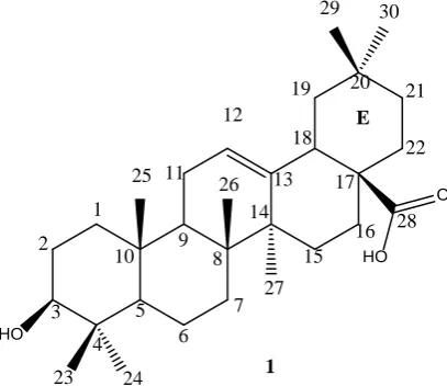
DOI: 10.4236/ijoc.2018.81011 161 International Journal of Organic Chemistry active points: C3, where hydroxyl is stored; the double bonds present in the ring between the C12 and C13 carbons; and C28, where carboxylic acid is stored (Figure 1); these points enable modifications, which lead to the creation of new chemical compounds [14] [15].
However, OA has limited bioavailability and does not provide a good plasma half-life because of its low solubility in water [16] [17]. Several studies have demonstrated that the structural alterations in OA can have a significant impact on biological activities [18]. The improvements in biological activities, such an-ti-inflammatory, antidiabetic, nephropathy, and cytotoxicity, have been achieved by changing some points in the OA structure [19] [20]. Terpenes complexes were very effective in several diseases [21].
Tabrizi et al. [22] showed that ruthenium (II) p-cymene complexes of naph-thoquinone derivatives worked powerfully to combat melanomas in humans. Ghosh et al. [23] demonstrated the antioxidant activity of the quercetin-magnesium complex, and similar benefits were recorded for the complexation of copper with quercetin. The use of the bactericidal properties of calcium and copper com-plexes against Gram-positive bacteria has also been demonstrated [24].
Taking into consideration the aforementioned and potential medical uses of OA and its derivatives, five new compounds were synthesized using the OA.
2. Experimental
2.1. General Procedures and Equipment
[image:2.595.271.475.527.703.2]The 1H and 13C experiments were performed on a Bruker 600-MHz NMR spec-trometer equipped with an AVANCE III console, and a DCH cryogenically cooled probe. The conditions for 1H experiments were as follows: Bruker pulse sequence = zg30; the number of acquisition points = 65,536; the number of ac-quisitions = 128; sweep width = 20.55 ppm; recycle delay = 2 s; frequency = 600.13 MHz. The conditions for 13C experiments were as follows: Bruker pulse sequence = zgpg30; the number of acquisition points = 65,536; the number of
Figure 1. Molecular structure of oleanolic acid.
O
HO
HO
E
1
2
3
4 5
6 7 8 9 10
11 12
13
14
15 16 17 18
19 20 21
22
28
29 30
26 25
24 23
27
DOI: 10.4236/ijoc.2018.81011 162 International Journal of Organic Chemistry acquisitions = 4096; sweep width = 238.9 ppm; recycle delay = 2 s; frequency = 150.9 MHz. Deuterated methanol, CD3OD, was used as the solvent.
The IR spectra were recorded on a Perkin Elmer Paragon 1000 FTIR spectro-photometer in the ATR mode over the range 400 - 4000 cm−1.
The analyses were performed on LC-MS-QTOF-6530 model L-1200 (Agilent brand) with the software Profinder series B.06.00; flow: 0.6 mL/min-C18 column - 1.8 μM, 3 mm × 100 mm. The mobile phase A: 0.1% formic acid in water, mobile phase B: 0.1% formic acid in acetonitrile.
2.2. Isolation of Oleanolic Acid
The leaves of Plumeria alba were collected and dried in a greenhouse, after dry-ing were submitted to extraction with ethanol in the ratio 6:1 solvent for the sample. After 7 days, the material was filtered, and the solvent was removed us-ing reduced pressure. The sample was subjected to the silica gel 60 (0.063 - 0.2 mm/70 - 230 mesh ASTM) filter column [25] [26] (Macherey-Nagel, Germany) and fractionated with one liter of each of the following eluents: hexane, dichlo-romethane, ethyl acetate, methanol, and water. The solutions obtained were concentrated and analyzed by CCD. The pre-selected extracts were subjected to classical silica gel chromatography and Sephadex® LH-20 for the isolation and purification of compounds.
2.3. Melting Temperature
The melting temperatures were determined on MQAPF 320 equipment from Microquímica Equipamentos Ltda.
2.4. Complex Reaction
A round bottom flask (500 mL) was charged with 1 equivalent of metal sulfate to 2 equivalent of OA and 300 mL methanol. The resulting reaction mixture was vigorously stirred at 57˚C for 6 hours. The solution was cooled to room temper-ature, and the methanol was removed using reduced pressure to obtain the compounds (Figure 1) [27]. The material was measured and calculated the yield the reaction.
2.5. RNA Extraction and qPCR
Total RNA was isolated from H4IIE cells or from tissue from rats maintained with liquid nitrogen, using the TRIzol reagent (Life Technologies). The total RNA was isolated from cells using TRIzol reagent (Life Technologies). RNA was quantified using Synergy H1/Take 3 plate setup (BioTek). The cDNAs were synthesized using 2 µg of RNA for each sample using high-capacity cDNA Re-verse Transcription kit (Life Technologies) on an ABI GeneAMP 9700 (Life Technologies).
DOI: 10.4236/ijoc.2018.81011 163 International Journal of Organic Chemistry were selected using the Primer Express version 2.0 software (Applied Biosys-tems, Foster City, CA) as follows: β-actin (housekeeping gene), forward primer: 5'-GGG AAA TCG TGC GTG ACA TT-3', reverse primer: 5'-GCG GCA GTG GCC ATC TC-3'; G6Pase, forward primer: 5'-TGT TCC TCT TAA TCC TGC CCA-3', reverse primer: 5'-CCA ACC TGC ACA AGT TCC CTT-3'. qPCR am-plifications were performed on an ABI 7500 Fast real-time PCR (Life Technolo-gies) using 1 cycle at 50˚C for 2 min and 1 cycle of 95˚C for 10 min, followed by 40 cycles of 15 s at 95˚C and 1 min at 60˚C. The dissociation curve was com-pleted with 1 cycle of 1 min at 95˚C, 30 s at 55˚C, and 30 s at 95˚C. mRNA ex-pression was analyzed using the ΔΔCT method, and normalized concerning the expression of the β-actin using ABI 7500 Fast System SDS Software v1.3.0 (Life Technologies). Amplification of specific transcripts was further confirmed by obtaining melting curve profiles. All results were expressed as fold change from the induced Dex-cAMP controls. For the assays, samples at the concentration of 5.0 mg/100 μl were used with 5.0 μl of the cDNA.
3. Results and Discussion
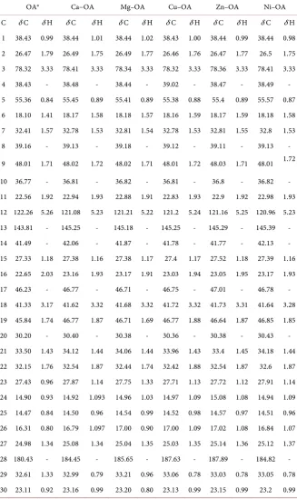
After the use of three chromatographic columns with different mobile phases and monitoring the separation with thin layer chromatography and with the de-velopers phosphomolybdic acid and vanillin, the oleanolic acid compound was obtained, where it was confirmed according to the 13C and 1H NMR data in Ta-ble 1.
The synthesized compounds were analyzed using the NMR technique, and the 13C and 1H data for OA were initially obtained for comparison with the NMR
data of the compounds.
The results in Table 1 show that C28 for the compounds with Ca, Mg, Cu, Zn, and Ni has a different displacement value compared to that obtained with OA. Considering C28 as one of the possible reaction points [14] [15], and the data obtained by the IR technique which show the absence of the band for carboxylic acid bound to C28, it is clear that the reaction is formed together with a carbox-ylic acid group. It is still possible to discern a small variation in the displacement of the C12 and C13 carbons and those carrying the double bond. This suggests that electron displacement is promoted by the withdrawal effect exerted by both metals when they are bound to the hydroxyl oxygen of the carboxylic acid group. The structures of the synthesized compounds are also supported by the fol-lowing data.
C30H48O3 (Oleanolic acid) White solid, 0.621 g of the pure compound, m.p. 307˚C - 309˚C, IR (ATR) νmax/cm−1: 3455, 3048, 2932, 2859, 1689, 1460, 1382, 1271, 1031, 954, 658.
C60H94CaO6 It was obtained in 71% yield (0.1150 g, 0.1209 mmol) via reac-tion of the 1.0 eq. of calcium sulfate to 2.0 eq. of oleanolic acid. White solid, IR (ATR) νmax/cm−1: 3370, 2928, 2865, 1537, 1465, 1387, 1136, 1029, 995. LC-MS [M+ H]+ calculate for C
DOI: 10.4236/ijoc.2018.81011 164 International Journal of Organic Chemistry Table 1. 1H and 13C NMR chemical shifts to OA and complexes (δ, ppm) a–oleanolic acid
isolated.
OAa Ca−OA Mg–OA Cu–OA Zn–OA Ni–OA
C δ C δ H δ C δ H δ C δ H δ C δ H δ C δ H δ C δ H 1 38.43 0.99 38.44 1.01 38.44 1.02 38.43 1.00 38.44 0.99 38.44 0.98 2 26.47 1.79 26.49 1.75 26.49 1.77 26.46 1.76 26.47 1.77 26.5 1.75 3 78.32 3.33 78.41 3.33 78.34 3.33 78.32 3.33 78.36 3.33 78.41 3.33 4 38.43 - 38.48 - 38.44 - 39.02 - 38.47 - 38.49 - 5 55.36 0.84 55.45 0.89 55.41 0.89 55.38 0.88 55.4 0.89 55.57 0.87 6 18.10 1.41 18.17 1.58 18.18 1.57 18.16 1.59 18.17 1.59 18.18 1.58 7 32.41 1.57 32.78 1.53 32.81 1.54 32.78 1.53 32.81 1.55 32.8 1.53 8 39.16 - 39.13 - 39.18 - 39.12 - 39.11 - 39.13 -
9 48.01 1.71 48.02 1.72 48.02 1.71 48.01 1.72 48.03 1.71 48.01 1.72
10 36.77 - 36.81 - 36.82 - 36.81 - 36.8 - 36.82 - 11 22.56 1.92 22.94 1.93 22.88 1.91 22.83 1.93 22.9 1.92 22.98 1.93 12 122.26 5.26 121.08 5.23 121.21 5.22 121.2 5.24 121.16 5.25 120.96 5.23 13 143.81 - 145.25 - 145.18 - 145.25 - 145.29 - 145.39 - 14 41.49 - 42.06 - 41.87 - 41.78 - 41.77 - 42.13 - 15 27.33 1.18 27.38 1.16 27.38 1.17 27.4 1.17 27.52 1.18 27.39 1.16 16 22.65 2.03 23.16 1.93 23.17 1.91 23.03 1.94 23.05 1.95 23.17 1.93 17 46.23 - 46.77 - 46.71 - 46.75 - 47.01 - 46.78 - 18 41.33 3.17 41.62 3.32 41.68 3.32 41.72 3.32 41.73 3.31 41.64 3.28 19 45.84 1.74 46.77 1.87 46.71 1.69 46.77 1.88 46.64 1.87 46.85 1.85 20 30.20 - 30.40 - 30.38 - 30.36 - 30.38 - 30.43 - 21 33.50 1.43 34.12 1.44 34.06 1.44 33.96 1.43 33.4 1.45 34.18 1.44 22 32.15 1.76 32.54 1.87 32.44 1.74 32.42 1.88 32.54 1.87 32.6 1.87 23 27.43 0.96 27.87 1.14 27.75 1.33 27.71 1.13 27.72 1.12 27.91 1.14 24 14.90 0.93 14.92 1.093 14.96 1.03 14.97 1.09 15.08 1.08 14.94 1.09 25 14.47 0.84 14.50 0.96 14.54 0.99 14.52 0.98 14.57 0.97 14.51 0.96 26 16.31 0.80 16.79 1.097 17.00 0.90 17.00 1.09 17.02 1.08 16.84 1.07 27 24.98 1.34 25.08 1.34 25.04 1.35 25.03 1.35 25.14 1.36 25.12 1.37 28 180.43 - 184.45 - 185.65 - 187.63 - 187.89 - 184.82 - 29 32.61 1.33 32.99 0.79 33.21 0.96 33.06 0.78 33.03 0.78 33.05 0.78 30 23.11 0.92 23.16 0.99 23.20 0.80 23.13 0.99 23.15 0.99 23.2 0.99
DOI: 10.4236/ijoc.2018.81011 165 International Journal of Organic Chemistry C60H94CuO6 It was obtained in 70% yield (0.0989 g, 0.1015 mmol) via reaction of the 1.0 eq. of copper sulfate to 2.0 eq. of oleanolic acid. Green solid, IR (ATR) νmax/cm−1: 3395, 2938, 2862, 1537, 1386, 1094, 995, 728. LC-MS [M+ H]+ calcu-late for C60H94CuO6: 973.6346, found 973.6344.
C60H94ZnO6 It was obtained in 68% yield (0.101 g, 0.1036 mmol) via reaction of the 1.0 eq. of zinc sulfate to 2.0 eq. of oleanolic acid. White solid, IR (ATR) νmax/cm−1: 3299, 2940, 2864, 1595, 1461, 1084, 736, 677. LC-MS [M+H]+ calculate for C60H94ZnO6: 974.6342, found 974.6341.
C60H94NiO6 It was obtained in 69% yield (0.0899 g, 0.0928 mmol) via reaction of the 1.0 eq. of nickel sulfate to 2.0 eq. of oleanolic acid. Green solid, IR (ATR) νmax/cm−1: 3320, 2928, 2863, 1534, 1453, 1373, 1029, 772. LC-MS [M+ H]+ calcu-late for C60H94NiO6: 968.6404, found 968.6401.
The shift in displacement promoted by the bonding between the metal (Ca/Mg/Zn/Ni/Cu) with the oxygen of the hydroxyl at C28 indicates the forma-tion of that new bond.
The stretch ν (C=O) at C28 at 1691 cm−1 (Figure 2) present to OA is absent in the IR spectra for all complex formed. Also shows the absence of characteristic stretching ν (C=O) band in the wave-number region between 1715 - 1680 cm−1, and the absence of band resulting from the angular deformation of the ν (OH) bond of the carboxylic acid in the region between 955 - 875 cm−1 [29]. However, these bands are present in the IR spectrum of OA and reinforce the formation of the complex; therefore, five new complexes with OA are created.
As shown (Figure 3) it is possible to identify the formation of the complexes with the metal and OA, the bond between the metal and the oxygen atom pro-motes a displacement of electrons, in this way the NMR values (Table 1) in-crease, such as the values obtained for C28. The same bond, suppresses the band obtained in 1689 cm−1 for OA in the IR experiments.
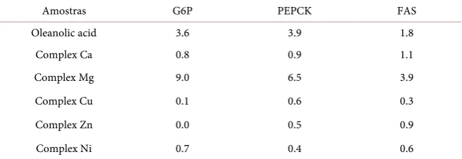
[image:6.595.254.493.525.702.2]The fold change values show the activity of the compounds isolated in trials where three different genes were used (Table 2). The compound synthesized
Figure 2. The infrared spectrum of oleanolic acid.
4000 3500 3000 2500 2000 1500 1000 500
84 86 88 90 92 94 96 98 100
Int
ens
ity
Wavenumber / cm-1
Oleanolic Acid
1689
1460 1382
1031
2932
3408
DOI: 10.4236/ijoc.2018.81011 166 International Journal of Organic Chemistry Figure 3. The molecular structure of the complex formed with calcium, magnesium, copper, zinc, and nickel with oleanolic acid.
Table 2. Results of PCR assays for the complexes of calcium, magnesium, and oleanolic acid.
Amostras G6P PEPCK FAS
Oleanolic acid 3.6 3.9 1.8
Complex Ca 0.8 0.9 1.1
Complex Mg 9.0 6.5 3.9
Complex Cu 0.1 0.6 0.3
Complex Zn 0.0 0.5 0.9
Complex Ni 0.7 0.4 0.6
from oleanolic acid and calcium showed little efficiency in the control of these genes, presenting lower values when compared to pure oleanolic acid, that is, there was a decrease in the biological activity of the synthesized compound. Dif-ferent results were obtained for the compound formed from oleanolic acid and magnesium; the values were higher than the result of the pure oleanolic acid and the compound formed with calcium. Greater success was achieved with the G6P gene reaching a fold change value close to 9.0, for the PEPCK gene, there is also a significant value. Not being so effective for the FAS gene [30] [31].
4. Conclusion
Five novel compounds were synthesized using OA as starting material, the novel and inedited structures were confirmed by NMR, IR and LC-MS experiments. The NMR, IR, and LC-MS data support the literature data for the passive points of reactions in the OA molecule. Among the five new compounds synthesized, the complex formed with magnesium, showed better results for the tests per-formed.
References
[1] Liu, J. (1995) Pharmacology of Oleanolic Acid and Ursolic Acid. Journal of Ethno-O
HO
1
2 3
4 5 6
7 8 9 10 11 12 13 14 15 16 17 18 19 20 21 22 28 29 30 26 25 24 23 27 O O OH 1 2 3 4 5 6 7
8 9 10
11 12 13 14 15 16 17 18 19 20 21 22 28 29 30 26 25 24 23 27 O Me
[image:7.595.207.539.311.425.2]DOI: 10.4236/ijoc.2018.81011 167 International Journal of Organic Chemistry pharmacology, 49, 57-68.https://doi.org/10.1016/0378-8741(95)90032-2
[2] Jäger, S., Trojan, H., Kopp, T., Laszczyk, M.N. and Scheffler, A. (2009) Pentacyclic Triterpene Distribution in Various Plants—Rich Sources for a New Group of Mul-ti-Potent Plant Extracts. Molecules, 14, 2016-2031.
https://doi.org/10.3390/molecules14062016
[3] Shanmugam, M.K., Dai, X., Kumar, A.P., Tan, B.K.H., Sethi, G. and Bishayee, A. (2014) Oleanolic Acid and Its Synthetic Derivatives for the Prevention and Therapy of Cancer: Preclinical and Clinical Evidence. Cancer Letters, 346, 206-216.
https://doi.org/10.1016/j.canlet.2014.01.016
[4] He, X. and Rui, H.L. (2007) Triterpenoids Isolated from Apple Peels Have Potent Antiproliferative Activity and May Be Partially Responsible for Apple’s Anticancer Activity. Journal of Agricultural and Food Chemistry, 55, 4366-4370.
https://doi.org/10.1021/jf063563o
[5] Gambhava, N.S., Ezhava, S.B., Rathod, I.S., Chhabria, M.T. and Patwari, A.H. (2013) Estimation of Ursolic Acid and Oleanolic Acid from Leaves of Plumeria Ob-tuse by HPTLC Method after Iodine Derivatization. Der Pharma Chemica, 5, 44-50. [6] Ovesna, Z., Kozics, K. and Slamenova, D. (2006) Protective Effects of Ursolic Acid and Oleanolic Acid in Leukemic Cells. Mutation Research/Fundamental and Mole-cular Mechanisms of Mutagenesis, 600, 131-137.
https://doi.org/10.1016/j.mrfmmm.2006.03.008
[7] Lee, W., Yang, E.J., Ku, S.K., Song, K.S. and Bae, J.S. (2013) Anti-Inflammatory Ef-fects of Oleanolic Acid on LPS-Induced Inflammation in vitro and in vivo. Inflam-mation, 36, 94-102.https://doi.org/10.1007/s10753-012-9523-9
[8] Hsu, H.Y., Yang, J.J. and Lin, C.C. (1997) Effects of Oleanolic Acid and Ursolic Acid on Inhibiting Tumor Growth and Enhancing the Recovery of Hematopoietic Sys-tem Postirradiation in Mice. Cancer Letters, 111, 7-13.
https://doi.org/10.1016/S0304-3835(96)04481-3
[9] Mengoni, F., Lichtner, M., Battinelli, L., Marzi, M., Mastroianni, C.M., Vullo, V., et al. (2002) In vitro Anti-HIV Activity of Oleanolic Acid on Infected Human Mono-nuclear Cells. Planta Medica, 68, 111-114.
https://doi.org/10.1055/s-2002-20256
[10] Dzubak, P., Hajduch, M., Vydra, D., Hustova, A., Kvasnica, M., Biedermann, D., et al. (2006) Pharmacological Activities of Natural Triterpenoids and Their Therapeu-tic Implications. Natural Product Reports, 23, 394-411.
https://doi.org/10.1039/b515312n
[11] Liu, J. (2005) Oleanolic Acid and Ursolic Acid: Research Perspectives. Journal of Ethnopharmacology, 100, 92-94.https://doi.org/10.1016/j.jep.2005.05.024
[12] Chen, J., Gong, Y., Liu, J., Hua, W., Zhang, L. and Sun, H. (2008) Synthesis and Bi-ological Evaluation of Novel-Pyrazolo [4,3-b]Oleanane Derivatives as Inhibitors of Glycogen Phosphorylase. Chemistry and Biodiversity, 5, 1304-1312.
https://doi.org/10.1002/cbdv.200890117
[13] Place, A.E., Suh, N., Williams, C.R., Risingsong, R., Honda, T., Honda, Y., etal. (2003) The Novel Synthetic Triterpenoid, CDDO-Imidazolide, Inhibits Inflamma-tory Response and Tumor Growth in vivo. Clinical Cancer Research, 9, 2798-2806. [14] Bednarczyk-Cwynar, B., Zaprutko, L., Ruszkowski, P. and Hładoń, B. (2012)
Anti-cancer Effect of A-Ring or/and C-Ring Modified Oleanolic Acid Derivatives on KB, MCF-7 and HeLa Cell Lines. Organic & Biomolecular Chemistry, 10, 2201-2205.
DOI: 10.4236/ijoc.2018.81011 168 International Journal of Organic Chemistry
[15] Garcia-Granados, A., López, P.E., Melguizo, E., Parra, A. and Simeó, Y. (2006) Reactivity of Chiral Sesquiterpene Synthons Obtained by the Degradation of Mas-linic Acid from Olive-Pressing Residues. Synthetic Communications, 36, 3001-3018.
https://doi.org/10.1080/00397910600773858
[16] Chen, H., Gao, Y., Wang, A., Zhou, X., Zheng, Y. and Zhou, J. (2015) Evolution in Medicinal Chemistry of Ursolic Acid Derivatives as Anticancer Agents. European Journal of Medicinal Chemistry, 92, 648-655.
https://doi.org/10.1016/j.ejmech.2015.01.031
[17] Zhang, H., Li, X., Ding, J., Xu, H., Dai, X., Hou, Z., et al. (2013) Delivery of Ursolic Acid (UA) in Polymeric Nanoparticles Effectively Promotes the Apoptosis of GASTRIC CANCER CELLs through Enhanced Inhibition of Cyclooxygenase 2 (COX-2). International Journal of Pharmaceutics, 441, 261-268.
https://doi.org/10.1016/j.ijpharm.2012.11.034
[18] Acebey-Castellon, I.L., Voutquenne-Nazabadioko, L., Doan Thi Mai, H., Roseau, N., Bouthagane, N., Muhammad, D., et al. (2011) Triterpenoid Saponins from Sym-plocos Lancifolia. Journal of Natural Products, 74, 163-168.
https://doi.org/10.1021/np100502y
[19] Masullo, M., Pizza, C. and Piacente, S. (2017) Oleanane Derivatives for Pharma-ceutical Use: A Patent Review (2000-2016). Expert Opinion on Therapeutic Patents, 27, 237-255.https://doi.org/10.1080/13543776.2017.1253680
[20] Suh, N., Wang, Y., Honda, T., Gribble, G.W., Dmitrovsky, E., Hickey, W.F., et al. (1999) A Novel Synthetic Oleanane Triterpenoid, 2-Cyano-3,12-Dioxoolean-1,9-Dien-28-Oic Acid, with Potent Differentiating, Antiproliferative, and Anti-Inflammatory Activi-ty. Cancer Research, 59, 336-341.
[21] Partal Ureña, F., Moreno, J.R.A. and López González, J.J. (2009) Conformational Study of (R)-(+)-Limonene in the Liquid Phase Using Vibrational Spectroscopy (IR, Raman, and VCD) and DFT Calculations. Tetrahedron: Asymmetry, 20, 89-97.
https://doi.org/10.1016/j.tetasy.2009.01.024
[22] Tabrizi, L. and Chiniforoshan, H. (2016) Ruthenium(II) p-Cymene Complexes of Naphthoquinone Derivatives as Antitumor Agents: A Structure Activity Relation-ship Study. Journal of Organometallic Chemistry, 822, 211-220.
https://doi.org/10.1016/j.jorganchem.2016.09.003
[23] Bukhari, S.B., Memon, S., Mahroof-Tahir, M. and Bhanger, M.I. (2009) Synthesis, Characterization and Antioxidant Activity Copper-Quercetin Complex. Spectro-chimica Acta Part A: Molecular and Biomolecular Spectroscopy, 71, 1901-1906.
https://doi.org/10.1016/j.saa.2008.07.030
[24] Jadeja, R.N., Vyas, K.M., Gupta, V.K., Joshi, R.G. and Ratna Prabha, C. (2012) Syn-theses, Characterization and Molecular Structures of Calcium(II) and Copper(II) Complexes Bearing O2-Chelate Ligands: DNA Binding, DNA Cleavage and
An-ti-Microbial Study. Polyhedron, 31, 767-778.
https://doi.org/10.1016/j.poly.2011.11.004
[25] Hench, L.L. and West, J.K. (1990) The Sol-Gel Process. Chemical Reviews, 90, 33-72.https://doi.org/10.1021/cr00099a003
[26] Tubert-Brohman, I., Sherman, W., Repasky, M. and Beuming, T. (2013) Improved Docking of Polypeptides with Glide. Journal of Chemical Information and Model-ing, 53, 1689-1699.
DOI: 10.4236/ijoc.2018.81011 169 International Journal of Organic Chemistry Quimica, 36, 107-127.https://doi.org/10.1590/S0100-46702011000200006
[28] Novotny, L., Abdel-Hamid, M.E., Hamza, H., Masterova, I. and Grancai, D. (2003) Development of LC-MS Method for Determination of Ursolic Acid: Application to the Analysis of Ursolic Acid in Staphylea holocarpa Hemsl. Journal of Pharmaceut-ical and BiomedPharmaceut-ical Analysis, 31, 961-968.
https://doi.org/10.1016/S0731-7085(02)00706-9
[29] Kalođera, Z. and Sofić, E. (2009) Identification and Isolation of Pharmacologically Active Triterpenes in Betula Cortex, Betula Pendula Roth., Betulaceae. Bosnian Journal of Basic Medical Sciences, 9, 31-38.
https://doi.org/10.17305/bjbms.2009.2853
[30] Kim, H.J., Jee, H.J. and Yun, J. (2011) DNA Damage Induces Down-Regulation of PEPCK and G6P Gene Expression through Degradation of PGC-1. Acta Biochimica et Biophysica Sinica, 43, 589-594.https://doi.org/10.1093/abbs/gmr053
[31] Nordgren, J., Nitiema, L.W., Sharma, S., Ouermi, D., Traore, A.S., Simpore, J., et al. (2012) Emergence of Unusual G6P Rotaviruses in Children, Burkina Faso, 2009-2010. Emerging Infectious Diseases, 18, 589-597.