PREDICTION OF TRANSVERSE CEREBELLAR DIAMETER AS A BETTER INDICATOR FOR
ESTIMATION OF GESTATIONAL AGE IN THIRD TRIMESTER
1
Renu Mishra,
1Department of Anatomy Saraswathi Institute of Medical Sciences, Pilkhuwa 2Department of Anatomy, Pandit
ARTICLE INFO ABSTRACT
Introduction:
the cornerstone of modern obstetrics.
measurements become less accurate but recent studies have shown that fetal transcerebellar diameter is one such parameter that has remained consistently superior in prediction GA in both singleton and twin gestation.
TCD & HC measurement in third trimester &correlated the prediction of gestational age by comparing transcerebellar diameter (TCD), Head circumference (HC) and LMP.
and Methods:
Radiodiagnosis, SIMS, Hapur
included antenatal women between 28 to 40 weeks of gestation by applying simple random technique.Ultrasonography (USG) was done between 28 to 4
PNDT form.
HC. Although correlation was found to be statistically significant in both TCD and HC groups but GA had better correlation with TCD than the
present study has revealed that TCD shows good correlation with the LMP for the prediction of gestational age in third trimester, however TCD has a better correlation with LMP than HC for the gestational age estimation.
Copyright©2019, Renu Mishra et al. This is an open access
distribution, and reproduction in any medium, provided
INTRODUCTION
Prediction of gestational age (GA) based on sonographic fetal parameters is the cornerstone of modern obstetrics.
determination of gestational age helps us to estimate the pregnancy and differentiates the normal from abnormal growth patterns. Thus it helps to distinguish between wrong estimate of gestational age and intrauterine growth retardation and to decrease the number of postdated pregnancies and various complications associated with it. The commonly employed method and the standard of care in monitoring gestation is antenatal ultrasound examination. Based on certain fetal parameters the gestational age is calculated and compared with period of gestation to look for fetal growth. Estimation of EDD based on LMP is a simple, low- cost method of estimating gestational age. Limitations associated with the use of menstrual based gestational age estimation inc
problems such as uncertainty regarding the LMP date, possible due to bleeding not associated with menses, as well as concerns about the incidence of delayed ovulation, which can result in invalid estimation, even for women with certain LMP.
ISSN: 0975-833X
Article History:
Received 28th April, 2019 Received in revised form 24th May, 2019
Accepted 29th June, 2019 Published online 31st July, 2019
Citation: Renu Mishra, Rajni, Manu Gupta and Raman khare
gestational age in third trimester”, International Journal of Current Research
Key Words:
Transverse Cerebellar Diameter (TCD), Gestational Age (GA),
Ultrasonography (USG), Period of Gestation( POG), Head Circumference (HC).
*Corresponding author: Rajni
RESEARCH ARTICLE
PREDICTION OF TRANSVERSE CEREBELLAR DIAMETER AS A BETTER INDICATOR FOR
ESTIMATION OF GESTATIONAL AGE IN THIRD TRIMESTER
Renu Mishra,
2, *Rajni,
1Manu Gupta and
1Raman khare
t of Anatomy Saraswathi Institute of Medical Sciences, Pilkhuwa Hapur U.P., India t of Anatomy, Pandit Deendayal Upadhyaya Medical College Churu Rajasthan
ABSTRACT
Introduction:Prediction of gestational age (GA) based on sonographic fetal parameters is the cornerstone of modern obstetrics. In the third trimester all the individu
measurements become less accurate but recent studies have shown that fetal transcerebellar diameter is one such parameter that has remained consistently superior in prediction GA in both singleton and twin gestation. Aims and Objective: To pre
TCD & HC measurement in third trimester &correlated the prediction of gestational age by comparing transcerebellar diameter (TCD), Head circumference (HC) and LMP.
and Methods: Present study was done on 100 pregnant
Radiodiagnosis, SIMS, Hapur from the month of October 2016 to March 2017. Study group included antenatal women between 28 to 40 weeks of gestation by applying simple random technique.Ultrasonography (USG) was done between 28 to 4
PNDT form. Result: Pearson’s correlation coefficient was 0.894 for TCD and 0.856 for HC. Although correlation was found to be statistically significant in both TCD and HC groups but GA had better correlation with TCD than the HC (p<0.001).
present study has revealed that TCD shows good correlation with the LMP for the prediction of gestational age in third trimester, however TCD has a better correlation with LMP than HC for the gestational age estimation.
access article distributed under the Creative Commons Attribution the original work is properly cited.
Prediction of gestational age (GA) based on sonographic fetal parameters is the cornerstone of modern obstetrics. Accurate determination of gestational age helps us to estimate the pregnancy and differentiates the normal from abnormal growth patterns. Thus it helps to distinguish between wrong estimate of gestational age and intrauterine growth retardation and to rease the number of postdated pregnancies and various The commonly employed method and the standard of care in monitoring gestation is antenatal ultrasound examination. Based on certain fetal is calculated and compared with Estimation of EDD cost method of estimating gestational age. Limitations associated with the use of menstrual based gestational age estimation include reporting problems such as uncertainty regarding the LMP date, possible due to bleeding not associated with menses, as well as concerns about the incidence of delayed ovulation, which can result in invalid estimation, even for women with certain LMP.
According to Hertz et al study only 18% of women give the exact date of their LMP. The commonly employed fetal parameters for estimating gestational age include biparietal diameter (BPD), head circumference (HC), abdominal circumference (AC) and femur
gestational age by measuring fetal parameters is maximum in first trimester and accuracy reduces as fetal age advances from second to third trimester. By the third trimester all the individual parameter measurements become less
recent studies have shown that fetal transcerebellar diameter is one such parameter that has remained consistently superior in prediction GA in both singleton and twin gestation. accurate measurement of these parameters depends a lot on fetal lie, shape of skull, location of placenta, flexion of fetal head and engagement, maternal obesity and multiplicity of gestation. More recently, transcerebellar diameter (TCD) has evolved as a promising indicator for assessing fetal growth and gestational age. The present prospective study was envisaged in order to explore the reliability of transcerebellar diameter (TCD) and Head circumference(HC) in third trimester of gestation respectively in Indian population.
International Journal of Current Research Vol. 11, Issue, 07, pp.5678-5682, July, 2019
DOI: https://doi.org/10.24941/ijcr.35940.07.2019
Renu Mishra, Rajni, Manu Gupta and Raman khare, 2019. “Prediction of Transverse cerebellar diameter as a better indicator for estimation of
International Journal of Current Research, 11, (07), 5678-5682.
PREDICTION OF TRANSVERSE CEREBELLAR DIAMETER AS A BETTER INDICATOR FOR
ESTIMATION OF GESTATIONAL AGE IN THIRD TRIMESTER
Raman khare
Hapur U.P., India Upadhyaya Medical College Churu Rajasthan, India
Prediction of gestational age (GA) based on sonographic fetal parameters is In the third trimester all the individual parameter measurements become less accurate but recent studies have shown that fetal transcerebellar diameter is one such parameter that has remained consistently superior in prediction GA in To predict gestational age using TCD & HC measurement in third trimester &correlated the prediction of gestational age by comparing transcerebellar diameter (TCD), Head circumference (HC) and LMP. Materials
done on 100 pregnant women in the Department of from the month of October 2016 to March 2017. Study group included antenatal women between 28 to 40 weeks of gestation by applying simple random technique.Ultrasonography (USG) was done between 28 to 40 weeks of POG, after filling Pearson’s correlation coefficient was 0.894 for TCD and 0.856 for HC. Although correlation was found to be statistically significant in both TCD and HC HC (p<0.001). Conclusion:The present study has revealed that TCD shows good correlation with the LMP for the prediction of gestational age in third trimester, however TCD has a better correlation with
License, which permits unrestricted use,
Hertz et al study only 18% of women give the The commonly employed fetal parameters for estimating gestational age include biparietal diameter (BPD), head circumference (HC), abdominal circumference (AC) and femur length (FL). The accuracy of gestational age by measuring fetal parameters is maximum in first trimester and accuracy reduces as fetal age advances from second to third trimester. By the third trimester all the individual parameter measurements become less accurate but recent studies have shown that fetal transcerebellar diameter is one such parameter that has remained consistently superior in prediction GA in both singleton and twin gestation. The accurate measurement of these parameters depends a lot on etal lie, shape of skull, location of placenta, flexion of fetal head and engagement, maternal obesity and multiplicity of transcerebellar diameter (TCD) has evolved as a promising indicator for assessing fetal growth and The present prospective study was envisaged in order to explore the reliability of transcerebellar diameter (TCD) and Head circumference(HC) in third trimester of gestation respectively in Indian population.
INTERNATIONAL JOURNAL OF CURRENT RESEARCH
Aims and objectives
1. To predict gestational age using TCD& HC measurement in third trimester.
2. To correlated the prediction of gestational age in third trimester by comparing transcerebellar diameter (TCD), Head circumference (HC) with LMP.
MATERIALS AND METHODS
Present study was conducted in the Department of Anatomy and Department of Radiodiagnosis in the Saraswathi Institute of Medical Sciences, Hapur, U.P. from the month of October 2016 to March 2017. Study group included 100 antenatal women of third trimester between 28 to 40 weeks of gestation by applying simple random technique. Ultrasonography (USG) was done between 28 to 40 weeks of POG, after filling PNDT form. Informed consent was obtained.
Inclusion criteria
1. Singleton pregnancy.
2. Gestational age between 28 to 40 weeks. 3. Patient sure of her last menstrual period (LMP) 4. Previous regular menstrual cycles.
Exclusion criteria
1. Patient not sure of her last menstrual periods. 2. Multiple pregnancies.
3. Previous irregular cycles
4. Fetuses having any gross congenital anomaly 5. Patients having any chronic medical illness.
The method of study
USG machine used was of model PHILIP HDI 4000. All the routine fetal parameters i.e. biparietal diameter (BPD), abdominal circumference (AC) femur length (FL) and head circumference (HC) were taken. Gestational age (GA) was then calculated using BPD, HC and by using TCD and the results were subjected to statistical analysis. In addition, the amount of liquor, placental localization, estimated fetal weight and any gross congenital abnormality (GCA) in the fetus were noted. USG was done mainly at 32 to 36 weeks of POG after filling PNDT form. All the fetal parameters including TCD were noted & the gestational age was calculated using HC and by using TCD and the GA were compared and analyzed statistically. The amount of liquor, placental localization estimated fetal weight any GCA was noted. GA was predicted in 3rd trimester using HC& TCD measurement by using the nomograms given by Chavez et al. in 2004 in both second and third trimester. The results were compared with the actual GA calculated from the LMP and statistical analysis was carried out. Correlation and regression equations were then applied to assess the concordance and the accuracy of these parameters.
Techniques of measurement
Transverse cerebellar diameter: TCD is mainly measured in
transcerebellar plane. The measurement of TCD was obtained by placing electronic calipers at outer to outer margins of cerebellum. The landmarks of thalami, cavum, septum pellucidum and third ventricle were identified thereby slightly
[image:2.595.308.561.131.360.2]rotating the transducer below the thalamic plane. The posterior fossa was revealed with the characteristic butterfly like appearance of cerebellum. In all cases cerebellum was seen as two lobules on either side of midline in the posterior cranial fossa. The measurement is obtained by positioning the calipers on the outer margins of the two hemispheres.
Figure 1. Measurement of Transverse Cerebellar Diameter
Head circumference
The head circumference (HC) is better gestational age evaluation if the head shape is flattened (dolichocephaly) or rounded (bracycephaly), this measurement is more reliable than the BPD. The fetal is assumed to be an ellipse. The ellipse circumference serves as an estimate of the fetal head circumference. HC is measured on the same plane as BPD,that is on an axial plane that traverses the thalami, cavum and septum pellucidum. Transducer must be perpendicular to the central axis of the head, and thus the hemispheres and calvaria should appear symmetric. Alternatively, HC may be calculated from BPD and occipito-frontal diameter (OFD) as: HC = 1.62 × (BPD + OFD).
Figure 2. Measurement of Head circumference
[image:2.595.307.559.562.774.2]OBSERVATIONS AND RESULT
[image:3.595.74.251.165.217.2]In this prospective study, 100 antenatal women coming to the Department of Radiodiagnosis, SIMS, Hapur, U.P, without high risk factor were selected. USG was performed between 28-40 weeks. The biometric parameters HC & TCD measured ultrasonographically.
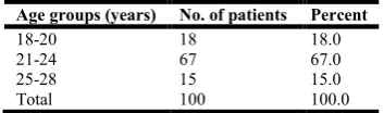
Table 1. Age distribution of the study group women
Age groups (years) No. of patients
18-20 18
21-24 67
25-28 15
Total 100
[image:3.595.36.289.259.362.2]67% of the study group women belonged to the age group of 21-24 years. Mean age in the study group was 22.5 years.
Table 2. Distribution of the study group women into according to their background
Residence No. of patients Percent
Rural 41 41.0
Urban 59 59.0
[image:3.595.303.564.306.362.2]Total 100 100.0
Table 3. Distribution of the study group women according to their parity
Parity No. of women Percent
G1 34 34.0
G2 43 43.0
G3 21 21.0
G4 2 2.0
Total 100 100.0
Majority of the patients (43%) in our study group were second gravida.
In this prospective study, 100 antenatal women coming to the Department of Radiodiagnosis, SIMS, Hapur, U.P, without selected. USG was performed between 40 weeks. The biometric parameters HC & TCD measured
Age distribution of the study group women
Percent
18.0 67.0 15.0 100.0
67% of the study group women belonged to the age group of 24 years. Mean age in the study group was 22.5 years.
Distribution of the study group women into according to
Percent
41.0 59.0 100.0
Distribution of the study group women according to their
Percent
study group were second
Table 4. Gestational age prediction by HC in USG POG (32-36 weeks)
Gestational according to LMP USG
POG 34.38±1.08
Mean period of gestation was 34 weeks 3 days by LMP. period of gestation predicted by HC was 32 weeks 3 days.
Table 5. Gestational age prediction by TCD in USG POG (32-36 weeks)
Gestational according to LMP USG
POG 34.38±1.08
Mean period of gestation was 34 weeks 3 days by LMP. Mean period of gestation predicted by HC was 33 weeks 4 days.
Table 6. Gestational age prediction by HC and TCD in USG POG (32-36 weeks)
Gestational according to LMP
POG by HC in weeks
USG
POG 34.38±1.087 32.45±1.04
Second USG was performed between 32
and gestational age was predicted by measuring HC and TCD. A comparison of estimated gestational age by
[image:3.595.41.286.386.565.2]biometric parameters and by LMP is depicted in Table 6.
Table 7. Statistical correlation of POG by HC and TCD with POG by LMP
Gestational according to LMP
Pearson Correlation Sig. (1-tailed) N
POG by HC in wks
Pearson Correlation Sig. (1-tailed) N
POG by TCD in wks
Pearson Correlation Sig. (1-tailed) N
The Pearson’s correlation coefficient was 0.894 for TCD and 0.856 for HC. Although correlation was found to be statistically significant in both TCD and HC groups but GA had better correlation with TCD than the HC.
Gestational age prediction by HC in USG POG 36 weeks)
Gestational according to LMP POG by HC in weeks P value
32.45±1.04 P
<0.001
Mean period of gestation was 34 weeks 3 days by LMP. Mean period of gestation predicted by HC was 32 weeks 3 days.
Gestational age prediction by TCD in USG POG 36 weeks)
Gestational according to LMP POG by HC in weeks P value
33.65±1.06 P<0.005
was 34 weeks 3 days by LMP. Mean period of gestation predicted by HC was 33 weeks 4 days.
Gestational age prediction by HC and TCD in USG POG 36 weeks)
POG by TCD in weeks
P value
33.65±1.06
LMP vs HC p <0.001 LMP vs TCD p <0.005 HC vs TCD p <0.001
Second USG was performed between 32-36 weeks of gestation and gestational age was predicted by measuring HC and TCD. A comparison of estimated gestational age by using these biometric parameters and by LMP is depicted in Table 6.
Statistical correlation of POG by HC and TCD with POG by LMP
POG by LMP in weeks
POG by HC in weeks
POG by TCD in weeks
Pearson Correlation 1 .856** .894**
.000 .000
100 100 100
Pearson Correlation .856** 1 .814**
.000 .000
100 100 100
Pearson Correlation .894** .814** 1
.000 .000
100 100 100
[image:3.595.308.562.432.597.2] [image:3.595.315.553.638.746.2] [image:3.595.35.293.707.805.2]DISCUSSION
Accurate determination of gestational age is fundamental to obstetric care and is important in a variety of situations. Fetal growth retardation or macrosomia may be missed owing to errors in gestational age assessment. Proper decisions regarding the presumed labour or postdated pregnancies are only possible when gestational age is accurately estimated. All methods of gestational age assessment have merits and demerits that deserve careful consideration. Primary methods of gestational age estimation: estimation based on last menstrual period (LMP), ultrasound-based gestational age estimation and neonatal estimating gestational age. Estimation of EDD based on LMP is a simple, low- cost method of estimating gestational age but its limitations include reporting problems such as uncertainty regarding the LMP date, possible due to bleeding not associated with menses, as well as concerns about the incidence of delayed ovulation, which can result in invalid estimation, even for women with certain LMP. A certain percentage of women comes to the obstetrician for the first time for their antenatal check up in the last trimester only without any previous visits or ultrasound. We faces problems regarding their gestational age estimation, ultrasound done in third trimester for these group of women will give a gestational age estimation with an error of 3 weeks by using of current parameters (HC, TCD). This is associated with the problems regarding the termination of pregnancy if required and issues related to prematurity, postmaturity and the perinatal problems associated with them. Obstetric management most appreciate this potential for error. If the gestational age from the average of all the parameters are similar, assignment of gestational age from the average of all of the parameters improves the accuracy. If gestational age estimates by the various parameters are quite different, averaging multiple parameters decrease the accuracy of the best predictors. a patient presenting in spontaneous labor at 33 ± 3 weeks gestation should be managed as if the pregnancy may be as little as 30 weeks gestation, rather than as advanced at 36 weeks gestation. The patient presenting for prenatal care at 39 ± 3 weeks gestation, should be managed for the potential of postdated pregnancy.
The present study is an endeavor to determine the accuracy of TCD and HC for gestational age estimation in third trimester antenatal women and the neonatal outcome associated with it. Doublet studied the reliability of second and third trimester fetal measurements for gestational age estimation and concluded that BPD was able to predict the GA with an error of 1.4 wks in 14 to 20 wks of POG. The observation of present study conquer with the results of the above investigator. Varol
et al., studied USG preformed in the third trimester between 32-36 weeks showed the mean GA calculated by LMP to be 34-48 wks (34 wk 3 days). The mean GA calculated by HC was 32.45 wks (32wk 3 days.) with an error of 2 wks. Benson C.B et al studied through the sonographic prediction of gestational age using HC found that the error in GA estimation was 3.4 wks (at 26-32 wks of POG) and was 3.8 wks (at 32-42 wks of POG). They found that HC was not a very good predictor of GA estimation in third trimester. Contrarily, the present study has revealed that GA estimated by using HC had and error of 2 weeks only. The difference may possibly be attributed to racial variation. Mean gestational age calculated by TCD was 33.65 wks (33wks 4 days), which mean that TCD was able to predict the GA with an error of 6 days. Chavez M.R et al studied the GA prediction using fetal TCD and found
out that predicted GA between 29-36 wks was within 5 days of the actual GA, which is quiet in agreement with the results of our study. Pearsons correlation coefficient (r) was calculated by applying statistical analysis and correlation was calculated for POG in third trimester by TCD & HC. Correlation of POG by TCD with POG by LMP was found to be 89.4% (r = .894, P < 0.001) which was statistically significant. Chavez et al. study on fetal TCD measurement for gestational age estimation also shown similar results. Their study has shown a significant correlation between POG by TCD and LMP. The concordance between the actual GA and the predicted GA was high (r=0.81, P< 0.001) in the third trimester. However the accuracy was superior in second trimester than in third trimester. Correlation of POG by HC with POG by LMP was found to be 85.6% (r=.856, p<0.001) in our study which was statistically significant. Accuracy for gestational age estimation by TCD and HC was predicted in third trimester using regression equations. These results have GA can be predicted accurately in 79.8% of the women using TCD alone in 3rd trimester. However GA can be predicted accurately in only 73.3% of the women using HC alone in 3rd trimester. Further if both parameters are used together, the accuracy was 84.5%, which shown that accuracy got improved by 4.7% when both parameters were used. Thus evident that TCD alone can be used to predict the GA accurately in around 80% of the patients, and accuracy could be improved by around 5% by using TCD as well as HC for gestational age estimation in third trimester. This improved the accuracy by around 5%. Hadlock et al. predicted the GA using fetal head circumference and found that the accuracy was only 61% in third trimester (30-36 wks.). However the present study has revealed that accuracy with HC was 73%. It is evident that HC alone does not serve as a very reliable parameter for estimation of gestational age in third trimester. Thus, in conclusion it can be stated that TCD as a more reliable parameter than BPD and HC gestational age prediction in third trimester. Moreover it can be used in cases where LMP is not known and in unbooked cases.
Conclusion
The present study was conducted with a view to explore the applicability of transcerebellar diameter along with head circumference and biparietal diameter for prediction of gestational age in Indian subjects &the findings concludes that the TCD is a better predictor of gestational age in third trimester as well an error of just 6 days when compared with HC which had an error of 2 wks. The accuracy of TCD for GA prediction is found to be 79.8% which is better than HC who had a predictive accuracy of just 73% in third trimester. However the accuracy improved by 5% when both HC and TCD are used together.
REFERENCES
Benson CB, Doubilet PM. 1991. Sonographic prediction of gestational age: accuracy of second- and third-trimester fetal measurements. AJR Am J Roentgenol., 157(6):1275-7. Berg AT. 1991. Menstrual cycle length and the calculation of
gestational age. Am J Epidemiol., 133(6):585-9.
Chavez MR, Ananth CV, Kaminsky LM, Smulian JC, Yeo L, Vintzileos AM. 2006. Fetal transcerebellar diameter measurement for prediction of gestational age in twins. Am J Obstet Gynecol., 195(6):1596-600.
Chavez MR, Ananth CV, Smulian JC, Vintzileos AM. 2007. Fetal transcerebellar diameter measurement for prediction of gestational age at the extremes of fetal growth. J
Ultrasound Med., 26(9):1167-71.
Chavez MR, Ananth CV, Smulian JC, Yeo L, Oyelese Y, Vintzileos AM. 2004. Fetal transcerebellar diameter measurement with particular emphasis in the third trimester: a reliable predictor of gestational age. Am J Obstet Gynecol., 191(3):979-84.
Hadlock FP, Deter RL, Harrist RB, Park SK. 1987. Computer assisted analysis of getal age in third trimester using multiple fetal growth parameters. J Clin Ultrasound.,
11:313-6.
Hadlock FP, Kent WR, Loyd JL, Harrist RB, Deter RL, Park SK. 1982. An evaluation of two methods for measuring fetal head and body circumferences. J Ultrasound Med.,
1(9):359-60.
Hertz RH, Sokol RJ, Knoke JD, Rosen MG, Chik L, Hirsch VJ. 1978. Clinical estimation of gestational age: rules for avoiding preterm delivery. Am J Obstet Gynecol.,
131(4):395.
Lee W, Barton S, Comstock CH, Bajorek S, Batton D, Kirk JS. 1991. Transverse cerebellar diameter: a useful predictor of gestational age for fetuses with asymmetric growth retardation. Am J Obstet Gynecol., 165(4 Pt 1):1044-50. L'ubuský M, Mícková I, Procházka M, Dzvincuk P, Malá K,
Cízek L, et al. 2006. Discrepancy of ultrasound biometric parameters of the head (HC--head circumference, BPD--biparietal diameter) and femur length in relation to sex of the fetus and duration of pregnancy. Ceska Gynekol.,
71(3):169-72.
Malik R, Panda VK, Srivastava P. 2003. Gestational age estimation using transcerebellar diameter with grading of
fetal cerebellum and evaluation of TCD/AC ratio as a gestational age independent parameter. IJRI, 13:95:97. Meyer WJ, Gauthier DW, Goldenberg B, Santolaya J, Sipos J,
Cattledge F. 1993. The fetal transverse cerebellar diameter/abdominal circumference ratio: a gestational age-independent method of assessing fetal size. J Ultrasound Med., 12(7):379-82.
Ott WJ. 1984. The use of ultrasonic fetal head circumference for predicting expected date of confinement. J Clin Ultrasound, 12(7):411-5.
Saito M, Yazawa K, Hashiguchi A, Kumasaka T, Nishi N, Kato K. 1972. Time of ovulation and prolonged pregnancy.
Am J Obstet Gynecol, 112(1):31-8.
Schwimer SR, Lebovic J. 1984. Transvaginal pelvic ultrasonography. J Ultrasound Med., 3(8):381-3.
Sharma M, Suri V, Vasishta K. Fetal transverse cerebellar diameter and abdominal circumference ratio in intrauterine growth retardation. J Obstet Gynecol Ind., 51:77-81. Snijders RJ, De Courcy-Wheeler RH, Nicolaides KH. 1994.
Intrauterine growth retardation and fetal transverse cerebellar diameter. Prenat Diagn., 14(12):1101-5.
Taipale P, Hiilesmaa V. 2001. Predicting delivery date by ultrasound and last menstrual period in early gestation.
Obstet Gynecol., 97(2):189-94.
Vantanasiri C, Saifon MB, Titapant V, Kanokongsakdi S, Phatithattakorn C, Sawasdimogkol S, et al. 2006. R$eference centile chart for transverse cerebellar diameter (TCD) in Thai fetuses throughout gestation. SMJ, 58:1010-1012.
Varol F, Saltik A, Kaplan PB, Kiliç T, Yardim T. 2001. Evaluation of gestational age based on ultrasound fetal growth measurements. Yonsei Med J., 42(3):299-303.