Cell Viability of
Bifidobacterium lactis
Strain in Long-Term
Storage Butter Assessed with the Plate Count
and Fluorescence Techniques
Magdalena Olszewska
1, Bogusław staniewski
2and Łucja Łaniewska-trOkenheiM
11Chair of industrial and Food Microbiology and 2Chair of Dairy science and Quality Management,
Faculty of Food science, University of warmia and Mazury in Olsztyn, Olsztyn, Poland
Abstract
Olszewska M., Staniewski B., Łaniewska-Trokenheim L. (2012): Cell viability of Bifidobacterium
lactis strain in long-term storage butter assessed with the plate count and fluorescence techniques.
Czech J. Food Sci., 30: 421–428.
Great interest in functional products containing bacterial strains displaying health-promoting properties is expressed worldwide and is as a result connected with a demand for developing new probiotic-based products, especially those containing bifidobacteria. The Bifidobacterium strains play a key role in gastrointestinal homeostasis, providing many health-related attributes, but as fastidious microorganisms require specific conditions (e.g. anaerobic environment, neutral pH) to survive in the long-term at the needed level above 106 CFu/g. In consequence, not every food product
guarantees optimal maintenance of Bifidobacterium viability. From this point of view, the objective of the study was to examine the survival of Bifidobacterium lactis strain in butter during long-term refrigerated storage. Two enu-meration techniques: microscopic LIVE/DEAD® and plating were compared by monitoring bifidobacterial counts for 4 weeks. The plate method was characterised by underestimation of the cell counts in relation to the results evalu-ated microscopically. However, the good survival exhibited by B. lactis was found with both techniques. Moreover, the microscopic LIVE/DEAD® method permitted to trace delicate changes in the viable/non-viable bifidobacterial population at the single-cell level.
Keywords: Bifidobacterium sp.; survival; food products; LIVE/DEAD® method; plate count method
Bacteria of the Bifidobacterium genus were dis-covered and described for the first time by Tissier in 1899 as microflora of breast-fed newborns. A natural environment for Bifidobacterium sp. is the human and animal gastrointestinal tracts. The population size of these bacteria in the gastroin-testinal tract is high, amounting to 108–109 CFu/g,
but it is subject to changes, depending mainly on the diet and the age of the organism (Soomro et al. 2002). Many health-related properties are at-tributed to these bacteria, including the regulation of intestine microbial homeostasis, inhibition of
pathogenic bacteria, modulation of immunological response, anticancerogenic effects, production of bacteriocins or bioconversion of the diet com-ponents into the so-called bioactive compounds (Soomro et al. 2002; Hernandez-Mendoza et al. 2007).
response to such demand is broadening and im-proving the range of products of therapeutic and prophylactic properties (Antunes et al. 2007). The concentration of probiotic bacteria required to gain health-promoting benefits is often valu-ated as > 106 CFu/ml in the small intestine and
> 108 CFu/ml in the large intestine (Sanders
2003; Bertazzoni Minelli & Benini 2008). In the light of this, one should consume an adequate number of live and active cells, but there is simply not yet enough evidence to recommend the most appropriate dosage that is valid to all probiotic bacteria. The dose required differs significantly depending on the strain and product. Some prod-ucts have proved to be efficient at lower levels, while some necessitate considerably higher ones, in the order of 10-fold or 100-fold in terms of CFu. The common dosage in humans is referred to as 107–109 CFu/ml (mg) per day (Bertazzoni
Minelli & Benini 2008). The role of the matrix in bringing functional effects also has to be taken into account – the viability of the strain in a par-ticular product that will remain up to the end of the shelf-life (Grand et al. 2003; Suvarna & Boby 2005; Hernandez-Mendoza et al. 2007). With this aim in view, monitoring the viability of intestine-derived bacteria in different types of products is required. Epifluorescence microscopy can be an advantage in this type of research. This method, after an appropriate sample preparation, makes it possible to determine relatively quickly the number of microorganisms in food products (Warmińska-Radyko et al. 2010). The plate count method, widely used in determining bacteria count, is based on the cell ability to proliferate and form colonies on a solid medium. Epifluorescence microscopy, depending on the staining technique applied, allows the users to specify the number of live cells on the basis of cytoplasmic membranes integrity, for example. One such method is LIVE/ DEAD® staining. This assay includes a set of two
fluorochromes: SYTO9, which penetrates the cell through the integrated, as well as damaged membrane, and propidium iodide (PI), which can penetrate only a damaged membrane (Breeuwand & Abee 2000; Warmińska-Radyko et al. 2010). In effect, live cells can be distinguished as visibly fluorescing green, and dead – as fluorescing red. The monitoring of subtle changes in the dynamics of the proliferation/decline of the population at the level of single cells is the basic advantage of its application in studies on the physiology of
micro-organisms (Joux & Lebaron 2000; Łaniewska-Trokenheim et al. 2010).
Therefore, a study has been undertaken into determining the viability of intestine-derivative Bifidobacterium lactis strain in butter during a four-week refrigerated storage. After microbiologi-cal analysis with the application of the fluorescence and plate methods, the size of the Bifidobacterium population assessed by those two techniques was compared.
MATERIAL AND METHODS
Preparation of butter product.
Bifidobacte-rium lactis B100.6 strain (CSK Food Enrichment, Toruń, Poland) was studied. The model butters were prepared in seven repetitions by inoculating churned, pasteurised cream (30% (v/v) fat) with > 106 CFu/mlof the given culture. The prepared
butters were stored at 6°C for 4 weeks. Sampling was performed at selected time intervals 0, 1, 2, 3, and 4 weeks.
Enumeration procedure. To estimate the live/
dead bifidobacterial counts, the samples were ex-amined by Direct Epifluorescent Filter Technique using LIVE/DEAD®BacLightTM Bacterial Viability
Kit (Molecular Probes, Eugene, uSA). The LIVE/ DEAD® BacLightTM was performed according to the
peptone (casitone) 20.0 g/l; cystin 0.4 g/l; lactose 20.0 g/l; CH3COONa·6H2O 10.0 g/l; MgSO4·7 H2O 1 g/l; Na2HPO4·12 H2O 4.66 g/l; KH2PO4 1.8 g/l; p-aminobenzoic acid 2.0 g/l; calcium pantothenate 2.0 g/l; biotin 20 µg; agar 1.8%; plus supplement g/100 cm3 ddH
2O: lithium chloride 6.0; nalidixic
acid 0.03; neomycin sulphate 0.2; paramycin sulphate 0.25) and incubated anaerobically (Anaerocult A; Merck, Darmstadt. Germany) at 37°C/72 hours.
Statistical analysis. After verifying the test
as-sumptions, the significance of differences of the results obtained was based on the following statis-tical analysis. Analysis of variance (ANOVA) with repeated measurements was used to verify that the population size of B. lactis evaluated by microscopic counts vary significantly over time. Anova Fried-man’s Rank Test was applied to resolve whether the bifidobacterial counts determined by the plate method change statistically during storage. Mann-Whitney u Test was used to compare the results of the number of bifidobacteria determined by two methods of quantitative analysis, and for this purpose the comparison of the results in each measurement point (P0,1,2,3,4) was made. The results of this test re-solve whether the numbers of cells determined using those methods differed significantly. The significance of differences was analysed with respect to P < 0.05. The calculations were performed using Statistica StatSoft® Vers. 9 (StatSoft Inc., Tulsa, uSA).
RESULTS AND DISCUSSION
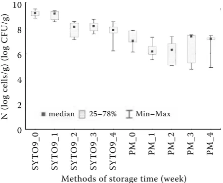
At each stage of butter storage, significant differ-ences were found between the counts of B. lactis assessed by the fluorescence method and by the plate method (P0,1,2,3: P = 0.002; P4: P = 0.04). The cell count determined by the plate method at each measurement point (P0,1,2,3,4) was underestimated in relation to the count of live cells determined by the microscopic SYTO9 method (Figure 1). The value of the so-called underestimation of the plate method depended on the storage stage, and it was the highest in the samples of butter at zero determination and after one week – the difference in the numbers of cells between the plate and the microscopic SYTO9 methods was 2 logarithmic units. The results of determination by the plate method were also characterised by a larger range in comparison to the results obtained by the microscopic SYTO9 method, except for the last measurement, in which minimum-maximum
values determined by the SYTO9 method affected the proximity of exceeding the criterion of the lack of differences between the determinations by both methods (P = 0.04).
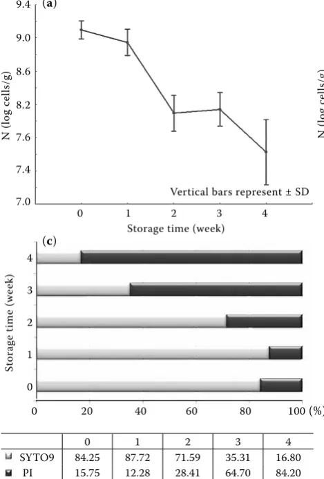
The variation analysis showed that the count of B. lactis assessed by the plate method did not signifi-cantly change during the butter storage (P = 0.14), maintaining a high level of about 7 logarithmic units of CFu/g (Figure 1). Discrepancies in the results concerning the cell count determined by the plate method were the reason for introducing additional statistical calculations, comparing the numbers of bacteria in arrangements: P0↔1; 1↔2; 2↔3; 3↔4 separately, and also here, no differences were found towards the established significance level. According to the determinations by the microscopic method with SYTO9, the count of live cells at the initial stage of storage was found at the level of 9 logarithmic units (Figure 2a). Afterwards, along with the but-ter storage time, it was observed that the number of viable cells revealed a tendency to decrease by more than one logarithmic cycle in the last week. A decreasing trend was confirmed by the results of statistical tests, indicating a significant influence of time on the size of the B. lactis population in butter (P = 0.002) (Figure 2a). The microscopic LIVE/DEAD BacLight® method proved a good
[image:3.595.304.530.527.712.2]viability of live cells of the B. lactis strain, preserv-ing the integrity of membranes in the examined butter, and it enabled to monitor the rate at which the cells die out during the storage period. As it was calculated directly after manufacturing, a significant number of dead cells were introduced
Figure 1. The comparison of Bifidobacterium lactis B100.6 counts in long-term storage butter obtained by micro-scopic method (SYTO9) and plate method (PM)
Methods of storage time (week)
SY T O 9_ 0 SY T O 9_ 1 SY T O 9_ 2 SY T O 9_ 3 SY T O 9_ 4 PM _0 PM _1 PM _2 PM _3 PM _4 N (l og c el ls /g ) ( lo g C Fu /g ) 10 8 6 4 2 0
together with live cells, accounting for about 16% of the population (Figure 2c). In subsequent weeks, dying-out of the cells was observed and the bal-ance between the numbers of live and dead cells was established between weeks 2 and 3. The cell dying process was slow – by week 4, the number of dead cells had increased by about one logarithmic unit, and according to the results of the statistical tests, it significantly increased during the storage period (P = 0.001) (Figure 2b). The LIVE/DEAD BacLight® method revealed that the population of
B. lactis was characterised by stability under the conditions assumed by the experiment, displaying subtle differences in the counts of live and dead cells. The count of B. lactis strain determined in butter, as regards both live and dead populations, significantly changed during the four-week storage of the product, which could not be demonstrated while applying the plate method. The proportions of live and dead cells of B. lactis calculated on the basis of the microscope data (LIVE/DEAD Bac-Light® method) graphically display both tenden-cies revealed on the basis of statistics, namely, a reduction in the live population and an increase
in the dead population during the refrigerated storage of butter (Figure 2c).
[image:4.595.63.298.86.433.2]On the basis of the results obtained, it was found that the plate method was characterised by underes-timation of the cell counts in relation to the values determined microscopically. The underestimation could result from the possibility of Bifidobacterium rods occurring in clusters and, consequently, from the doubtful origin of an individual colony from an individual cell (Moreno et al. 2006; Warmińska-Radyko et al. 2010). Another cause for inaccuracies in determining their number by the plate method could be the choice of the culture medium and in-cubation conditions, resulting from high nutritive and environmental requirements of Bifidobacte-rium and, consequently, from the uncertainty of their optimisation for the species and even the strain (Lahtinen et al. 2006). In addition, other things to consider should include the occurrence in the population of cells incapable of growing on the culture media, known as VBNC (viable but non-culturable) (Colwell 2000; Joux & Lebaron 2000; Oliver 2005; Olszewska & Łaniewska-Trokenheim 2011). This concerns the bacteria
Figure 2. The estimation of (a) viable population – method SYTO9, non-viable population – method PI (propidium iodide) (b), and (c) the percentage of live/dead (SYTO9/PI) cells – microscopic method (LIVE/DEAD®) of B. lactis B100.6 in long-term storage butter assessed with micro-scopic method (SYTO9)
0 1 2 3 4
SYTO9 84.25 87.72 71.59 35.31 16.80 PI 15.75 12.28 28.41 64.70 84.20
0 20 40 60 80 100 (%)
St
or
ag
e
tim
e
(w
ee
k)
4
3
2
1
0
Vertical bars represent ± SD Vertical bars represent ± SD 0 1 2 3 4
Storage time (week)
0 1 2 3 4 Storage time (week)
9.4
9.0
8.6
8.2
7.6
7.4
7.0
N
(l
og
c
el
ls
/g
)
9.4
9.0
8.6
8.2
7.6
7.4
7.0
N
(l
og
c
el
ls
/g
)
(a) (b)
cells in the products stored in the long-term as well as in starter cultures (Breeuwand & Abee 2000; Bunthof & Abee 2002; Lahtinen et al. 2005; Gatti et al. 2006; Lahtinen et al. 2006; Rault et al. 2007; Warmińska-Radyko et al. 2010). The cell population in the dormant state is identified as a result of various unfavourable conditions, such as e.g. temperature shock, starvation caused by the lack or exhaustion of nutrients, the presence of various bacteriostatic agents (Barer & Har-wood 1999; Joux & Lebaron 2000; Bunthof et al. 2001; Guchte et al. 2002; Ben Amor et al. 2007). Lahtinen et al. (2006), while examining the viability of two strains of Bifidobacterium in an oat beverage stored in refrigerated conditions for 30 days, demonstrated a lower ability of the B. longum strain to stay alive in comparison to B. lactis, as assessed by the plate method and the LIVE/DEAD BacLight® method. However, they also
demonstrated a statistically significant reduction in the number of B. longum during the product storage, from the level of 7.8 to 0.4 logarithmic units when determining their number by the plate method, and to 7.2 by the LIVE/DEAD BacLight® method. The count of the B. lactis assessed with the plate method changed within a month from the level of 7.2 to 6.3. LIVE/DEAD BacLight® results
did not reveal this pattern, where the level of 7.2 (at the beginning) and 7.0 (at the end) was established – only in this case it was not a significant change of the count with time. The researchers, on the basis of those discrepancies in the determinations between the methods applied, suggest that those strains lost their ability to proliferate and part of the population (particularly large in the case of B. longum) entered the dormant state. Auty et al. (2001) also published results proving higher counts of lactobacillus paracasei NFBC 338, Bifidobacte-rium sp. uCC 35612, and BifidobacteBifidobacte-rium sp. uCC 401 in milk and in fermented milk as determined by the direct cell count method based on LIVE/ DEAD BacLight® staining, in relation to the plate method – and the differences presented depended on the strain and the product in which they were quantitatively examined. Kramer et al. (2009), studying the viability of e.g. B. lactis BB-12 strain in a freeze-dried product during a three-month storage period, found a lack of significant changes in the number of those cells over the entire period of research, and the usefulness of the LIVE/DEAD BacLight® staining method in the determination of
their count, being a supplement to the plate
tech-nique, which is characterised by the limitation of determining only the culture part of the population. Other researchers (Collado et al. 2005) took up the task of determining the viability of 10 strains of Bifidobacterium isolated from commercially available products, in in vitro model of the gastro-intestinal conditions, quantitatively determined, as above. The individual strains differed in their resistance to stress factors. Nevertheless, it is worth emphasizing that microbiological analysis with the use of the two techniques revealed differences of about 1 logarithm, with the highest count always determined by the LIVE/DEAD method, leading to conclusions about the identification of the VBNC status. Ben Amor et al. (2002) undertook the as-sessment of viability of B. lactis and B. adolescentis cells in the presence of bile salt. The first strain proved to be more resistant, which in the environ-ment of the bile salt concentration of about 0.1% demonstrated viability on a level between about 50% (plate method) to about 70% (fluorescence method), while the other strain revealed viability between close to zero (plate method) to about 40% (fluorescence method). Again, the determi-nation results obtained by both techniques were not equal, which confirms the complexity of the induced physiological changes in those cells under the influence of stress factors.
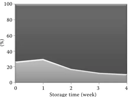
of the dormant and/or damaged cells determined at the first stage resulted from a reduced activity of bacterial inoculum cells due to the form of a deeply frozen concentrate used for direct inocu-lation during butter working phase of churning. The percentage of cells in the physiological status characterised by non-proliferation, preserving the membrane integrity, is maintained during the first week of storage, and structure stabilisation is then gradually lost by the not fully active cells, the pro-portion of the dormant population being reduced. Bifidobacterium ssp. require an anaerobic envi-ronment and neutral pH to survive and maintain levels greater than 6 log CFu/g or ml (Boylston et al. 2004). Therefore, not all foodstuffs provide the most favourable conditions for the growth and survival of the Bifidobacterium genus. Scientific research demonstrates that the Bifidobacterium genus is characterised by a lower viability during storage in acidic foods such as yoghurt and cul-tured milk (pH of 3.7–4.3) (Barreto et al. 2003; Gueimonde et al. 2004; Antunes et al. 2005) which, in such cases, does not apply to butter made of sweet cream with a pH close to neutral (pH of 6.6). Moreover, Boylston et al. (2004) presented evidence that cheese (pH level between 4.8–5.6) provides an appropriate environment for a long-term survival of Bifidobacterium, proposing the effective incorporation of cells into cheese as the main criterion for the maintenance of bifidobacteria viability. The data indicated that, in spite of the fastidious characteristics of the Bifidobacterium genus, the count of the examined strain in butter
for the entire period of storage was maintained at a high level of > 7 log units which could be de-termined by either of the methods applied. The plate method demonstrated a lack of significant changes in the cell count, the population of which was maintained at a stable level. The microscopic method also demonstrated a good viability of the B. lactis strain cells maintaining the integrity of membranes in the examined butter but, at the same time, it provided the possibility of monitoring the changes in the counts of live and dead cells dur-ing storage. This method provides an interestdur-ing alternative to the conventional method by basing the cell detection criterion on other viability deter-minants than proliferation. However, a combina-tion of techniques based on various approaches to the assessment of the physiological status of cells can become an advantage while analysing the multidimensionality of a population and enriching knowledge of the physiology of microorganisms.
The obtained results of B. lactis viability in but-ter encourage the continuation of model research for such a matrice, in order to expand the range of products of health-related properties. unless the maintenance of the B. lactis viability for a whole examined period was observed, other factors like the evaluation of health claims of this specific strain should be also taken into account. While considering therapeutic aspects of intestinal microflora, accord-ing to FAO/WHO and WGO the term ‘probiotic’ should be used cautiously, since they involve the requirement to carry out clinical trials confirming their effects. What is more, EFSA highlights the relation between the health claim and the number of bacteria consumed with the food dose. It was seen that B. infantis was considered effective in lower-ing the symptoms of Irritable Bowel Syndrome at min 8 log CFu/day. As a result, the daily dose of probiotics ranges from 8 to 9 log CFu with daily amount of 100 g infant formula. From the functional point of view butter should be considered as an additional source of probiotics and contribute to the diversity on the probiotic markets.
References
[image:6.595.65.284.82.247.2]Antunes A.E.C., Cazetto T.F., Bolini H.M.A. (2005): Viability of probiotic micro-organism during storage, post acidification and sensory analysis of fat-free yogurts with added whey protein concentrate. International Journal of Dairy and Technology, 58: 169–173.
Figure 3. The percentage of the dormant population estimated on proportion between ■ ‘viable cells’ (deter-mined by the SYTO9 method) and ■ ‘culturable cells’ (determined by the plate method) at each of the butter storage stages
0 1 2 3 4 Storage time (week)
100
80
60
40
20
0
(%
Auty M.A.E., Gardiner G.E., McBrearty S.J., O’Sullivan E.O., Mulvihill D.M., Collins J.K., Fitzgerald G.F., Stanton C., Ross R.P.M. (2001): Direct in situ viability assessment of bacteria in probiotic dairy products using viability staining in conjunction with confocal scanning laser microscopy. Applied Environmental Microbiology, 67: 420–425.
Barer M.R., Harwood C.R. (1999): Bacterial viability and culturability. Advances in Microbial Physiology, 41: 93–137.
Barreto G.P.M., Silva N., Silva E.N., Botelho L., Yim D.K., Almeida C.G., Saba G.L. (2003): Quantificação de lactobacillus acidophilus, bifidobactérias e bactérias láticas totais em produtos probióticos comercializados no Brasil. Brazilian Journal of Food Technology, 6: 119–126. Ben Amor K., Vaughan E.E., De Vos W.M. (2007): Ad-vanced molecular tools for the identification of lactic acid bacteria. Journal of Nutrition, 137: 741S–747S.
Ben Amor K., Breeuwer P., Verbaarschot P., Rombouts F.M., Akkermans A.D.L., De Vos W.M., Abee T. (2002): Multiparametric flow cytometry and cell sorting for the assessment of viable, injured, and dead Bifidobacte- rium cells during bile salt stress. Applied and Environ-mental Microbiology, 68: 5209–5216.
Bertazzoni Minelli E., Benini A. (2008): Relationship between number of bacteria and their probiotic effects. Microbial Ecology in Health and Disease, 20: 180–183. Boylston T.D., Vinderola C.G., Ghoddusi H.B.,
Rein-heimer J.A. (2004): Incorporation of bifidobacteria into cheeses: challenges and rewards. International Dairy Journal, 14: 375–387.
Breeuwand P., Abee T. (2000): Assessment of viability of microorganisms employing fluorescence techniques. International Journal of Food Microbiology, 55: 193–200. Bunthof C., Bloemen K., Breeuwer P., Rombouts F.,
Abee T. (2001): Flow cytometric assessment of viability of lactic acid bacteria. Applied and Environmental Mi-crobiology, 67: 2326–2335.
Bunthof C.J., Abee T. (2002): Development of a flow cy-tometric method to analyze subpopulations of bacteria in probiotic products and dairy starters. Applied and Environmental Microbiology, 68: 2934–2942.
Collado M.C., Moreno Y., Hernandez E., Cobo J.M., Hernandez M. (2005): in vitro viability of Bifidobacte-rium strains isolated from commercial dairy products exposed to human gastrointestinal conditions. Food Sci-ence and Technology International, 11: 307–314. Colwell R.R. (2000): Viable but nonculturable bacteria: a
survival strategy. Journal of Infection and Chemotherapy, 6: 121–125.
Decker E.M. (2001): The ability of direct fluorescence based, two-colour assays to detect different physiological
states of oral streptococci. Letters in Applied Microbiol-ogy, 33: 188–192.
European Food Safety Authority (EFSA): Available at www. efsa.europa.eu/cs/Satellite
Gatti M., Bernini V., Lazzi C., Neviani E. (2006): Fluo-rescence microscopy for studying the viability of micro-organisms in natural whey starters. Letters in Applied Microbiology, 42: 338–343.
Guchte M., Serror P., Chervaux C., Smokvina T., Ehrlich S., Maguin E. (2002): Stress responses in lactic acid bacteria. Antonie van Leeuwenhoek, 82: 187–216.
Gueimonde M., Delgado S., Mayo B., Ruas-Madiedo P., Margolles A., Reyes-Gavilán C.G. (2004): Viability and diversity of probiotic lactobacillus and Bifidobac-terium populations included in commercial fermented milks. Food Research International, 37: 839–850. Grand M., Küffer M., Baumgartner A. (2003):
Quanti-tative analysis and molecular identification of bifidobac-teria strains in probiotic milk products. European Food Research and Technology, 217: 90–92.
Hermida M., Taboada M., Menendes S., Rodriguez-Otero J.L. (2000): Semi-automated direct epifluorescent filter technique for total bacterial count in raw milk. AOAC International, 83: 1345–8.
Hernandez-Mendoza A., Robles V.J., Angulo J.O., De La Cruz J., Garcia H.S. (2007): Preparation of a whey-based probiotic product with lactobacillus reuteri and
Bifidobacterium bifidum. Food Technology and Biotech-nology, 45: 27–31.
Khurana K., Kanawjia S.K. (2007): Recent trends in development of fermented milks. Current Nutrition and Food Science, 3: 91–108.
Kramer M., Obermajer N., Bogovič Matijašić B., Ro- gelj J., Kmetec V. (2009): Quantification of live and dead probiotic bacteria in lyophilised product by real-time PCR and by flow cytometry. Applied Microbiology and Biotechnology, 84: 1137–1147.
Joux F., Lebaron F. (2000): use of fluorescent probes to assess physiological functions of bacteria at single-cell level. Microbes and Infection, 2: 1523−1535.
Lahtinen S., Gueimonde M., Ouwehand A., Reini-kainen J., Salminen S. (2005): Probiotic bacteria may become dormant during storage. Applied and Environ-mental Microbiology, 71: 1662–1663.
Lahtinen S., Gueimonde M., Ouwehand A., Reini-kainen J., Salminen S. (2006): Comparison of methods to enumerate probiotic bifidobacteria in a fermented food product. Food Microbiology, 23: 571–577.
strains during long-term incubation in milk. Journal of Microbiology, 48: 445–451.
Moreno Y., Collado M., Ferrus M., Cobo J., Hernan-dez E., HernanHernan-dez M. (2006): Viability assessment of lactic acid bacteria in commercial dairy products stored at 4°C using LIVE/DEAD BacLight staining and conven-tional plate counts. Internaconven-tional Journal of Food Science and Technology, 41: 275–280.
Oliver J.D. (2005): The viable but nonculturable state in bacteria. Journal of Microbiology, 43: 93–100.
Olszewska M., Łaniewska-Trokenheim Ł. (2011): Study of the nonculturable state of lactic acid bacteria cells under adverse growth conditions. Veterinary Medicine, 67: 105–109.
Rault A., Beal C., Ghorbal S., Ogier J.-C., Bouix M. (2007): Multiparametric flow cytometry allows rapid
as-sessment and comparison of lactic acid bacteria viability after freezing and during frozen storage. Cryobiology, 55: 35–43.
Sanders M.E. (2003): Probiotics: considerations for human health. Nutrition Reviews, 61: 91–9.
Soomro A.H., Masud T., Anwaar K. (2002): Role of lac-tic acid bacteria (LAB) in food preservation and human health – A review. Pakistan Journal of Nutrition, 1: 20–24. Suvarna V.C., Boby V.u. (2005): Probiotics in human health: A currant assessment. Current Science, 88: 1744–1748. Warmińska-Radyko I., Olszewska M., Mikš-Krajnik
M. (2010): Effect of temperature and sodium chloride on the growth and metabolism of lactococcus strains in long-term incubation of milk. Milchwissenschaft, 65: 32–35.
Received for publication August 12, 2011 Accepted after corrections November 9, 2011
Corresponding author:
Magdalena Olszewska, university of Warmia and Mazury in Olsztyn, Faculty of Food Science, Chair of Industrial and Food Microbiology, Pl. Cieszyński 1, 10-726 Olsztyn, Poland