RECENT ADVANCES IN IMAGE PROCESSING
TECHNIQUES USING USG AND MRI OF LIVER
Garima Sharma
1, Pooja Sharma
2, Rupali Bharti
3, Sonam Dubey
41,2,3,4
Student, M.Tech (CS), Banasthali Vidyapith, Rajasthan (India)
ABSTRACT
Magnetic resonance imaging (MRI) is playing an increasing role in the assessment of patients with liver disease due to its high soft tissue resolution, lack of ionizing radiation and ability to provide functional data. There are various types of diseases in the liver. So, to identify these diseases and to get a clear image of the damaged part various imaging techniques are used mainly Sonography and MR based techniques. These techniques are easy to implement across different MRI platforms, and results in enhanced disease detection and characterization. Diffusion Weighted Imaging (DWI) is used to identify the lesion in the liver, Perfusion Weighted Imaging (PWI) is used to identify the volume and functioning of the cirrhotic liver.
Keywords: ANOVA, Chronic Liver Disease (CLD), Dynamic hepatocyte-specific contrast-enhanced
MRI (DHCE-MRI), Diffusion Weighted Magnetic Resonance Imaging (DWMRI), Kruskal-Wallis
test, RLE (Relative Liver enhancement)
I. INTRODUCTION
There are various imaging techniques which are prevailing now a day in the assessment of patients with liver
diseases. One of the widely used techniques is Diffusion Weighted Magnetic Resonance Imaging (DWMRI).It
is a functional imaging technique.DW imaging is increasingly used in the abdomen, particularly in the liver with
promising results for liver lesion detection and characterization [7]. We calculate Apparent Diffusion
Coefficient (ADC) values in it and the calculated ADC values can be displayed as an image and quantitative
analysis can be performed by placing measuring the mean value within a Region of Interest (ROI). Earlier,
Mono-exponentially fitted ADC values were calculated which was contaminated by micro-perfusion so
Bi-exponential model was required and data was compared using ANOVA and Kruskal-Wallis test. DW Imaging
can be easily implemented in clinical protocols, as it can be performed relatively quickly and does not require
contrast agent injection [1].
Several, studies have reported that ADC can contribute to the differential diagnosis of benign and malignant
focal lesion in the liver. In particular, the combination of DWI and PWI of the liver may supply additional tools
to assess liver function, providing information concerning both the soft-tissue characteristics and the vascularity
of the lesions. By diagnosing both MR perfusion can improve the sensitivity and specificity of diagnostic liver
imaging [2].
Gd-EOB-DTPA is a contrast agent developed for MRI. We use dynamic hepatocyte-specific contrast-enhanced
Then gadoxetic acid-enhanced 3T MR Imaging was performed and RLE (Relative Liver enhancement) was
calculated. Liver failure was defined according to 50-50 criteria and ISGLS classification. RLE was inversely
related to the probability of liver failure according to 50-50 and ISGLS criteria. RLE was independently
associated with a higher probability of liver failure according to ISGLS classification [4].
In all above techniques, we described about liver cirhossis.Now, Grey Relational Analysis (GRA) is proposed to
recognize fatty livers in B-scan ultrasonic images. Main diagnostic methods for fatty liver are B-mode
ultrasound, followed by a CT-scan and MRI. Ultrasonography was based on brightness of the image where an
echo is created that is, returning signal [6].
II. IMAGING TECHNIQUES
Since morphologic alterations and features of portal hypertension are present only in advanced Chronic Liver
Disease (CLD), routine examinations by ultrasound (US), computed tomography (CT) and magnetic resonance
imaging (MRI) could produce specific findings, but with very limited sensitivity. CT offers improved resolution
of early morphological changes with cirrhosis but has low accuracy in fibrosis detection. MRI identify specific
features of cirrhosis such as hepatic vein narrowing, caudate to right lobe ratio, and expanded gallbladder fossa,
but remains lacking in earlier stages of fibrosis. Hence, assiduous efforts have been made to search for
technological developments [9].
2.1 Sonography Based Techniques for Assessment of Liver Fibrosis
Recently, diverse Sonography–based techniques have been used in assessment of liver fibrosis, including
Transient Elastography, Real-Time Elastography, and Acoustic Radiation Force Imaging sonoelastography.
2.1.1 Transient Elastography
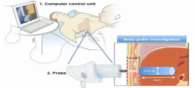
Transient Elastography (TE) (Fibro Scan®, Echosens, and Paris, France) is a new imaging modality for
detecting hepatic fibrosis. The measuring instrument comprises a computer driven control unit and a probe with
an ultrasound transducer, which is located at the end of a vibrating piston. TE measures liver stiffness in a
volume that approximates a cylinder 1 cm wide and 4 cm long, between 2.5 cm and 6.5 cm below the skin
surface [10].
Figure 1 Illustration of the two different constituent of the measuring instrument and the
The advantages of TE are that the results are immediately available, and the procedure is painless, rapid (~3
minutes per patient), and easy to perform. One of the important aspects of liver stiffness measurements is the
cut-off values that are adopted for different stages of fibrosis, with higher cut-off levels corresponding to higher
fibrosis stages [11-13]. There are some physical limitations of TE, such as obesity (particularly the fatness of the
chest wall), narrow intercostals space and ascites [11].
2.1.2 Real-Time Elastography
Real-Time Elastography (RTE) is an alternative method for measurement of tissue elasticity integrated in a
Sonography machine developed by Hitachi Medical Systems. Ophir et al. [14] first described the principle of
this technique in 1991. To reduce the time-consuming calculations, Pesavento etal.developed a fast
cross-correlation technique that is the basis for RTE. The difference in hardness between diseased and surrounding
tissue can be detected by RTE based on the physical properties of the tissue. The calculation of tissue elasticity
distribution is assessed in real-time ultrasound imaging and depicted as color-coded images with the
conventional B-mode image in the background [15, 16]. The color scale includes the following colors: red (soft
tissue), green (intermediate, normal tissue), and blue (anelastic, hard tissue).
Figure 2 Example of tissue elasticity distribution in a healthy subject represented as color-coded
images over conventional B-mode image [17].
As for TE even for RTE obesity, narrow intercostals space and ascites are potential physical limitations. More
number of samples about chronic hepatitis with assessment by RTE is needed to perform to certify its
advantages.
2.1.3 Acoustic Radiation Force Impulse Elastography
Acoustic Radiation Force Impulse (ARFI) imaging is a novel ultrasound-based Elastography method that is
integrated in a conventional Ultrasound machine enabling the exact localization of measurement site. Unlike
conventional B-mode Sonography, which provides anatomical details based on differences in 3–4 on a scale of
0–6 arbitrary units [9].
2.2 MR Imaging Based Techniques for Assessment of Liver Fibrosis
In the last decade, the development of MRI scanner with high-performance magnetic field gradients made the
near-isotropic voxels (1–3 mm in all three-dimensions) through the entire liver can be achieved in a single
breath-hold or using respiratory triggering. In detail, several technological advances have been made for
assessment of fibrosis, including Conventional MRI, Double contrast-material enhanced MRI,
Diffusion-weighted MRI, and MR Elastography, perfusion MRI, and MR spectroscopy [9].
2.2.1 Unenhanced MRI
In patients with precirrhotic stages of liver fibrosis as well as patients with early cirrhosis, the liver parenchyma
usually has a normal appearance or may reveal only subtle, generic heterogeneity on unenhanced MRI [18].
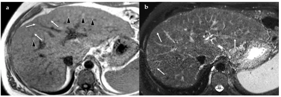
Figure 3 Unenhanced MR imaging in a in a 61-year-old man with alcohol-related cirrhosis.
Unenhanced T1-weighted image (a) shows hypo intense reticulations (arrows) and numerous
regenerative nodules (arrowheads), which are isotohyperintense. Unenhanced T2-weighted
fat-saturated image (b) allows a clearer visualization of the reticulations throughout the liver
parenchyma visible as hyper intense septa (arrows)[19].
2.2.2 Contrast-enhanced MRI
The detection of liver fibrosis is improved by the administration of contrast agents. Three contrast agents are
currently commercially available: gadolinium-based contrast agents; super paramagnetic iron oxide particles;
Gd-EOB-DTPA. Gadolinium-based contrast agents cause T1 shortening and signal enhancement on
T1-weighted images [20]. Most gadolinium-based contrast agent formulations freely equilibrate with the
extracellular compartment and accumulate in tissues with large extracellular volumes such as liver fibrosis.
Super paramagnetic iron oxide particles (SPIO) are reticulo-endothelial-specific particulate MRI contrast agents
which are cleared from the blood through phagocytosis and accumulate in the cells of the reticule-endothelial
system of the liver, spleen, and bone marrow, with approximately 80% taken up by the liver [9].
2.2.3 Double-contrast enhanced MRI
Double-contrast MRI (DC-MRI) using extracellular contrast agents in combination with SPIO particles was
shown to sensitively detect liver fibrosis and depict HCC in cirrhotic livers [21]. The consequence is high image
contrast between the low-signal-intensity liver parenchyma and high-signal-intensity fibrotic reticulations (Fig
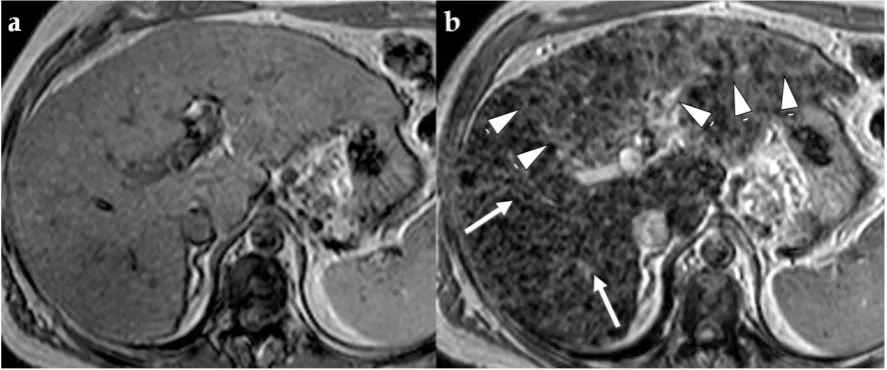
Figure 4 Advanced fibrosis and infiltrative HCC in a 46-year-old man with HCV-related
cirrhosis. T2*-weighted gradient-echo images obtained before (a) and after (b) intravenous
SPIO injection. After injection, fibrotic reticulations in the right lobe have diminished Kupffer
cell density, do not accumulate iron oxides, - hence appear relatively hyper intense (arrows in
b). The left lobe is expanded and shows a wedge-shaped mass with heterogeneous hyper
intensity (arrowheads in b) in the hepatocellular phase, suggestive for infiltrative HCC Aguirre
et al. [23] examined 101 CLD patients who underwent DC-MRI to detect hyper intense
reticulations, which are postulated to represent septal fibrosis, and hypo intense nodules.
2.2.4 MR Elastography
A new option for assessing shear stiffness in various tissue types, including liver fibrosis, is MR Elastography
(MRE) [24]. MRE uses a modified phase contrast technique to sensitively image the propagation characteristics
of acoustic shear waves that are generated with the organ of interest [25]. A specialized phase-contrast MRI
sequence is then used to image the propagating waves in the liver [9].
2.2.5 MR Spectroscopy
MR spectroscopy (MRS) enables the non-invasive measurement of concentrations of different chemical
components within tissues, which are displayed as a spectrum with peaks consistent with the various chemicals
detected [9].
III. CONCLUSION AND FUTURE SCOPE
A fast, safe and reliable technique to assess fibrosis of CLD and to follow up progression or regression of
fibrosis during treatment is required. Ultrasound is still a widespread, low cost, user-friendly, and accurate
technique [9]. An advantage of DC-MRI is that it works on routine imaging units and does not require
specialized equipment. Computer-based texture analysis techniques may assess texture abnormalities
qualitatively or quantitatively [9]. The primary advantage of the GRA method is that it is based on an algorithm
which precisely compares with own liver image [6]. Gadoxetic acid–enhanced MR imaging can help with the
assessment of the risk for liver failure after major liver resection[4]. In conclusion, to date, the most promising
detecting severe fibrosis and future developments promise to increase the reliability and accuracy of staging of
hepatic fibrosis. In the future, MRI technical development and new contrast agents could permit imaging of
fibro genesis.
REFERENCES
[1] Hildebrand Dijkstra&Paul Baron & Peter Kappert &Matthijs Oudkerk & Paul E. Sijens .Effects of
microperfusion in hepatic diffusion weighted imaging. Eur Radiol (2012) 22:891–899.DOI
10.1007/s00330-011-2313-1 open access at Springerlink.com .11-Dec-11.
[2] F. Donati, P. Boraschi, S. Salemi, R. Gigoni, C. Romei, C.Bartolozzi, F. Falaschi; Pisa/IT. Focal nodular
hyperplasia of the liver: Diffusion and perfusion MR imaging findings. Poster No.: C-0102Congress:
ECR 2010 Type: Scientific Exhibit Poster By:- European Society of Radiology(ESR) 2010
[3] Nilsson H, Blomqvist L, Douglas L, Nordell A, Janczewska I, Naslund,E Jonas
Gd-EOB-DTPA-enhanced MRI for the assessment of liver function and volume in liver cirrhosis doi:
10.1259/bjr.20120653 Br J Radiol 2013;86:20120653 31-Jan-13
[4] Andreas Wibmer, Alexander M. Prusa, Richard Nolz, Thomas Gruenberger, Martin Schindl, Ahmed
Ba-Ssalamah Liver Failure after Major Liver Resection: Risk Assessment by Using Preoperative Gadoxetic
Acid–enhanced 3-T MR Imaging Wibmer et al Radiology: Volume 269:Number 3—December 2013
radiology.rsna.org 15-May.
[5] Fernanda D. Gonzalez-Guindalini, MD • Marcos P. F. Botelho, MD • CarlaB. Harmath, MD • Kumaresan
Sandrasegaran, MD • Frank H. Miller, MD Riad Salem, MD, MBA • Vahid Yaghmai, MD Assessment of
Liver Tumor Response to Therapy: Role of Quantitative Imagingradiographics.rsna.org RadioGraphics
2013; 33:1781–1800 Published online 10.1148/rg.336135511.7-may-2013.
[6] Semra İçer & Abdulhakim Coşkun & Türkan İkizceli. Quantitative Grading Using Grey Relational
Analysis on Ultrasonographic Images of a Fatty Liver. J Med Syst,J Med Syst (2012) 36:2521–2528
DOI 10.1007/s10916-011-9724-z. 28-Apr-11.
[7] Bachir Taouli. Diffusion-weighted MR Imaging for Liver Lesion Characterization: A Critical Look.
Radiology: Volume 262: Number 2—February 2012, Published online 10.1148/radiol.11112417
Radiology 2012; 262:378–380. 6-Dec-2015.
[8] Timothy G. St. Pierre, Paul R. Clark, Wanida Chua-anusorn, Adam J. Fleming, Gary P. Jeffrey, John
K.Olynyk, Pensri Pootrakul, Erin Robins, and Robert Lindeman. Noninvasive measurement and imaging
of liver iron concentrations using proton magnetic resonance.The American Society of Hematology.
3-Aug-15.
[9] Luca Macarini and Luca P. Stoppino. Radiologic Assessment of Liver Fibrosis – Present and
Future..http://dx.doi.org/10.5772/55164.2013.
[10] Roulot D, Czernichow S, Le Clesiau H, et al. Liver stiffness values in apparently healthy subjects:
Influence of gender and metabolic syndrome. J Hepatol 2008;48:606-613.
[11] Fraquelli M, Rigamonti C, Casazza G, et al. Reproducibility of transient elastography in the evaluation of
[12] Sandrin L, Fourquet B, Hasquenoph JM, et al. Transient elastography: A new noninvasive method for
assessment of hepatic fibrosis. Ultrasound Med Biol 2003;29:1705-1713.
[13] Konate A, Boursier J, Reaud S, et al. Liver stiffness measurement by transient elastography: predictive
factors of accuracy, success and reproducibility. J Hepatol 2006;44:S195.
[14] Ophir J, Cespedes I, Ponnekanti H, Yazdi Y, Li X. Elastography: a quantitative method for imaging the
elasticity of biological tissues. Ultrason Imaging 1991;13:111-134.
[15] Frey H. Realtime elastography. A new ultrasound procedure for the reconstructionof tissue elasticity
Radiologe 2003; 43:850-855
[16] Srinivasan S, Kallel F, Ophir J. The effects of digitization on the elastographic signalto-noise ratio.
Ultrasound Med Biol 2002; 28:1521-1534.
[17] Wang J, Guo L, Shi X, et al. Real-time elastography with a novel quantitative technology for assessment
of liver fibrosis in chronic hepatitis B. Eur J Radiol 2012;81:e31-e36.
[18] Martin DR. Magnetic resonance imaging of diffuse liver diseases. Top Magn Reson Imaging
2002;13:151-163.
[19] Ito K, Mitchell DG, Siegelman ES. Cirrhosis: MR imaging features. Magn Reson Imaging Clin N Am
2002;10:75-92.
[20] Balci NC, Semelka RC. Contrast agents for MR imaging of the liver. Radiol Clin North Am
2005;43:887-898.
[21] Macarini L, Marini S, Milillo P, et al. Double-contrast MRI (DC-MRI) in the study of the cirrhotic liver:
Utility of administering Gd-DTPA as a complement to examinations in which SPIO liver uptake and
distribution alterations (SPIO-LUDA) are present and in the identification and characterisation of focal
lesions. Radiol Med 2006; 111:1087-1102.
[22] Hughes-Cassidy F, Chavez AD, Schlang A, et al. Superparamagnetic iron oxides and low molecular
weight gadolinium chelates are synergistic for direct visualization of advanced liver fibrosis. J Magn
Reson Imaging 2007;26:728-737.
[23] Aguirre DA, Behling CA, Alpert E, et al. Liver fibrosis: noninvasive diagnosis with double contrast
material enhanced MR imaging. Radiology 2006;239:425-437.
[24] Manduca A, Oliphant TE, Dresner MA, et al. Magnetic resonance elastography: noninvasive mapping of
tissue elasticity.Med Image Anal 2001;5:237-254.
[25] Kruse SA, Smith JA, Lawrence AJ, et al. Tissue characterization using magnetic resonance elastography:
preliminary results. Phys Med Biol 2000;45:1579-1590.
[26] Colombo S, Buonocore M, Del Poggio A, et al. Head-to-head comparison of transient elastography (TE),
real-time tissue elastography (RTE), and acoustic radiation force impulse (ARFI) imaging in the
diagnosis of liver fibrosis. J Gastroenterol 2012;47:461-469.
[27] Wang QB, Zhu H, Liu HL, Zhang B. Performance of Magnetic Resonance Elastography and
Diffusion-Weighted Imaging for the Staging of Hepatic Fibrosis: A Meta-Analysis. Hepatology 2012;56:239-247.
[28] Godfrey EM, Patterson AJ, Priest N, et al. A comparison of MR elastography and 31P MR spectroscopy
![Figure 2 Example of tissue elasticity distribution in a healthy subject represented as color-coded images over conventional B-mode image [17]](https://thumb-us.123doks.com/thumbv2/123dok_us/9246027.1460607/3.595.73.542.332.525/figure-example-elasticity-distribution-healthy-subject-represented-conventional.webp)