R E S E A R C H
Open Access
Comparison of biological characteristics of
mesenchymal stem cells derived from
maternal-origin placenta and Wharton
’
s
jelly
Gecai Chen
1, Aihuan Yue
2, Zhongbao Ruan
1, Yigang Yin
1, Ruzhu Wang
1, Yin Ren
1and Li Zhu
1*Abstract
Introduction:Although mesenchymal stem cells (MSCs) from different sources share many similar characteristics, they also exhibit individual properties. In this study, we compared MSCs derived from Wharton’s jelly in the umbilical cord with those derived from the decidual basalis in the maternal part of the placenta to better understand the similarities and differences between these two cell types.
Method:The morphology, immunophenotype (as assessed using flow cytometry), and multi-lineage differentiation potential were analyzed. Karyotype analysis was carried out to determine the origin of the MSCs. Growth kinetics were evaluated using analysis of the population doubling time and cell cycle. Immunosuppressive function was analyzed using mixed lymphocyte culture.
Results:MSCs from Wharton’s jelly and the decidua basalis exhibited similar morphology, immunophenotype, and differentiation potential to osteogenesis and adipogenesis. The percentage of MSCs in the G0/G1 phase was higher in the case of Wharton’s jelly than in the case of the decidua basalis (P <0.05). Decidual MSCs displayed more remarkable immunosuppressive effects on phytohemagglutinin-stimulated T-cell proliferation (P <0.05). Conclusion:MSCs from both sources had similar basic biological properties, but decidual MSCs had slower proliferation and stronger immunosuppressive function.
Keywords:Mesenchymal stem cells (MSCs), Decidua basalis, Wharton’s jelly, Immunosuppression, Cell cycle, T-cell proliferation
Introduction
Mesenchymal stem cells (MSCs) not only possess the basic characteristics of stem cells, including self-renewal and multi-lineage differentiation potential, but also ex-hibit hematopoietic [1, 2] and immunomodulatory func-tion [3–6]. Neonatal tissue is rich in MSCs derived from Wharton’s jelly in the umbilical cord and from the de-ciduae, which form the maternal part of the placenta. The placentome is customarily discarded as a medical waste, and there is no ethical controversy in obtaining MSCs from this tissue. There may be many similarities
between MSCs from the above two sources. Neverthe-less, they play different roles during fetal development, and so have their own characteristics. The placenta and fetal membranes function as immunological barriers be-tween the mother and the developing fetus during preg-nancy. The placenta can be conceptually divided into the fetal side, consisting of the amnion and chorion, and the maternal side, consisting of the decidua. As placental tis-sues are conventionally discarded after delivery, these tissues are readily available for research and clinical ap-plications. The decidua is a membrane of maternal ori-gin that plays an important role in immune tolerance, since maternal and fetal immune cells come into direct contact with each other at this site [5]. Wharton’s jelly is the embryonic mucous connective tissue found between
* Correspondence:[email protected]
1
Department of Cardiology, Taizhou Renmin Hospital, Taizhou, Jiangsu Province, China
Full list of author information is available at the end of the article
the amniotic epithelium and the umbilical vessels; it is a rich source of MSCs [7]. MSCs from Wharton’s jelly (WJ-MSCs) exhibit greater proliferation than adult MSCs from the bone marrow [6].
Most often MSCs are transplanted for tissue repair and regeneration. Due to their immunomodulatory properties, MSCs have garnered increasing research at-tention in recent years. MSCs have been used for treat-ing graft-versus-host disease [5, 8–10]. MSCs from the bone marrow, which were first described by Fridenstein et al. [11] in 1976, were the earliest stem cells to be de-tected and, currently, are the most used stem cells in clinical trials. However, their limited availability hindered their development in research and clinical applications. The use of neonatal tissue can overcome this shortcom-ing. In our study, we compared MSCs derived from Wharton’s jelly in the umbilical cord and from the de-cidual stroma in the maternal-origin placenta to under-stand their similarities and differences. The morphology and immunophenotype (assessed using flow cytometry) were analyzed. Karyotype analysis was carried out to de-termine the origin of the MSCs. Growth kinetics were evaluated using the population doubling time (PDT) and cell cycle. Immunosuppressive function was analyzed using mixed lymphocyte culture.
Materials and methods
Isolation and culture of MSCs from Wharton’s jelly and decidua
Ten human placentae and umbilical cords were obtained from healthy, full-term, naturally delivered, male new-borns. Peripheral blood samples were obtained from vol-untary blood donors. Written informed consent was obtained from the mothers and the donors. The study protocols were reviewed and approved by the Taizhou Renmin Hospital review board and ethics committee of Taizhou Renmin Hospital. We selected donors who tested negative for hepatitis B surface antigen, hepatitis B core antibody, hepatitis C virus antibody, hepatitis C virus RNA, HIV-I and -II antibodies, HIV-1 RNA, cytomegalovirus IgM, and anti-Treponema pallidum
antibody.
WJ-MSCs were separated and cultured according to previously published reports [11, 12]. MSCs from the cidua basalis (DB-MSCs) were separated from the de-cidua basalis of the placenta. The dede-cidua basalis tissue was sliced into small fragments of 1 mm3, washed twice with physiological saline, digested with collagenase for 1 h, and cultured in serum-free MesenCult-XF medium (Stemcell, Vancouver, Canada).
Karyotype analysis
Karyotype analysis was carried out at passage 0 (P0) to confirm that the cells were derived from the maternal
decidua basalis. For this purpose, 2 × 10⁶cells were har-vested, and 0.1–0.4 μg/mL colchicine (Gibco, Grand Island, USA) was added to the culture medium. After 12 h, 0.075 M KCl was added to the culture, and the cells were incubated in a water bath at 37 °C. Then, 1 mL of fixative (methanol/acetic acid mixture at 1:3) was added, and the samples were incubated for 30 min at 37 °C and centrifuged. A further 8 mL of fixative was added, and the cells were dried for 10 min with 10 % Giemsa, and then washed with distilled water. The fixed cells were observed under an electron microscope (IX71; Olympus, Tokyo, Japan). Chromosome analysis was car-ried out by applying G-bands, according to the guide-lines of the International System for Chromosome Nomenclature 2013. On average, 20 metaphase samples were evaluated for each passage [13].
Immunophenotype analysis by flow cytometry
At P3, MSCs from both sources (1 × 10⁷ cells) were digested with trypsin and washed twice with phosphate-buffered saline. The cell concentration was adjusted to 2 × 10⁶cells/mL, and cells were stained with the follow-ing fluorescent antibody conjugates: CD45-fluorescein isothiocyanate (FITC), CD34-phycoerythrin (PE), CD73-PE, CD14-FITC, CD79a-APC, the human major histo-compatibility complex (MHC) class II molecule HLA-DR-(PE), CD90-allophycocyanin (APC) (BD Biosciences, MD, USA), and CD105-PE (eBioscience, CA, USA). We also tested for the co-inhibitory molecule B7-H1(FITC) and the positive co-stimulatory factors CD80-PE, CD83-APC, and CD86-FITC. Surface staining was detected using flow cytometry (Diva software 6.0, FACScantoII, BD Biosciences).
Growth kinetics analysis
The proliferation of MSCs from both sources at P3, P5, P8, and P10was assessed. WJ-MSCs and DB-MSCs were plated on a 60-mm wide dish at a density of 7–10 × 105 cells/well, and the cells were counted until they reached 100 % confluency. The PDT was calculated using the fol-lowing formula:
PDT = (CT × ln2)/ln(Nf/Ni), where CT is the cell cul-ture time, Niis the initial number of cells, and Nfis the final number of cells [14].
Cell cycle analysis of MSCs from both sources by flow cytometry
incubated for 20 min at 4 °C, and their fluorescence was measured using flow cytometry. The data were analyzed using ModFit software.
Mixed lymphocyte reaction
We gently mixed 10 mL peripheral blood with 10 mL saline. Next, 10 mL lymphocyte separation medium 1.077 was poured into a 5.0-mL tube. Then, 20 mL cell suspension was carefully added on top of the separation medium without disturbing the interphase. The tube was centrifuged at 440g for 40 min. Most of the super-natant was then aspirated without disturbing the layer of mononuclear cells in the interphase. The mononuclear cells were then aspirated from the interphase, washed with saline, and centrifuged at 360gfor 10 min. The ex-cess red blood cells and plasma were removed.
Mixed lymphocyte reaction was carried out in 96-well plates. WJ-MSCs and DB-MSCs from 10 donors at P3 were irradiated with 60Co (20 Gy). Next, 1.0 × 10⁵ re-sponder cells were co-cultured with 1.0 × 10⁵stimulator cells in serum-free MesenCult-XF medium for 6 days at 37 °C in humidified air containing 5 % CO2. The cells were divided into eight groups: group A, 1.0 × 10⁶ per-ipheral blood mononuclear cells (PBMCs); group B, 1.0 × 10⁶PBMCs + phytohemagglutinin (PHA; 10 ug/mL); group C, 1.0 × 10⁵ DB-MSCs; group D, 1.0 × 10⁵ DB-MSCs + PHA; group E, 1.0 × 10⁶ PBMCs + 1.0 × 10⁵
DB-MSCs + PHA (10 μg/mL); group F, 1.0 × 10⁵ WJ-MSCs; group G, 1.0 × 10⁵ WJ-MSCs + PHA; group H, 1.0 × 10⁶PBMCs + 1.0 × 10⁵WJ-MSCs + PHA. For each group, three replications were used. Cell proliferation rates were assessed using (3H)-thymidine incorporation. The interferon (IFN)-γ levels in the co-culture super-natant were detected using an enzyme-linked immuno-sorbent assay (ELISA) kit (eBioscience). The optical density of each well was evaluated at 450/630 nm, and IFN-γcontent was calculated using a standard curve.
Statistical analysis
Data were expressed as mean ± SEM. The different groups were compared using analysis of variance. PDT was compared using thet-test. A 5 % probability (P< 0.05) was used as the level of statistical difference.
Results Morphology
The morphology of MSCs from both sources was assessed using light microscopy. We observed the cells at every passage. All cells retained a fibroblast-like morphology (Fig. 1).
Karyotype analysis
To ensure all cells in culture were derived from the ma-ternal placenta, the cytogenetic karyotypes of the cells at
Fig. 1Photomicrographs of mesenchymal stem cells from Wharton’s jelly (WJ-MSCs) and the decidua basalis (DB-MSCs) from donor 2 are shown.
P0 were analyzed. The sex chromosomes XX, not XY, were detected in the cells (Fig. 2).
Immunophenotype
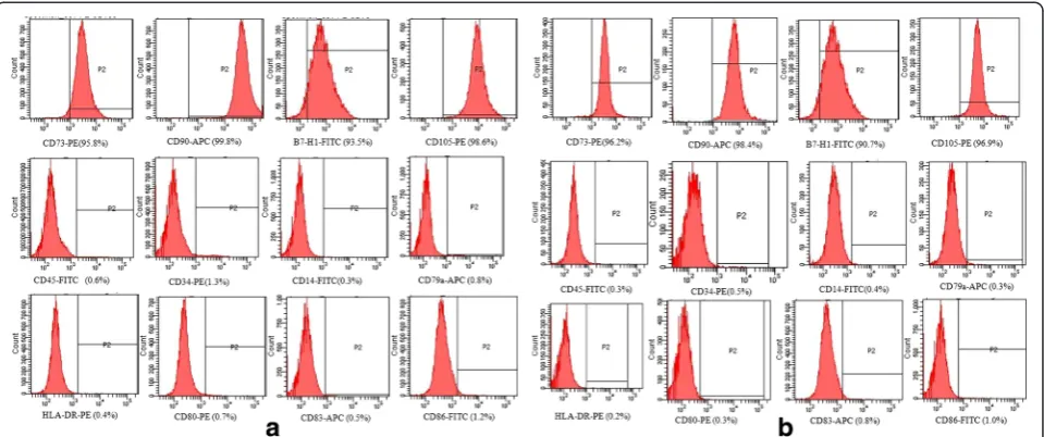
We investigated MSC immunophenotype at P3by stain-ing for cell surface markers, which were detected usstain-ing flow cytometry according to the International Society for Cellular Therapy standards [15]. MSCs from both sources highly expressed the typical MSC markers CD105, CD73, and CD90 and the co-inhibitory molecule B7-H1. In addition, the cells showed low expression of the hematopoietic markers CD45, CD14, and CD34, the MHC class II molecule HLA-DR, and the positive co-stimulatory factors CD80, CD83, and CD86. There was no difference between the two types of MSC in terms of immunophenotype (Fig. 3).
PDT of MSCs
DB-MSCs and WJ-MSCs from the same donor showed different proliferative capacities at the same culture pas-sage. The PDT of WJ-MSCs was 34.7 ± 3.4 h, 38.8 ± 3.3 h, 44.8 ± 4.1 h, and 56.8 ± 3.6 h at P3, P5, P8, and P10, respect-ively. The PDT of DB-MSCs was 47.5 ± 4.0 h, 51.8 ± 3.8 h, 60.7 ± 4.7 h, and 71.1 ± 3.0 h at P3, P5, P8, and P10, respect-ively. The PDT of DB-MSCs and WJ-MSCs from the same donor increased with an increase in the number of pas-sages (Fig. 4).
Cell cycle analysis
The cell cycles of DB-MSCs and WJ-MSCs from the 10 donors were assessed at P3. In the case of the DB-MSCs, the mean proportions of cells in the G0/G1 phase, S phase, and G2/M phase were 76.60 ± 2.34 %, 15.76 ± 2.11 %, and 7.64 ± 1.48 %, respectively. The corresponding proportions in the case of WJ-MSCs were 65.615 ± 2.91 %, 20.50 ± 1.96 %, and 13.89 ± 2.78 %. The differences in the
distribution of cells in the G0/G1 and G2/M phases be-tween DB-MSCs and WJ-MSCs were statistically signifi-cant (P< 0.05; Fig. 5).
Immunomodulatory properties of MSCs from both sources
To compare the immunomodulatory properties of MSCs from both sources, PBMCs were stimulated with PHA in the presence of WJ-MSCs or DB-MSCs for 6 days. Allogeneic PBMC proliferation rates were then assessed using (3H)-thymidine incorporation in the four groups. DB-MSCs showed stronger immunosuppression proper-ties than did WJ-MSCs (P< 0.05). IFN-γ content of the supernatant was tested using ELISA. The IFN-γ level in the supernatant was lower in the DB-MSC group than in the WJ-MSC group (P< 0.05; Fig. 6).
Discussion
In the present study, we compared two populations of MSCs derived from the decidua basalis and Wharton’s jelly. Although DB-MSCs and WJ-MSCs share global properties, such as morphology, plastic adherence, and multi-lineage differentiation potential [16], significant differences exist between them in terms of growth rate and immunomodulatory function.
During pregnancy, the maternal and fetal immune cells come into direct contact with each other in the decidua, which functions as an immunological barrier between the mother and the developing fetus [5]. Karyotype analysis showed that DB-MSCs are of maternal origin, since the sex chromosomes in these cells were XX, not XY. Pro-grammed cell death (PD)-L1 and PD-L2 are members of the B7 family, and are the ligands for the PD-1 receptor. PD-L1, also called B7-H1, is expressed on antigen-presenting cells, including IFN-γ-stimulated monocytes, and activated human and murine dendritic cells. PD-L1 is
Fig. 2Karyotyping. To ensure all cells in culture were derived from the maternal placenta, the cytogenetic karyotypes of cells at P0were
also expressed on placental trophoblasts, myocardial endothelium, cortical thymic epithelial cells, and on most carcinomas. Studies show overlapping functions of PD-L1 and PD-L2, and indicate an important role for the PD-L– PD-1 pathway in regulating T-cell responses [17]. The co-inhibitory molecule B7-H1 was highly expressed in DB-MSCs and WJ-DB-MSCs. This molecule may be related to the regulatory function of the cells [18]. Neither cell type expressed the surface MHC class II molecule HLA-DR or positive co-stimulatory molecules, such as CD83, CD80, and CD86. This is consistent with the results of previously published papers [19, 20].
DB-MSCs and WJ-MSCs from the 10 donors exhibited different proliferation rates, and the PDT greatly varied among cells obtained from different donors at the same passage. Shaer et al. [21] compared MSCs from the pla-cental decidua basalis, umbilical cord Wharton’s jelly, and amniotic membrane. The doubling times for WJ-MSCs were 21 ± 8 h at P3and 30 ± 5 h at P10, which are shorter than the times determined in this study. This dif-ference may have been caused by the use of different culture systems, i.e., serum-free versus serum-containing cultures. The authors of the above study also reported that the proliferative potential of WJ-MSCs tended to be
Fig. 3Flow cytometric analysis of the expression of surface markers on (a) WJ-MSCs and (b) DB-MSCs. The immunofluorescence analysis was
conducted at the 3rd passage and showed the immunofluorescence of cells obtained from donor 3. There was no difference between the two types of MSCs in terms of immunophenotype (n= 10).APCAllophycocyanin,FITCFluorescein isothiocyanate,PEPhycoerythrin
Fig. 4Analysis of the mean population doubling time (PDT) of mesenchymal stem cells from Wharton’s jelly (WJ-MSCs) and the decidua basalis
higher than that of the cells from the other two sources. Overall, WJ-MSCs exhibited higher growth rates than did DB-MSCs under the same conditions. The results of cell cycle assessments agreed with those of the PDT analysis.
The fetal–maternal interface seems to be immunologically special to enable maternal acceptance of the fetal allograft [22]. The human placenta, besides supporting fetal develop-ment, may also function as an immune regulator. MSCs are anti-proliferative to T cells and suppress the secretion of IFN-γin mixed lymphocyte reaction cultures [23]. Karlsson et al. [24] compared stromal cells obtained from term fetal membrane, umbilical cords, and placental villi, and found that the stromal cells obtained from term fetal membrane
had stronger immunosuppressive capacity than those from umbilical cords and placental villi. DB-MSCs produced sig-nificantly lower levels of IFN-γ than did WJ-MSCs. The mechanisms of T-cell immunosuppression by MSCs has al-ways been an issue of dispute. Toll-like receptors are consid-ered to play a key role in this process [25–27]. MSCs immunoregulate T-cell proliferation independent of heme oxygenase-1 [28].
Conclusion
In this study, we compared the essential biological char-acteristics of DB-MSCs and WJ-MSCs. Although the two cell types share global properties, such as morph-ology, plastic adherence, and multi-lineage differentiation
Fig. 5The cell cycles of mesenchymal stem cells from Wharton’s jelly (WJ-MSCs) and the decidua basalis (DB-MSCs) obtained from the 10 donors
were assessed at P3. The differences in G0/G1 and G2/M phase distribution between DB-MSCs and WJ-MSCs were statistically significant (*P< 0.05)
Fig. 6Mesenchymal stem cells from the decidua basalis (DB-MSCs) show strong immunosuppressive capacity.aThe significant difference
between groups A and B indicated that phytohemagglutinin (PHA) stimulated peripheral blood mononuclear cell (PBMC) proliferation (**P< 0.01,
potential, WJ-MSCs exhibited higher growth rates, and DB-MSCs had stronger immunomodulatory function. Bet-ter treatment effects may be obtained if the characBet-teristics of MSCs from different sources and the aim of the clinical application are considered.
Abbreviations
APC:Allophycocyanin; DB-MSC: Mesenchymal stem cell from the decidua basalis; ELISA: Enzyme-linked immunosorbent assay; FITC: Fluorescein isothiocyanate; IFN: Interferon; MHC: Major histocompatibility complex; MSC: Mesenchymal stem cell; P: Passage; PBMC: Peripheral blood mononuclear cell; PD: Programmed cell death; PDT: Population doubling time; PE: Phycoerythrin; PHA: Phytohemagglutinin; WJ-MSC: Mesenchymal stem cell from Wharton’s jelly.
Competing interests
The authors declare that they have no competing interests.
Authors’contributions
GC collected umbilical cord, placenta and peripheral blood samples, performed the experiments and analyzed the data. AY carried out isolation and culture of MSCs from Wharton’s jelly and deciduas, immunophenotype analysis by flow cytometry, growth kinetics analysis and cell cycle analysis of MSCs from both sources. ZR and YY participated in the Karyotype analysis. RW and YR participated in the design of the study and performed the statistical analysis. LZ and GC conceived of the study. All authors participated in its design and coordination and helped to draft the manuscript. All authors read and approved the final manuscript.
Funding
The authors have no support or funding to report.
Author details
1Department of Cardiology, Taizhou Renmin Hospital, Taizhou, Jiangsu
Province, China.2Stem Cell Research Center, Taizhou, Jiangsu Province, China.
Received: 2 July 2015 Revised: 21 October 2015 Accepted: 29 October 2015
References
1. Li O, Tormin A, Sundberg B, Hyllner J, Le Blanc K, Scheding S. Human embryonic stem cell-derived mesenchymal stroma cells (hES-MSCs) engraft in vivo and support hematopoiesis without suppressing immune function: implications for off-the shelf ES-MSC therapies. Plos One. 2013;8(1), e55319. 2. Nakao N, Nakayama T, Yahata T, et al. Adipose tissue-derived mesenchymal
stem cells facilitate hematopoiesis in vitro and in vivo: advantages over bone marrow-derived mesenchymal stem cells. Am J Pathol. 2010;177(2):547–54.
3. Hass R, Kasper C, Böhm S, Jacobs R. Different populations and sources of human mesenchymal stem cells (MSC): a comparison of adult and neonatal tissue-derived MSC. Cell Commun Signal. 2011;9:12.
4. Pezato R, de Almeida DC, Bezerra TF, Silva Fde S, Perez-Novo C, Gregório LC, et al. Immunoregulatory effects of bone marrow-derived mesenchymal stem cells in the nasal polyp microenvironment. Mediators Inflamm. 2014;2014:583409.
5. Ringdén O, Erkers T, Nava S, Uzunel M, Iwarsson E, Conrad R, et al. Fetal membrane cells for treatment of steroid-refractory acute graft-versus-host disease. Stem Cells. 2013;31:592–601.
6. Li X, Bai J, Ji X, Li R, Xuan Y, Wang Y. Comprehensive characterization of four different populations of human mesenchymal stem cells as regards their immune properties, proliferation and differentiation. Int J Mol Med. 2014;34(3):695–704.
7. Nekanti U, Mohanty L, Venugopal P, Balasubramanian S, Totey S, Ta M. Optimization and scale-up of Wharton’s jelly-derived mesenchymal stem cells for clinical applications. Stem Cell Res. 2010;5:244–54.
8. Wernicke CM, Grunewald TG, Hendrik J, Kuci S, Kuci Z, Koehl U, et al. Mesenchymal stromal cells for treatment of steroid-refractory GvHD: a review of the literature and two pediatric cases. Int Arch Med. 2011;4:27.
9. Amorin B, Alegretti AP, Valim V, Pezzi A, Laureano AM, da Silva MA, et al. Mesenchymal stem cell therapy and acute graft-versus-host disease: a review. Hum Cell. 2014;27(4):137–50.
10. Resnick IB, Barkats C, Shapira MY, Stepensky P, Bloom AI, Shimoni A, et al. Treatment of severe steroid resistant acute GVHD with mesenchymal stromal cells (MSC). Am J Blood Res. 2013;3(3):225–38.
11. Chen G, Yue A, Ruan Z, Yin Y, Wang R, Ren Y, et al. Monitoring the biology stability of human umbilical cord-derived mesenchymal stem cells during long-term culture in serum-free medium. Cell Tissue Bank. 2014;15:513–21. 12. Chen G, Yue A, Ruan Z, Yin Y, Wang R, Ren Y, et al. Human umbilical
cord-derived mesenchymal stem cells do not undergo malignant transformation during long-term culturing in serum-free medium. PLoS One. 2014;9(6), e98565.
13. Shaffer L, McGowan-Jordan J, Schmid M. ISCN, an international system for human cytogenetic nomenclature. Seoul: Cytogenetic and Genome Research; 2013.
14. Redaelli S, Bentivegna A, Foudah D, Miloso M, Redondo J, Riva G, et al. From cytogenomic to epigenomic profiles: monitoring the biological behavior of in vitro cultured human bone marrow mesenchymal stem cells. Stem Cell Res Ther. 2012;3:47.
15. Dominici M, Le Blanc K, Mueller I, Slaper-Cortenbach I, Marini F, Krause D, et al. Minimal criteria for defining multipotent mesenchymal stromal cells. The International Society for Cellular Therapy position statement. Cytotherapy. 2006;8:315–7.
16. Chen G, Yue A, Ruan Z, Yin Y, Wang R, Ren Y, et al. Potential of 5-azacytidine induction decidual stromal cells from maternal human term placenta towards cardiomyocyte-like cells in serum-free medium. Cell Tissue Bank. 2015;16(3):477-85. doi:10.1007/s10561-015-9493-x.
17. Luz-Crawford P, Noël D, Fernandez X, Khoury M, Figueroa F, Carrión F, et al. Mesenchymal stem cells repress Th17 molecular program through the PD-1 pathway. PLoS One. 2012;7(9), e45272.
18. Yagi H, Soto-Gutierrez A, Parekkadan B, Kitagawa Y, Tompkins RG, Kobayashi N, et al. Mesenchymal stem cells: mechanisms of immunomodulation and homing. Cell Transplant. 2010;19(6):667–79.
19. Terada S, Matsuura K, Enosawa S, Miki M, Hoshika A, Suzuki S, et al. Inducing proliferation of human amniotic epithelial (HAE) cells for cell therapy. Cell Transplant. 2000;9(5):701–4.
20. Wang M, Yang Y, Yang D, Luo F, Liang W, Guo S, et al. The immunomodulatory activity of human umbilical cord blood-derived mesenchymal stem cells in vitro. Immunology. 2009;126(2):220–32. 21. Shaer A, Azarpira N, Aghdaie MH, Esfandiari E. Isolation and characterization
of human mesenchymal stromal cells derived from placental decidua basalis; umbilical cord Wharton’s jelly and amniotic membrane. Pak J Med Sci. 2014;30(5):1022–6.
22. Bieback K, Brinkmann I. Mesenchymal stromal cells from human perinatal tissues: from biology to cell therapy. World J Stem Cells. 2010;2(4):81–92. 23. Magatti M, De Munari S, Vertua E, Gibelli L, Wengler GS, Parolini O. Human
amnion mesenchyme harbors cells with allogeneic T-cell suppression and stimulation capabilities. Stem Cells. 2008;26:182–92.
24. Karlsson H, Erkers T, Nava S, Ruhm S, Westgren M, Ringdén O. Stromal cells from term fetal membrane are highly suppressive in allogeneic settings in vitro. Clin Exp Immunol. 2012;167(3):543–55.
25. Haddad R, Saldanha-Araujo F. Mechanisms of T-cell immunosuppression by mesenchymal stromal cells: what do we know so far? Biomed Res Int. 2014;2014:216806.
26. Tomchuck SL, Zwezdaryk KJ, Coffelt SB, Waterman RS, Danka ES, Scandurro AB. Modulation of adult mesenchymal stem cells activity by toll-like receptors: implications on therapeutic potential. Stem Cells. 2008; 26(1):99–107. 27. DelaRosa O, Lombardo E. Modulation of adult mesenchymal stem cells
activity by toll-like receptors: implications on therapeutic potential. Mediators Inflamm. 2010;2010:865601.