R E S E A R C H
Open Access
Deciphering functional diversification
within the lichen microbiota by meta-omics
Tomislav Cernava
1,2*†, Armin Erlacher
1†, Ines Aline Aschenbrenner
1†, Lisa Krug
1, Christian Lassek
3,
Katharina Riedel
3, Martin Grube
4and Gabriele Berg
1Abstract
Background:Recent evidence of specific bacterial communities extended the traditional concept of fungal-algal lichen symbioses by a further organismal kingdom. Although functional roles were already assigned to dominant members of the highly diversified microbiota, a substantial fraction of the ubiquitous colonizers remained unexplored. We employed a multi-omics approach to further characterize functional guilds in an unconventional model system.
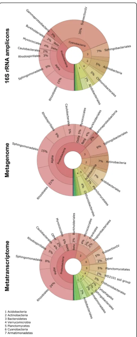
Results:The general community structure of the lichen-associated microbiota was shown to be highly similar irrespective of the employed omics approach. Five highly abundant bacterial orders—Sphingomonadales,
Rhodospirillales,Myxococcales,Chthoniobacterales, andSphingobacteriales—harbor functions that are of substantial importance for the holobiome. Identified functions range from the provision of vitamins and cofactors to the degradation of phenolic compounds like phenylpropanoid, xylenols, and cresols.
Conclusions:Functions that facilitate the persistence ofLobaria pulmonariaunder unfavorable conditions were present in previously overlooked fractions of the microbiota. So far, unrecognized groups likeChthoniobacterales
(Verrucomicrobia) emerged as functional protectors in the lichen microbiome. By combining multi-omics and imaging techniques, we highlight previously overlooked participants in the complex microenvironment of the lichens.
Keywords:Metagenomics, Metaproteomics, Metatranscriptomics, Amplicon sequencing, Lichen symbiosis,Lobaria pulmonaria
Background
Lichens were among the first life forms to conquer life on land already in the Lower Devonian [35]. The drought-tolerant symbiosis evolved an intricate symbiotic architec-ture, also known as the lichen thallus, which comprises peripheric fungal structures to shelter photosynthetic part-ners. The long-living lichen thalli also provide microhabi-tats for diverse microorganisms, in particular fungi and bacteria [5, 27, 28, 31–33, 46, 65]. This new knowledge suggested the emendation of the classic concept of lichens as a bipartite partnership of fungi and algae, especially if the additional partnerships contribute to the global func-tioning of the symbiosis. First evidence in this direction is
seen in protective functions against biotic as well as abi-otic stresses in the bacterial microbiome [15, 28]. Various further roles were assigned to prominent members of the symbiosis; a holistic picture remains to be generated. In our work on the lichen-associated microbial communities, we focused on the lung lichen Lobaria pulmonaria (L.) Hoffm., as a model organism. The leaf-like morphology of this lichen comprises a fungus that encloses a layer of green algae and intermingled clusters of cyanobacteria (so-called internal cephalodia, functional analogues of leg-ume rhizobial nodules; [19]). This complex microenviron-ment makes this lichen an ideal object for further explorations of complex host-microbe interplay. Especially in life forms that are experimentally difficult, current omics approaches provide ideal tools for studies of func-tional diversity. These approaches yield also comprehen-sive baseline information for future experimental studies. For instance, a combined omics approach delivered im-portant information on nitrogen fixation strategies
* Correspondence:[email protected] †Equal contributors
1
Institute of Environmental Biotechnology, Graz University of Technology, Petersgasse 12, 8010 Graz, Austria
2Austrian Centre of Industrial Biotechnolgy GmbH, Petersgasse 14, 8010 Graz, Austria
Full list of author information is available at the end of the article
employed by cyanobacteria within the lichen symbiosis [34].
So far, the application of meta-omics and amplicon sequencing facilitated the identification of the highly diversified bacterial microbiota of lichens, which seem commonly dominated by Alphaproteobacteria [3, 28, 31–33, 62]. We hypothesize that the lichen microbiota is composed of functional guilds, which fulfill distinct roles in the holobiome. By a first in-depth analysis, focusing on the predominant group of Rhizobiales, we detected signatures of nitrogen fixation, as well as synthetic po-tential of phytohormone and vitamin production [22]. Within the same taxonomic group, a so far unknown lineage of lichen-associatedRhizobialeswas identified in a preceding study [31]. This major group represents po-tential “feeders” in the holobiome, while other smaller groups, e.g., the generaBurkholderia,Paenibacillus, and Pseudomonas, were identified as“protectors”[15, 16]. So far, little attention was paid to other bacterial lineages. We nevertheless hypothesize that besides their unrecov-ered diversity, such lineages could also contribute functionally to the adaptability and versatility of the symbiosis. Five bacterial orders with high occurrence in available meta-omics datasets were selected for the present study. These groups were repeatedly found in lichen microbiomes during the past years but remained unexplored in terms of their potential functions. Only recently, Aschenbrenner et al. [3] reported about the presence ofSpartobacteria(Verrucomicrobia) in lichens, a poorly investigated group earlier found to be abundant in soil and aquatic environments [9, 37, 71]. New data reinforced the hypothesis that specific members of Chthoniobacterales (Spartobacteria) and Sphingobacter-iales(Bacteroidetes) could potentially be more important colonizers of L. pulmonaria [4]. A major obstacle in studying the as yet uncultivable Chthoniobacteria was their absence in former releases of public sequence data-bases due to their rather low occurrence in terrestrial habitats and thus low presence in most environmental samples. The ecological significance of this and other abundant groups (Sphingomonadales, Rhodospirillales, Myxococcales, and Sphingobacteriales) will be assessed here by comparative analysis of a newly obtained meta-transcriptomics dataset with metagenomic, metaproteo-mic, and 16S ribosomal RNA (rRNA) gene sequencing amplicon data.
Methods
Sampling ofL. pulmonaria
Lichen thalli of L. pulmonaria were sampled in the Austrian Alps (Johnsbach, N 47° 32′35″, E 14° 37′38″; 1175 m above sea level) from a rich population on mountain maple bark (Acer pseudoplatanus) on 28 June 2014. Samplings were conducted in the late morning
hours, when thalli were humidified (rather than dry) to ensure sufficient metabolic activity for transcriptome analysis. The samples were collected with sterile tweezers, cleaned from macroscopic contaminations (e.g., moss, bark, and insects), and immediately trans-ferred into RNAlater®Stabilization solution (Ambion, Life Technologies, Germany) and stored at −20 °C until fur-ther processing.
Meta-omics-based evaluations of bacterial community structures and functioning
16S rRNA gene amplicon and metagenomic datasets of the Lobaria-associated bacterial communities were re-trieved from already published studies for comparative analyses with the two newly generated metatranscriptomic datasets. The available datasets were utilized for comple-menting analyses of the lichen microbiome [4, 28]. All datasets employed in this study were obtained withL. pul-monaria samples from the same sampling site. The 16S rRNA gene amplicon dataset consists of 24 barcoded sam-ples while the utilized metagenome is based on one com-posite sample. Additional information about the different omics studies are listed in Additional file 1: Table S1.
Metatranscriptome—sample preparation and sequencing
paired-end sequencing was performed by GATC Biotech AG (Konstanz, Germany).
Taxonomic analysis
Downstream sequence analysis for taxonomical assign-ments was done using QIIME 1.9.0 [12]. The 16S rRNA gene fragment sequences were filtered from the genomic and transcriptomic datasets (deposited and publicly available on the metagenomics analysis server MG-RAST; IDs mgm4583748.3, mgm4745782.3) with Sort-MeRNA [42] based on the integrated bacterial 16S rRNA database (SILVA SSU Ref NR v.119; [50]). These sequences as well as the 16S rRNA gene amplicon sequences were clustered at 97% similarity based on the “pick_closed_reference_OTUs.py” script. The SILVA database (release 119; [57]) was used as reference sequence set for taxonomical assignment [56]. Mito-chondrial and chloroplast sequences were removed from datasets with the implemented QIIME “filter_taxa_fro-m_otu_table.py”script to omit non-bacterial reads. Each dataset was normalized to 5720 sequences, which equals the lowest sequence number in the limiting dataset. Taxonomic composition for each omic approach was vi-sualized as Krona charts [52].
Functional analysis
Functional analysis of the lichen-associated bacterial com-munities was performed with MG-RAST based on metage-nomic and metatranscriptomic data. Sequences were compared to GenBank using a maximum e value of 1e−5 and a minimum identity cutoff of 70% [8]. All reads assigned to the orders Sphingobacteriales (Bacteroidetes), Chthonio-bacterales (Verrucomicrobia), Myxococcales, Sphingomona-dales, and Rhodospirillales (Proteobacteria) were extracted for further analysis. The short DNA/cDNA reads were aligned to the protein reference database NCBI-NR (version 05/2015) using DIAMOND (version 0.7.9; [10]). Functional assignment was performed with MEGAN5 [36] based on SEED classification [53]. The abundances of function-assigned sequences of the specified taxonomic orders were subsampled (1000 times randomly subsampled; default set-tings in MEGAN5) for comparison.
Database search and metaproteome data analysis
The raw files were converted to mgf files by the Prote-ome Discoverer software (Thermo Scientific V1.3) and searched with the Mascot search engine (version 2.2.04, Matrix Science Inc.) with the following parameters: par-ent mass tolerance 10 ppm, fragmpar-ent mass tolerance 0.5 Da, maximum missed cleavages 2, charge state 1+, and oxidation of methionine as variable modification. In order to improve the identification of high-confidence peptide sequence matches, a two-step database search was performed, similar to an approach described
previously [38]. To this end, mass spectra were searched in a primary step against the NCBInr protein database (version 2014.06.25, 44,828,108 entries). Hits from the primary search were filtered according to the following parameters: 80% protein probability, 95% peptide prob-ability, and a minimum of one uniquely identified pep-tide). Subsequently, protein identifiers fulfilling the above-described criteria were extracted. Based on these identifiers, a subset target-decoy database (34,344 en-tries) was constructed using an in-house script for local data deposition (“database creator”). Results from the second database search were filtered applying more stringent parameters (99% protein probability, 99% pep-tide probability, and a minimum of one uniquely identi-fied peptide,) including a replicate filter, i.e., a protein had to be identified in two out of three technical repli-cates. Functional classification and taxonomic assign-ment of the protein sequences have been accomplished by the in-house developed metaproteome analyses pipe-line “Prophane 2.0” (http://www.prophane.de, [62]). Relative protein quantification was based on normalized spectral abundance factor (NSAF) values [72], only con-sidering spectral counts that have been uniquely identi-fied for a specific protein.
DNA isolation
Total DNA of each sample was extracted using the MoBioPowerSoil® DNA Isolation Kit (Carlsbad, USA) ac-cording to the manufacturer’s protocol with modifica-tions from Aschenbrenner et al. [3]. Briefly, the samples were ribolyzed three times for 30 s at 5.5 m × s−1and kept 5 min on ice in between. DNA from control strains (Escherichia coli, Staphylococcus aureus, Pseudomonas aeruginosa) was extracted following an ethanol precipi-tation protocol.
Probe design, evaluation, andVerrucomicrobia-specific SSCP
Single-strand conformation polymorphism (SSCP) experi-ments were employed to validate the presence of Verruco-microbia populations on lichens with a classic, molecular method. To target Verrucomicrobia, the oligonucleotide primers VMB537f and VMB1295r were initially used as in previous studies [51]. However, the specificity and coverage of the primers was not suitable for our approach (30% Ver-rucomicrobia, 17% Spartobacteria) after in silico tests against the Arb-Silva database using the TestPrime tool (http://www.arb-silva.de/search/testprime/). We therefore designed a new primer set. The forward primer (Verruco f) was designed based on the Verrucomicrobia FISH probe
(EUB338III; 5′-GCTGCCACCCGTAGGTGT-3′; [20]).
on the Spartobacteria FISH probe [20]. The annealing position is located at about 680 bp upstream yielding in 350-bp fragments. According to TestPrime, 0.84% of all bacteria are amplified using these two primers Verruco f and 806r. This fraction includes 84% of the phylum Verru-comicrobiawithin 89% of the classSpartobacteria respect-ivelyChthoniobacterales.
To assess the specificity of our primers, we produced Verrucombicrobia-specific fingerprints with various Lobaria samples (Additional file 1: Fig. S1). No band patterns were retrieved with control strains (E. coli, S. aureus, P. aeruginosa), which represented the closest probeBase mismatches. Selected bands were sequenced and taxonomies were assigned by blast searches against the NCBI 16S ribosomal RNA sequence database. Using the novel primer approach, no additional taxa beyond Verrucomicrobia were detected within the selected se-quencing range (Additional file 1: Fig. S2).
SSCP analysis of the total community DNA of four dif-ferent samples, the type strain, and internal controls was carried out according to Schwieger and Tebbe [63] and Bassam et al. [31]. The PCR was performed using a total volume of 60μl containing 12μl of Taq&Go (QBiogen), 6μl of the purified DNA, 1.5μl of each primer (10μM), and 39μl of ultrapure water (95 °C, 5 min; 30 cycles of 95 °C, 20 s; 56.8 °C, 15 s; 72 °C, 30 s; final elongation at 72 °C, 10 min). Control strains (closest database mis-matches according to probeBase; http://probebase.csb.u-nivie.ac.at; E. coli, S. aureus, P. aeruginosa) were also amplified. The PCR products were purified using Wizard® SV Gel and PCR Clean-Up System (Promega, Madison, USA), prior to λ-exonuclease digestion and DNA single-strand folding [47]. Polyacrylamide gel electrophoresis was carried out on a TGGE platform (Biometra, Göttingen, Germany) at 26 °C and 400 V for 26 h using 8% (wt vol−1) acrylamide gel.
Since all sequences of the verrucomicrobial class Sparto-bacteriawere assigned toChthoniobacterales, we checked for specific FISH (fluorescent in situ hybridization) probes for the orderChthoniobacteralesin probeBase (http://pro-bebase.csb.univie.ac.at). As so order-specific probe exists, we used the probe SPA714 to detect specifically bacteria within Spartobacteria. However, this probe only targets about 66% (2553 out of 3874 hits) of all Chthoniobacter-alessequences according to RDP probe match. Addition-ally, we evaluated the FISH probe EUB338III, which is supposed to specifically target the order Verrucomicro-biales (class Verrucomicrobiae) besides the non-target taxon Chloroflexi. Sequence alignments to the Probe-match database [18] revealed that this probe does not only match to Verrucomicrobiae (6930/7266) but also covers the verrucomicrobial classes Subdivision 3 (3064/3647), Opitutae (4223/4460), and Spartobacteria (4441/4734). According to our data, about 89% (mean) of the
verrucomicrobial sequences were assigned to the class Spartobacteria, followed by Opitutae (7%). Hence, this FISH probe is also suitable to detect Spartobacteria, and in a wider sense, Chthoniobacterales in the case of L. pulmonaria-associated bacterial community.
FISH/CLSM
Fluorescence in situ hybridization (FISH) was performed to specifically visualize the colonization pattern of Chthoniobacterales(class Spartobacteria, phylum Verru-comicrobia) among other eubacteria. L. pulmonaria thalli were fixed with 4% paraformaldehyde/phosphate-buffered saline (PBS) with a ratio of 3:1 at 4 °C for at least 4 h. Prior to in situ hybridization according to Car-dinale et al. [13], thallus cross-sections were prepared. For the detection of Spartobacteria as well as Verruco-microbia, the Cy5-labled probes SPA714 (42 °C, 35% formamide; [2]) and EUB338III (42 °C, 15% formamide) were used. Other eubacteria were detected with an equi-molar mixture of the Cy3-labeled probes EUB338, EUB338II, and EUB338III (42 °C, 15% formamide; [1, 20]). Additionally, NONEUB probes [69] labeled with the respective fluorochromes were used as negative con-trols. Details about oligonucleotide probes are available on probeBase [48]. To suppress photobleaching of the fluorescently labeled probes, SlowFade Diamond antifade reagent (Molecular Probes, Eugene, USA) was used. Tar-geted Chthoniobacterales and all other eubacteria were visualized with a Leica TCS SPE confocal laser-scanning microscope (Leica Microsystems, Mannheim, Germany) and confocal stacks were processed with Imaris 7.3 (Bitplane, Zurich, Switzerland).
Results
Comparative composition of the lichen microbiome
bacterial community: Sphingomonadales (10.2 ± 3.3%; mean values and SD of the utilized omics and amplicon studies are provided),Rhodospirillales(5.8% ± 2.5%), Myxo-coccales (4.7 ± 0.8%, Proteobacteria), Chthoniobacterales (4.3 ± 0.7%, Verrucomicrobia), and Sphingobacteriales (5.8 ± 2.8%, Bacteroidetes). The composition and relative abundances of these orders at family level were visualized in Fig. 2. Within the order Sphingobacteriales, two main families were identified: Sphingobacteriaceae and Chitino-phagaceae. TheChitinophagaceae were more abundant in the amplicon dataset when compared with the metagen-ome, whereas Sphingobacteriaceae were less abundant in the former dataset. Chthoniobacteraleswas represented by the familiesChthoniobacteraceaeand Xiphinematobactera-ceae. Moreover, groups within Chthoniobacteraceae accounted for a lower fraction in the amplicon dataset than in the corresponding meta-omics data. Highly abundant families identified within the alphaproteobacterial orders Sphingomonadales were represented by Sphingomonada-ceae and Erythrobacteraceae, as well as Acetobacteraceae andRhodospirillaceaeinRhodospirillales.Both Sphingomo-nadaceae and Acetobacteraceae were a minor fraction in the amplicon data set in comparison with the metagenome data. The order Myxococcales was represented by various distinct families, whereasPolyangiaceae,Phaselicystidaceae, and Cystobacteraceae had a higher relative abundance in the metatranscriptome than in the other two meta-omics approaches. Other detected families within this order were HaliangiaceaeandSandaracinaceae.
Functional contribution of abundant bacterial community members to the lichen symbiosis
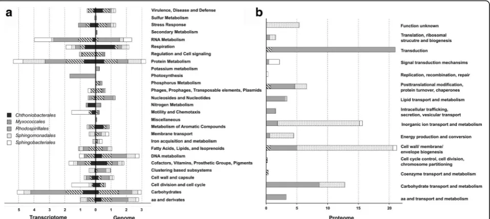
Together with metagenomic and metaprotemic data, the transcriptome information corroborated the functional potential on lichen-bacteria and provided expression data for the bacterial ordersSphingomonadales, Rhodos-pirillales, Myxococcales, Chthoniobacterales, and Sphin-gobacteriales. Functional assignments for each bacterial group based on SEED and COG classification are visual-ized in Fig. 3. During the analysis, we primarily focused on functions with a direct implication for the symbiosis or such that modulate interactions with the adjacent environment.
Provision of micronutrients (inkl. N, S, P, K, Fe metabolism)
In the metagenome, genes attributed to potassium and nitrogen metabolism were both predominantly (96%) assigned to the alphaproteobacterial order Rhodospiril-lalesand to a minor extent toSphingobacteriales (potas-sium metabolism) and Sphingomonadales (nitrogen metabolism). Rhodospirillalescontributed at a high pro-portion (94 and 96%) to the assimilation of ammonia and potassium uptake and transport, respectively. There were also functional assignments to nitrate and nitrite Fig. 1Bacterial community structures of the Lobaria-associated
ammonification and the production of nitric oxides. Sulfur and iron metabolism was predominately represented by Sphingobacteriales. Regarding iron metabolism, not only all Sphingobacteriales-specific reads contributed to the TonB-dependent receptor of Gram-negative bacteria but also all three proteobacterial taxa were involved in this iron acqui-sition strategy. Other identified iron transport mechanisms were systems based on siderophores or hemin. Functional genes within the phosphate metabolism were mainly assigned toProteobacteria, especiallyMyxococcales. In con-trast, nitrogen metabolism (ammonia assimilation) was mainly detected forChthoniobacteralesand only to a minor extent forRhodospirillales in the metatranscriptome data-set. Also expressed genes related to both, iron acquisition and metabolism, were only detected forSphingobacteriales and Sphingomonadales(including hemin transport system and a TonB-dependent receptor). With respect to iron transport, an outer membrane receptor protein detected in the metaproteome was also assigned toSphingomonadales.
Aromatic compounds metabolism
All examined taxa were involved in the metabolism of aromatic compounds according to the analyzed meta-genome. The majority of corresponding functions was assigned to various degradation mechanisms. Chthonio-bacterales was the primary contributor to the degrad-ation of n-phenylalkanoic acid. Alphaproteobacteria were involved in the degradation of phenylpropanoid, xylenols, and cresols (mainly by Rhodospirillales) and chloroaromatic compounds (Myxococcales). Especially, Proteobacteria were identified to potentially synthesize the plant hormone auxin. Rhodospirillales was also found to be involved in phenazine biosynthesis. In the metatranscriptome data, phenazine and auxin produc-tion were not present for any of the investigated groups. Instead, degradation and transport mechanisms of vari-ous compounds were found, such as biphenyl, carbazol, and benzoate (Sphingomonadales) as well as gentisate and salicylate (Rhodospirillales,Myxococcales).
Fig. 2Relative sequence abundances of families within the highly abundant ordersSphingobacteriales,Rhodospirillales,Sphingomonadales,
Cofactors, vitamins, prosthetic groups, and pigments
Chthoniobacterales encoded genes involved in the syn-theses of the vitamins riboflavin and biotin, whereas, genes for the thiamin, pyridoxine, and folate syntheses were predominantly assigned to Proteobacteria. How-ever, transcriptomic data revealed biotin and folate syn-thesized by Chthoniobacterales. The vitamins riboflavin, pyridoxine, and thiamin were mainly produced by Rho-dospirillales. Moreover,Sphingomonadalesexpressed the cobalamin-adenosyltransferase PduO (EC 2.5.1.17), which is involved in the biosynthesis of vitamin B12. Quinone cofactors were found to be synthesized by Sphin-gobacteriales (menaquinone) and Myxococcales, whereas the latter one expressed 4-hydroxyphenylpyruvate dioxy-genase (EC 1.13.11.27) which is involved in the biosyn-thesis of plastoquinone and tocopherol.
Stress response
Function-related genes within the category stress re-sponse were mainly assigned to oxidative stress and heat shock. According to the metagenome, the latter one was predominantly represented by Myxococcales and Sphin-gobacteriales encoding for the chaperones DnaK and GroEL. DnaK was also found to be expressed by Myxo-coccales and GroEL byRhodospirillales and Sphingomo-nadalesaccording to transcriptomic and proteomic data, respectively. Additionally, various sigma factors could be detected either based on RNA or protein sequences: RpoH (Rhodospirillales) for heat shock, RpoN (Sphingo-bacteriales) for nitrogen-limitation, and a homolog of the exocytoplasmic heat stress sigma factor RpoE
(Myxococcales). Notably, all of the observed taxa encoded to a certain extent defense mechanisms against oxidative stress. In particular, Chthoniobacterales encoded for a redox-sensitive transcription regulator and Rhodospirillales for rubrerythrin, which is involved in oxidative stress tolerance in anaerobic bacteria. All Pro-teobacteria facilitated glutathione-dependent protection against ROS-induced (reactive oxygen species) oxidative damage. Although the catalase EC 1.11.1.6 was found to be encoded in all observed Proteobacteria, transcripts could be only detected for Myxococcales. According to transcriptomic data, Chthoniobacterales, Myxococcales, and Rhodospirillales were involved in the syntheses of glutaredoxin-related proteins, glutaredoxin, and rubrery-thrin and glutathione for non-redox reactions. Moreover, hits for iron/manganese superoxide-dismutases were found in the proteome. Resistance mechanisms against acid stress (arginine decarboxylase EC 4.1.1.19) were additionally detected forMyxococcales.
Virulence, disease, and defense
in the transcriptome could be also detected for Sphingo-bacteriales,MyxococcalesandChthoniobacterales. The lat-ter one exhibited additionally resistance to acriflavin. Beta-lactamases were detected especially for Alphaproteobac-teria and Sphingobacteriales, whereas multidrug resist-ance, e.g., via efflux pumps, were found for all taxa, except for Myxococcales. Proteomic data revealed no hits for functions related to resistance mechanisms.
Membrane transport
Various types of transport systems were found in the metagenomic dataset, such as protein secretion system types II, III, and IV. Secretion system type IV was found in all observed taxa except forChthoniobacterales. Addition-ally, Rhodospirillales encoded for the sec-independent twin-arginine translocation pathway to transport folded proteins as well as manganese, zinc, nickel, and cobalt. Functional assignments to Ton- and Tol-dependent trans-port systems were found for all observed taxa. Metatran-scriptomic data displayed the expression of Ton and Tol transport systems forSphingomonadalesand Sphingobac-teriales, as well as hits for parts of the type IV secretion system inRhodospirillales. TonB-dependent receptor pro-teins (SphingomonadalesandSphingobacteriales) and pro-tein export chaperone SecB (Rhodospirillales) were also found in the metaproteome.
Motility and chemotaxis
Proteobacteria, and Myxococcales especially, were in-volved in chemotaxis encoding the proteins methyltrans-ferase CheR (EC 2.1.1.80) and the methylesterase CheB (EC 3.1.1.61). According to the metatranscriptome, only the signal transduction histidine kinase CheA was de-tected for Myxococcales. Rhodospirillales predominantly encoded for flagellar motility. Contrarily, in the meta-transcriptome, the main contributor to flagellar motility was Sphingobacteriales, followed by Chthoniobacterales andMyxococcales.
Occurrence of phages, prophages, transposable elements, and plasmids
Functional assignments within this category were only found for Sphingomonadales and Sphingobacteriales, including transposable elements like Tn552 encoding for a beta-lactamase or the staphylococcal phi-Mu50B-like prophages. In the metatranscriptome analysis, tran-scripts for the staphylococcal pathogenicity island SaPI were found forSphingobacterialesandMyxococcales.
Verrucomicrobia-specific microbial fingerprints (PCR-SSCP) and sequencing revealed several lineages of
Chthoniobacterales
PCR amplicons retrieved usingVerrucomicrobia-specific primer were separated by SSCP (single-strand conform-ation polymorphism). The phylogenetic analysis of se-quences obtained from excised products revealed three lineages in Chthoniobacteriales. One of these is related to Chthoniobacter, a second represents relatives of Udaeobacter, and a third lineage has no relatives among known genera in the order.
Visualization ofChthoniobacteraleswithin the lichen symbiosis
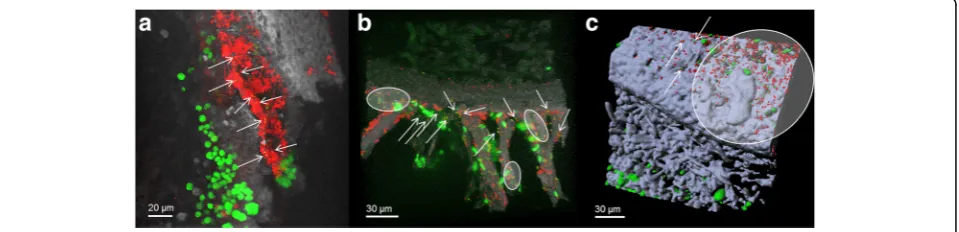
The microscopic localization ofChthoniobacteraleswas uti-lized to explore colonization patterns and potential accu-mulations of these previously underexplored colonizers. Fluorescence in situ hybridization with Spartobacteria-spe-cific probes revealed consistent colonization of both the upper and the lower cortex ofL. pulmonaria (Fig. 4a, b). Volume rendering of the confocal stacks clearly visualized a general tendency ofSpartobacteriato be dispersed on the lichen as single small colonies between biofilm-like struc-tures of other eubacteria (which were not specified further). Although there were also small areas with higher cell dens-ities (Fig. 4b), no larger colonies of Spartobacteria were detected. Micrographs with the Verrucomicrobia-specific probe confirmed these colonization patterns (Fig. 4c).
Fig. 4Colonization patterns ofSpartobacteria(a,b) andVerrucomicrobia(c) on the lichen thallus ofL. pulmonariastained by fluorescence in situ hybridization (FISH).Green: algaeDictyochloropsis reticulata;grayorblue/purple: lichenized fungusL. pulmonaria;yellow:SpartobacteriaorVerrucomicrobia;
red, other eubacteria.a,bVolume rendering of confocal stacks.cThree-dimensional model reconstruction visualized as isosurface and spheres.Arrows
Discussion
In the present study, we show how the addition of tran-scriptomic data to genomic and proteomic information contributes to the detection of so far unknown active bacterial players in the lichen symbiosis. This approach is backed up by their complementary microscopic FISH/ CLSM visualization. Lichens have complex and function-ally diverse microbiota. As some may serve in nutrition of the host while others provide protective functions, or growth regulating functions to the hosts, we may generally distinguish feeders and protectors. The ob-tained data indicates that members of the identified functional guild are found in both roles and primarily serve as probiotics and detoxifiers in the Lobaria holobiome. Similarly, Hodkinson et al. [34] employed a metatranscriptomics-based approach to explore
detailed functioning of nitrogen fixation by lichen-symbiotic cyanobacteria.
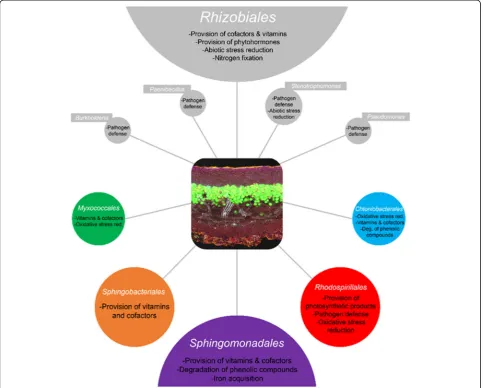
In the current study, specific functional contributions to the overall symbiosis were explored in five abundant bacter-ial orders, namely Sphingomonadales, Rhodospirillales, Myxococcales (allProteobacteria),Chthoniobacterales (Ver-rucomicrobia), and Sphingobacteriales(Bacteroidetes) (Fig. 5). Both metagenomic and metatranscriptomic analyses re-vealed various strategies of associated bacteria to survive under stressful conditions. Lichens are known to produce extracellular reactive oxygen species (ROS) including super-oxide, hydroxyl radicals, and hydrogen peroxide [6], which are produced at high rates after desiccation-rehydration events. According to Beckett et al. [7], extracellular ROS production might also help defending against bacterial and fungal pathogens in this lineage of lichenized fungi, which
largely lacks crystals of potentially antimicrobial lichen compounds in the surface layers. In general, fast-growing bacterial pathogens are not known to degrade lichens and many bacterial associates rather represent stress-tolerant commensals or even beneficials, which contribute various functions to the lichen meta-organism [22, 28]. These bac-teria require efficient mechanisms to withstand periodic desiccation/rehydration cycles with associated oxidative bursts. Metagenomic data support this hypothesis as cor-responding mechanisms against oxidative stress, such as catalases and low-molecular-weight antioxidants (e.g., glutathione) are encoded across bacterial groups. These functions were found also for the active fraction in the metatranscriptome.
Oxidative stress conditions also induce the expression of various heat shock proteins [68]. In particular, the chap-erones GroEL and DnaK, which were highly abundant in our data, can be induced by various oxygen species such as superoxide or hydrogen peroxide, respectively [23], or by the presence of toxic compounds like antibiotics, heavy metals, and aromatic compounds [68]. Several functions, which protect against such toxic compounds, were also reflected in our data, e.g., those contributing to resistances to the toxic metals arsenic, cadmium, cobalt, or zinc. In addition, resistance mechanisms for antibiotics such as fluoroquinolones were also found like beta-lactamases and efflux pumps for multidrug resistances. As secondary me-tabolites (including depsides, depsidones, and
dibenzofu-rans) produced by the lichenized fungus have
antimicrobial activities to defend the host against fungal and bacterial pathogens [43], lichen- and plant-associated bacteria likely developed different defense mechanisms against these specific aromatic compounds. Our data con-firmed the presence of potential functions to degrade these metabolites such as phenylpropanoid, which is the carbon skeleton of a wide range of polyphenols. In con-trast, there were also hints for the production of phenolic compounds by bacteria themselves. Especially, Rhodospir-illales was found to be involved in the production of phenazines, which are known to inhibit the bacterial and fungal growth [49] thereby increasing its own competitive-ness and ecological fitcompetitive-ness [54]. This could also have a positive effect for the lichen itself as these broad-specificity antibiotics might control fast-growing bacterial and fungal pathogens.
According to our omics datasets, the observed taxa encode and express various transport machineries such as Ton- and Tol-dependent transport systems as well as different secretion systems. In this context, the type IV secretion system (T4SS) is also interesting, as it is not only involved in the DNA uptake from or the release to the surrounding environment or in the conjugal DNA transfer but also in the translocation of effector mole-cules to eukaryotic target cells [21]. These
trans-kingdom transfers include fungi, plants, and mammalian cells [11, 14, 70]. We hypothesize that T4SS might be also used for inter-kingdom“cross-talking” between the lichen and members of the associated microbiome, analogous to the Rhizobia-plant interaction [26, 64].
In this work, we showed the metabolic contribution of the hitherto overlooked phylum Verrucomicrobia. After the discovery of Chthoniobacter flavus in soil [60], only a few studies so far focused on Spartobacteriaas a bac-terial class of Verrucomicrobia [39, 61]. According to genomic data, Spartobacteria have roles in the carbon cycle by in degradation of various complex carbohy-drates (such as cellulose and xylan; [30]). With newly de-signed oligonucleotides, we were now able to visualize the presence of Chthoniobacterales for the first time in lichens. Transcriptomics data suggest their involvement in the metabolism of aromatic compounds (degradation of phenolic substances), production of various vitamins, and defense against antibiotics (fluoroquinolone) and oxidative stress. Interestingly, no information on this particular group was found in the metaproteome, which might be due to the lack of data in incomplete reference databases.
The new transcriptomic data of the lichen microbiome validate and corroborate the evidence for particular functions, but appropriate integration of different omics approaches nevertheless require further optimization of lab procedures and bioinformatic analyses. Variation may occur during sample preparation for individual omics approaches [66]. Primer selection has an impact on representation of taxonomic composition in ampli-con sequencing [25, 41, 55], which may be further com-plicated due to gene copy number variations in the rRNA operons [40, 58]. Community composition in metatranscriptomic data is influenced by bacteria-specific differences of metabolic activity. Similar applies also to representation of the metaproteome, which is also sensitive to the preparation of the protein fraction. In contrast, the metagenome may also comprise a fraction of inactive or dead bacteria, which were recently visualized by Cernava et al. [17]. Since predominant members of the lichen microbiota remain fairly stable [27], we exclude that different sampling dates are of sig-nificant influence.
the functionally diverse communities of bacteria on the surfaces of the lichens provide additional benefits beside the algal partners. Our vision is to use the insights from this natural symbiotic system also as inspirations for novel biotechnological applications, which surpass the era of axenic culture.
Conclusions
The present study allowed further advancements in the exploration of the lichen holobiome. Previously unex-plored functional guilds of this unconventional model were captured with independent methods and further characterized in an integrative approach. It was shown that Sphingomonadales, Rhodospirillales, Myxococcales, Chthoniobacterales, and Sphingobacteriales account for consistent fractions of theL. pulmonariamicrobiome ir-respective of the utilized meta-omics tool. Deepening analyses provided insights into functional contributions of this bacterial consortium, which was then brought into an ecological context. By combining various meta-omic techniques, deficiencies of the particular methods were bypassed. Cost-efficient amplicon sequencing stud-ies are often preferred when microbial community struc-tures are explored in novel habitats. However, they deliver less comprehensive information compared to metagenomic and metatranscriptomic datasets. The combination of the advantages of various NGS methods provides generally a higher accuracy for a holistic study. Taken together, we successfully applied a combination of state-of-the-art tools for a deepening exploration of a complex multi-partner network.
Additional file
Additional file 1: Table S1. Metadata and basic information related to the DNA/RNA-based omic approaches.Table S2:Primer sets for eukaryotic and prokaryotic rRNA gene fragment amplification. Bold: T7 promoter sequence; blue: enhancer sequence.Table S3:Taxonomical assignments.Fig. S1: Molecular fingerprints of 16S rRNA gene fragments from total DNA (Tot DNA) from the lichen, isolates from the lichen thalli (L. pulmonaria) and control strains (E. coli, S. aureus, P. aeruginosa).Fig. S2:Phylogenetic tree of bacteria-strains within the phylumVerrucomicrobia. The tree was constructed from an evolutionary distance matrix based on the neighbor-joining method [59]. The phylogenetic tree was constructed with 1000 seeds and 1000 bootstraps with the neighboring-joining method [59] using clustalX2 [45] and phylip [24].Fig. S3:Common and unique OTUs among three omics approaches based on 16S rRNA gene fragment sequences with a genetic distance of 3%.
Acknowledgements
We want to thank Alexander Mahnert (Graz) for the helpful discussions during the bioinformatic analyses. We also appreciate the help of Melanie Marek (Graz) and Tanja Nottendorfer (Graz) during the wet lab experiments.
Funding
This work was supported by a grant from the Austrian and German Science Foundation to GB, KR, and MG (FWF-DACH Project I882).
Availability of data and materials
All details related to the utilized data and the accession numbers are presented in Table S1 of Additional file 1.
Authors’contributions
TC, IAA, and GB designed the study. MG provided specific knowledge related to the lichen symbiosis. TC, IAA, and AE performed the bioinformatic analyses based on DNA and RNA sequencing datasets. CL and KR contributed to the metaproteomic analyses and corresponding expertise in the data evaluation. AE and LK performed the SSCP and FISH/CLSM experiments and the corresponding data evaluation. TC, IAA, AE, KR, MG, and GB wrote the manuscript. All authors read and approved the final version of the manuscript.
Ethics approval and consent to participate Not applicable.
Consent for publication Not applicable.
Competing interests
The authors declare that they have no competing interests.
Publisher’s Note
Springer Nature remains neutral with regard to jurisdictional claims in published maps and institutional affiliations.
Author details
1Institute of Environmental Biotechnology, Graz University of Technology, Petersgasse 12, 8010 Graz, Austria.2Austrian Centre of Industrial Biotechnolgy GmbH, Petersgasse 14, 8010 Graz, Austria.3Institute of Microbiology, Ernst-Moritz-Arndt University of Greifswald, Friedrich-Ludwig-Jahn Strasse 15, 17489 Greifswald, Germany.4Institute of Plant Sciences, University of Graz, Holteigasse 6, 8010 Graz, Austria.
Received: 14 February 2017 Accepted: 6 July 2017
References
1. Amann RI, Binder BJ, Olson RJ, Chisholm SW, Devereux R, Stahl DA. Combination of 16S rRNA-targeted oligonucleotide probes with flow cytometry for analyzing mixed microbial populations. Appl Environ Microbiol. 1990;56:1919–25.
2. Arnds J, Knittel K, Buck U, Winkel M, Amann R. Development of a 16S rRNA-targeted probe set for Verrucomicrobia and its application for fluorescence in situ hybridization in a humic lake. Syst Appl Microbiol. 2010;33:139–48. 3. Aschenbrenner IA, Cardinale M, Berg G, Grube M. Microbial cargo: do
bacteria on symbiotic propagules reinforce the microbiome of lichens? Environ Microbiol. 2014;16:3743–52.
4. Aschenbrenner IA, Cernava T, Erlacher A, Berg G, Grube M. Differential sharing and distinct co-occurrence networks among spatially close bacterial microbiota of bark, mosses and lichens. Mol Ecol. 2017;26:2826–38. 5. Bates ST, Cropsey GW, Caporaso JG, Knight R, Fierer N. Bacterial
communities associated with the lichen symbiosis. Appl Environ Microbiol. 2011;77:1309–14.
6. Beckett RP, Minibayeva FV, Laufer Z. Extracellular reactive oxygen species production by lichens. Lichenologist. 2005;37:397.
7. Beckett RP, Minibayeva FV, Vylegzhanina NN, Tolpysheva T. High rates of extracellular superoxide production by lichens in the suborder Peltigerineae correlate with indices of high. Plant Cell Environ. 2003;26:1827–37. 8. Benson DA, Karsch-Mizrachi I, Lipman DJ, Ostell J, Rapp BA, Wheeler DL.
GenBank. Nucleic Acids Res. 2000;28:15–8.
9. Bergmann GT, Bates ST, Eilers KG, Lauber CL, Caporaso JG, Walters WA, et al. The under-recognized dominance of Verrucomicrobia in soil bacterial communities. Soil Biol Bioch. 2011;43:1450–5.
10. Buchfink B, Xie C, Huson DH. Fast and sensitive protein alignment using DIAMOND. Nat Meth. 2015;12:59–60.
12. Caporaso JG, Kuczynski J, Stombaugh J, Bittinger K, Bushman FD, Costello EK, Fierer N, Gonzalez Pena A, Goodrich JK, Gordon JI, Huttley GA, Kelley ST, Knights D, Koenig JE, Ley RE, Lozupone CA, McDonald D, Muegge BD, Pirrung M, Reeder J, Sevinsky JR, Turnbaugh PJ, Walters WA, Widmann J, Yatsunenko T, Zaneveld J, Knight R. QIIME allows analysis of high-throughput community sequencing data. Nat Meth. 2010;7:335–6. 13. Cardinale M, De Castro JV, Müller H, Berg G, Grube M.In situanalysis of the
bacterial community associated with the reindeer lichenCladonia arbusculareveals predominance ofAlphaproteobacteria. FEMS Microbiol Ecol. 2008;66:63–71. 14. Cascales E, Christie PJ. The versatile bacterial type IV secretion systems. Nat
Rev Microbiol. 2003;1:137–49.
15 Cernava T, Müller H, Aschenbrenner IA, Grube M, Berg G. Analyzing the antagonistic potential of the lichen microbiome against pathogens by bridging metagenomic with culture studies. Front Microbiol. 2015a;6:620. 16. Cernava T, Aschenbrenner IA, Grube M, Liebminger S, Berg G. A novel assay
for the detection of bioactive volatiles evaluated by screening of lichen-associated bacteria. Front Microbiol. 2015b;6:398.
17. Cernava T, Berg G, Grube M. High life expectancy of bacteria on lichens. Microb Ecol. 2016;72:510–3.
18. Cole JR, Chai B, Farris RJ, Wang Q, Kulam SA, McGarrell DM, et al. The Ribosomal Database Project (RDP-II): sequences and tools for high-throughput rRNA analysis. Nucleic Acids Res. 2005;33:294–6. 19. Cornejo C, Scheidegger C. New morphological aspects of cephalodium
formation in the lichen Lobaria pulmonaria (Lecanorales, Ascomycota). Lichenologist. 2013;45:77–87.
20. Daims H, Brühl A, Amann R, Schleifer KH, Wagner M. The domain-specific probe EUB338 is insufficient for the detection of all bacteria: development and evaluation of a more comprehensive probe set. Syst Appl Microbiol. 1999;22:434–44.
21. Ding Z, Atmakuri K, Christie PJ. The outs and ins of bacterial type IV secretion substrates. Trends Microbiol. 2003;11:527–35.
22. Erlacher A, Cernava T, Cardinale M, Soh J, Sensen CW, Grube M, Berg G. Rhizobiales as functional and endosymbiontic members in the lichen symbiosis ofLobaria pulmonariaL. Front Microbiol. 2015;6:53. 23. Farr SB, Kogoma T. Oxidative stress responses inEscherichia coliand
Salmonella typhimurium. Microbiol Rev. 1991;55:561–85. 24. Felsenstein J. Evolutionary trees from DNA sequences: a maximum
likelihood approach. J Mol Evol. 1981;17:368–76.
25. Gonzalez JM, Portillo MC, Belda-Ferre P, Mira A. Amplification by PCR artificially reduces the proportion of the rare biosphere in microbial communities. PLoS One. 2012;7:e29973.
26. Gourion B, Berrabah F, Ratet P, Stacey G. Rhizobium–legume symbioses: the crucial role of plant immunity. Trends Plant Sci. 2015;20:186–94.
27. Grube M, Cardinale M, Vieira de Castro J, Müller H, Berg G. Species-specific structural and functional diversity of bacterial communities in lichen symbiosis. ISME J. 2009;3:1105–15.
28. Grube M, Cernava T, Soh J, Fuchs S, Aschenbrenner I, Lassek C, Wegner U, Becher D, Riedel K, Sensen CW, Berg G. Exploring functional contexts of symbiotic sustain within lichen-associated bacteria by comparative omics. ISME J. 2015;9:412–24.
29. Hays SG, Patrick WG, Ziesack M, Oxman N, Silver PA. Better together: engineering and application of microbial symbioses. Curr Opin Biotechnol. 2015;36:40–9.
30. Herlemann DP, Lundin D, Labrenz M, Jürgens K, Zheng Z, Aspeborg H, Andersson AF. Metagenomic de novo assembly of an aquatic representative of the verrucomicrobial class Spartobacteria. MBio. 2013;4:e00569–12. 31. Hodkinson BP, Lutzoni F. A microbiotic survey of lichen-associated bacteria
reveals a new lineage from the Rhizobiales. Symbiosis. 2009;49:163–80. 32. Hodkinson BP. A Phylogenetic, ecological, and functional characterization of
non-photoautotrophic bacteria in the lichen microbiome. Durham, North Carolina: Doctoral Dissertation, Duke University; 2011.
33. Hodkinson BP, Gottel NR, Schadt CW, Lutzoni F. Photoautotrophic symbiont and geography are major factors affecting highly structured and diverse bacterial communities in the lichen microbiome. Environ Microbiol. 2012;14:147–61. 34. Hodkinson BP, Allen JL, Forrest LL, Goffinet B, Sérusiaux E, Andrésson ÓS,
Miao V, Bellenger JP, Lutzoni F. Lichen-symbiotic cyanobacteria associated with Peltigera have an alternative vanadium-dependent nitrogen fixation system. Eur J Phycol. 2014;49:11–9.
35. Honegger R, Edwards D, Axe L. The earliest records of internally stratified cyanobacterial and algal lichens from the Lower Devonian of the Welsh Borderland. New Phytol. 2013;197:264–75.
36. Huson DH, Mitra S, Weber N, Ruscheweyh H, Schuster SC. Integrative analysis of environmental sequences using MEGAN4. Genome Res. 2011;21:1552–60. 37. Janssen PH, Costa KC, Hedlund BP. Class III. Spartobacteria. In: Krieg NR,
Ludwig W, Whitman WB, Hedlund BP, Paster BJ, Staley JT, Ward N, Brown D, Oarte A, editors. Bergey’s manual of systematic bacteriology, vol. 4. 2nd ed. New York: Springer Verlag; 2011. p. 834–41.
38. Jagtap P, Goslinga J, Kooren JA, McGowan T, Wroblewski MS, Seymour SL, Griffin TJ. A two‐step database search method improves sensitivity in peptide sequence matches for metaproteomics and proteogenomics studies. Proteomics. 2013;13:1352–57.
39. Kant R, Van Passel MW, Palva A, Lucas S, Lapidus A, del Rio TG, Smidt H. Genome sequence of Chthoniobacter flavus Ellin428, an aerobic heterotrophic soil bacterium. J Bacteriol. 2011;193:2902–3.
40. Klappenbach JA, Dunbar JM, Schmidt TM. rRNA operon copy number reflects ecological strategies of bacteria. Appl Environ Microbiol. 2000;66: 1328–33.
41. Klindworth A, Pruesse E, Schweer T, Peplies J, Quast C, Horn M, Glöckner FO. Evaluation of general 16S ribosomal RNA gene PCR primers for classical and next-generation sequencing-based diversity studies. Nucleic Acids Res. 2013; 41:1–11.
42. Kopylova E, Noé L, Touzet H. SortMeRNA: fast and accurate filtering of ribosomal RNAs in metatranscriptomic data. Bioinformatics. 2012; doi:10. 1093/bioinformatics/bts611.
43. KosanićM, RankovićB. Lichen secondary metabolites as potential antibiotic agents. In: RankovićB, editor. Lichen secondary metabolites. Bioactive properties and pharmaceutical potential. Berlin: Springer Verlag; 2015. p. 81–104. 44. Kukutla P, Steritz M, Xu J. Depletion of ribosomal RNA for mosquito gut
metagenomic RNA-seq. J Vis Exp. 2013;74:e50093.
45. Larkin MA, Blackshields G, Brown NP, Chenna R, McGettigan PA, McWilliam H, et al. Clustal W and Clustal X version 2.0. Bioinformatics. 2007;23:2947–8. 46. Lawrey JD, Diederich P. Lichenicolous fungi : interactions, evolution, and
biodiversity. Bryologist. 2003;106:81–120.
47. Lieber A, Kiesel B, Babel W. Microbial diversity analysis of soil by SSCP fingerprinting technique using TGGE Maxi System. In: Merbach W, Hütsch BW, Augustin J, editors. Ökophysiologie des Wurzelraumes. Stuttgart, Leipzig, Wiesbaden: Teubner Verlag; 2003. p. 61–5.
48. Loy A, Maixner F, Wagner M, Horn M. probeBase—an online resource for rRNA-targeted oligonucleotide probes: new features 2007. Nucleic Acids Res. 2007;35:D800–4.
49. Mavrodi DV, Peever TL, Mavrodi OV, Parejko JA, Raaijmakers JM, Lemanceau P, Mazurier S, Heide L, Blankenfeldt W, Weller DM, Thomashow LS. Diversity and evolution of the phenazine biosynthesis pathway. Appl Environ Microbiol. 2010;76:866–79.
50. Meyer F, Paarmann D, D’Souza M, Olson R, Glass EM, Kubal M, et al. The metagenomics RAST server—a public resource for the automatic phylogenetic and functional analysis of metagenomes. BMC Bioinformatics. 2008;9:386.
51. O’Farrell KA, Janssen PH. Detection of verrucomicrobia in a pasture soil by PCR-mediated amplification of 16S rRNA genes. Appl Environ Microbiol. 1999;65:4280–4.
52. Ondov BD, Bergman NH, Phillippy AM. Interactive metagenomic visualization in a web browser. BMC Bioinformatics. 2011;12:385. 53. Overbeek R, Begley T, Butler RM, Choudhuri JV, Chuang HY, Cohoon M, de
Crécy-Lagard V, et al. The subsystems approach to genome annotation and its use in the project to annotate 1000 genomes. Nucleic Acids Res. 2005;33: 5691–702.
54. Pierson LS III, Pierson EA. Metabolism and function of phenazines in bacteria: impacts on the behavior of bacteria in the environment and biotechnological processes. Appl Microbiol Biotechnol. 2010;86:1659–70. 55. Pinto AJ, Raskin L. PCR biases distort bacterial and archaeal community
structure in pyrosequencing datasets. PLoS One. 2012;7:e43093. 56. Pruesse E, Quast C, Knittel K, Fuchs BM, Ludwig WG, Peplies J, Glöckner FO.
SILVA: a comprehensive online resource for quality checked and aligned ribosomal RNA sequence data compatible with ARB. Nucl Acids Res. 2007; 35:7188–96.
57. Quast C, Pruesse E, Yilmaz P, Gerken J, Schweer T, Yarza P, Peplies J, Glöckner FO. The SILVA ribosomal RNA gene database project: improved data processing and web-based tools. Nucleic Acids Res. 2013;41:D590–6. 58. Rajendhran J, Gunasekaran P. Microbial phylogeny and diversity: small
59. Saitou N, Nei M. The neighbor-joining method: a new method for reconstructing phylogenetic trees. Mol Biol Evol. 1987;4:406–25. 60. Sangwan P, Chen X, Hugenholtz P, Janssen PH.Chthoniobacter flavusgen.
nov., sp. nov., the first pure-culture representative of subdivision two,
Spartobacteriaclassis nov., of the phylumVerrucomicrobia. Appl Environ Microbiol. 2004;70:5875–81.
61. Sangwan P, Kovac S, Davis KE, Sait M, Janssen PH. Detection and cultivation of soil Verrucomicrobia. Appl Environ Microbiol. 2005;71:8402–10. 62. Schneider T, Schmid E, de Castro JV Jr, Cardinale M, Eberl L, Grube M, Berg
G, Riedel K. Structure and function of the symbiosis partners of the lung lichen (Lobaria pulmonariaL. Hoffm.) analyzed by metaproteomics. Proteomics. 2011;11:2752–6.
63. Schwieger F, Tebbe CC. A new approach to utilize PCR– single-strand-conformation polymorphism for 16S rRNA gene-based microbial community analysis. Appl Environ Microbiol. 1998;64:4870–6. 64. Soto MJ, Sanjuán J, Olivares J. Rhizobia and plant-pathogenic bacteria:
common infection weapons. Microbiol. 2006;152:3167–74. 65. Spribille T, Tuovinen V, Resl P, Vanderpool D, Wolinski H, Aime MC,
Schneider K, Stabentheiner E, Toome-Heller M, Thor G, Mayrhofer H, Johannesson H, Cutcheon JP. Basidiomycete yeasts in the cortex of ascomycete macrolichens. Science. 2016;353:488–92.
66. Stach JEM, Bathe S, Clapp JP, Burns RG. PCR-SSCP comparison of 16S rDNA sequence diversity in soil DNA obtained using different isolation and purification methods. FEMS Microbiol Ecol. 2001;36:139–51.
67. Stewart FJ, Ottesen EA, DeLong EF. Development and quantitative analyses of a universal rRNA-subtraction protocol for microbial metatranscriptomics. ISME J. 2010;4:896–907.
68. Susin MF, Baldini RL, Gueiros-Filho F, Gomes SL. GroES/GroEL and DnaK/ DnaJ have distinct roles in stress responses and during cell cycle progression in Caulobacter crescentus. J Bacteriol. 2006;188:8044–53. 69. Wallner G, Amann R, Beisker W. Optimizing fluorescent in situ hybridization
with rRNA-targeted oligonucleotide probes for flow cytometric identification of microorganisms. Cytometry. 1993;14:136–43.
70. Waters VL. Conjugation between bacterial and mammalian cells. Nat Genet. 2001;29:375–6.
71. Zwart G, Huismans R, van Agterveld MP, Van de Peer Y, De Rijk P, Eenhoorn H, Muyzer G, van Hannen EJ, Gons HJ, Laanbroek HJ. Divergent members of the bacterial division Verrucomicrobiales in a temperate freshwater lake. FEMS Microbiol Ecol. 1998;25:159–69.
72. Zybailov B, Mosley AL, Sardiu ME, Coleman MK, Florens L, Washburn MP. Statistical analysis of membrane proteome expression changes in Saccharomyces cerevisiae. J Proteome Res. 2006;5:2339–47.
• We accept pre-submission inquiries
• Our selector tool helps you to find the most relevant journal • We provide round the clock customer support
• Convenient online submission • Thorough peer review
• Inclusion in PubMed and all major indexing services • Maximum visibility for your research
Submit your manuscript at www.biomedcentral.com/submit