© Indian Journal of Medical Research and Pharmaceutical Sciences http://www.ijmprs.com/
[34]
A MORPHOLOGICAL AND MORPHOMETRIC STUDY OF HUMAN CALCANEI AND THEIR ARTICULAR FACETS
Vijay Laxmi, RituMehra* & Ravikant Sharma
Department of Anatomy, Government Medical College, Amritsar, Punjab, India
*Department of Anatomy, Government Medical College, Amritsar, Punjab, India Department of Anatomy, Government Medical College, Amritsar, Punjab, India
Abstract
Keywords:
Calcaneum, talar articular facets, subtalar joint
Introduction: Calcaneum is the longest, strongest and biggest of all the tarsal bones of the proximal row.Anterior and posterior articulations between the calcaneus and talus form a functional unit termed the talocalcaneal or subtalar joint. Talocalcaneal joint maintains eversion and inversion of the foot. Differences with respect to race, as well as individual characteristics, suggest that the articular facets play a key role in both static and dynamic kinetics of the foot and ankle.
Material and method: The present study was carried out with 50 calcanei, 25 bones of right side and 25 bones of left side of unknown sex which were obtained from the Department of Anatomy, Government Medical College, Amritsar. Any calcaneum looking pathological on general examination was discarded from the study. All the parameters were taken by using standard digital vernier calliper which is capable of measuring to the nearest of 0.01mm.
Aim: The study seeks to observe the variations in the morphology and morphometry of the talar articular facets on the superior surface of dry calcaneal bones of adult human
Results: Type I - Fused anterior and middle talar facet with a separate posterior facet in 33 cases - 66% (Rt 18 cases - 36%, Lt 15 cases - 30%), Type II - Separate anterior and middle talar facet in 10 cases - 20%(Rt 5 cases - 10% , Lt 5 cases - 10%), ,with a separate posterior talar facet, Type III –Absence of anterior articular facet in 2 cases - 4% (Rt 1 case – 2%, Lt 1 case - 2%) andType IV – All the three facets i.e. anterior, middle and posterior facets were seen on the superior surface of the calcaneus but anterior and middle facets incompletely separated from each other in 5 cases - 10%
(Rt 1 cases - 2%, Lt 4 cases - 8%).
Conclusion: The individual and racial differences of the anatomic construction of calcaneal talar articular facets influence the static and kinetic dynamics of foot
Introduction
Calcaneum is the longest, strongest and biggest of all the tarsal bones of theproximal row.1,2 It is also referred to as heel bone and forms a major component of theskeleton of the hindfoot and prominence of the heel. This bone is unique, since it is the first tarsal bone to ossify.The purpose of the calcaneus is to transmit the weight of the body to the ground and act as a strong lever for the calf muscles.3,4It is well designed to sustain high tensile, bending and compressive forces. However, high instantaneous loads often result in fracture.5 It is located posteroinferior to the talus, providing support to the ankle joint. It measures about 3.5 inches in length and about 1.5 inch at it’s widest point.6,7Calcaneum fractures are also known as “Lover’s Fracture,” or “Don Juan Fracture.” The name lover’s fracture is derived from the fact that a lover may jump from great heights while trying to escape from the lover’s spouse.8 If untreated, they can interfere with normal coupled motion of ankle and subtalar joint and result in permanent pain, loss
© Indian Journal of Medical Research and Pharmaceutical Sciences http://www.ijmprs.com/
[35]
of motion and deformity.9 The morphology of human calcanei and their articular facets is of interest to anatomists, but more importantly the relationship is critical in anthropometery, kinesiology, orthopaedic surgery, physical therapy and rehabilitation. Differences with respect to race, as well as individual characteristics, suggest that the articular facets play a key role in both static and dynamic kinetics of the foot and ankle.10,11,12 The present study was designed to attain a complete knowledge about morphology and morphometry of talar articular facets.
Material and methods
Material for the present study comprised of 50 (25 right and 25 left) calcanei which were obtained from the Department of Anatomy, Government Medical College, Amritsar.
Any calcaneum looking pathological on general examination was discarded from the study. These were labelled from 1-50 with suffix R(Right) or L(Left). Study will be conducted after taking approval of Institutional Ethics Committee, Government Medical College, Amritsar.
AB: Length CD: Breadth
EF: Height
Figure: 1Mesurements of calcaneum
Each calcaneum was carefully examined for various types of articulating facets for talus and were categorised into four types.
Type I - Fused anterior and middle talar facet with a separate posterior facet Sub type cn – The fused facet was constricted
Sub type NC - The fused facet was not constricted Type II - Separate anterior and middle talar facet
Sub type A - With narrow separation <2mm Sub type B- With moderate separation 2- 5 mm Sub type C - With wide separation >5mm Type III - Absence of anterior articular facet
Type IV - All the three facets i.e. anterior, middle and posterior facets were seen onthe superior surface of the calcaneus but anterior and middle facets incompletely separated from each other.
Results
The frequency of distribution, mean and standard deviation of the different calcaneal measurements were derived. An independent t-test was used to determine the differences in the measurement oflength, height and breadth of right and left calcaneus at level of significance.
© Indian Journal of Medical Research and Pharmaceutical Sciences http://www.ijmprs.com/
[36]
Table no. 1 gross anatomical dimensions of calcanei (50)
DIMENSIONS Right n = 25
Left n = 25
Total n=50
t- value
p- value Mean±
S.D.
Mean±
S.D.
Mean±
S.D.
1.Length 67.6±
6.05
70.28±
5.39
68.94±
1.89
1.98 0.05 2.Breadth 36.84±
3.48
40.56±
6.1
38.70±
2.63
2.64 0.01 3.Height 42.40±
8.32
45.08±
7.04
43.24±
4.00
1.22 0.22
It was found that there were no statistically significant differences in the average values of right and left calcanei in their gross anatomical dimensions i.e length, breadth and height.
Table no.2 shape and surface area of fused anterior and middle talar facets
Shape Right Left Total
N % Surface area (mm2)
N % Surface area (mm2)
N % Elongate
d (El)
12 24 260.84±124 .92
12 24 194.45±39.2 3
24 48
Elongate d constrict ed (Elcn)
8 16 233.82±82.
53
8 16 233.5±68.04 16 32
Total 20 40 --- 20 40 --- 40 80
In the present study, the most common shape of fused anterior and middle facets was elongated (El) in total 24 cases – 48% (Rt 12 cases – 24%, Lt 12 cases – 24%). Elongated constricted (Elcn) was present in 16 cases – 32% (Rt 8 cases – 16%, Lt 8 cases – 16%). The surface area of right side fell in the range of 260.84±124.92 mm2(Elongated) and on left side was 194.45±39.23mm2 (Elongated).
Table no.3 shape and surface area of middle talar facet Shap
e
Right Left Total
N % Surface Area (mm2)
N % Surface area (mm2)
N %
Oval (O)
4 20 101.25±34.08 5 10 114±11.5 3
9 18 Roun
d (R)
1 2 50.00 0 0 --- 1 2
Total 5 10 --- 5 10 --- 10 20
In the present study, the most common shape of middle talar facet was oval (O) in 9 cases – 18% (Rt 4 cases – 20%, Lt 5 cases – 10%). The least common shape was round (R) in one case (2%). The surface area of facet was large in oval (O) shaped (Rt101.25±34.08 mm2, Lt 114±11.53mm2) and small in round (R) shaped (Rt 50.00mm2).
© Indian Journal of Medical Research and Pharmaceutical Sciences http://www.ijmprs.com/
[37]
Table no. 4 shape and surface area of anterior talar facet
Shape
Right Left Total
N % Surface Area (mm2)
N % Surface area (mm2)
N %
Irregular
(Irr) 0 0 --- 1 2 0 1 2
Oval
(O) 1 2 30 0 0 --- 1 2
Oval Irregular (OIrr)
1 2 86 1 2 34 2 4
Round
(R) 3 6 44±120.6
5 3 6 64.5±3.54 6 12
Total 5 10 --- 5 10 --- 10 20
In the present study, the most common shape of anterior talar facet was found to be round (R) in 6 cases – 12% (Rt 3 cases – 6%, Lt 3 cases – 6%) while the least common shape was irregular (Irr) with a incidence of 2%.
Figure 2– showing type I (CN) fused anterior and middle talar facets with a constriction in between
Figure 3– showing type I (NC) fused anterior and middle talar facets
© Indian Journal of Medical Research and Pharmaceutical Sciences http://www.ijmprs.com/
[38]
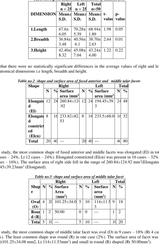
Figure 4 – showing type II subtype a with narrow separation <2mm
Figure 5 – showing type II subtype b with moderate separation 2-5 mm
Figure 6 – showing type II subtype c with wide separation >5 mm
© Indian Journal of Medical Research and Pharmaceutical Sciences http://www.ijmprs.com/
[39]
Figure 7 –showing type III absence of anterior articular facet
Figure 8 – showing type IV – all the three facets i.e. anterior, middle and posterior talar facets were seen on the superior surface of the calcaneus but anterior and middle talar facets incompletely separated from each other
Discussion
The racial and sexual variations in the morphology of talar articular facets of calcaneum is well documented by earlier researchers. Four different pattern types as described by Jha and Singh13, Gupta et al14, Saadeh et al15 and Kullar J S et al16. Three different types were described by Campos and Pellico17, Bunning and Barnett12 andDrayer-Verhagen18. Two facet configuration was documented by Testut19, Laidlaw20, Sharrafian S K21 and Padmanabhan22.
However, we chose the four different pattern grouping as it best categorizes the patterns of the talar facets observed in the present study.
Table no. 5 comparison of types of articular facets of calcanei
No Workers Year N (%) of types of calcanei
I Cn NC II A B C III IV V
1 Bunning and Barnett12
1965 194 33 -- -- 67 -- -- -- -- -- --
2 Jha and Singh13
1972 1600 59.5 33.7 5
25.7 5
37.5 -- -- -- 2.87 0.12 -- 3 Gupta et
al14
1977 401 67 -- -- 26 -- -- -- 5 2 --
© Indian Journal of Medical Research and Pharmaceutical Sciences http://www.ijmprs.com/
[40]
4 Drayer- Verhagen1
8
1993 191 54.45 -- -- 26.7 -- -- -- 18.8
5
-- --
5 Campos and Pellico17
1989 176 54 29 25 40 16 24 -- 6 -- --
6 Saddeh et al21
2000 300 63 -- -- 30.3 20.3 6.7 3.4 4.7 2 --
7 Muthuku maravel et al23
2011 237 65.82 -- -- 33.3
3
-- -- -- -- 0.42 0.42
8 Garg R et al24
2013 310 72.26 -- -- 24.5
2
-- -- -- 1.3 1.6 0.32
9 Chavan et al25
2014 60 68.33 -- -- 25 -- -- -- 6.66 -- --
10 Kori et al26
2015 600 73.9 58.8 15.1 21.5 9 11.3 1.2 3.6 0.3 0.6 11 Kullar J S
et al16
2015 200 72.5 30.0 42.5 25.5 10.5 15 -- 1.5 1.5 -- 13 Rajshree
et al2
2016 45 66.6 -- -- 31.3 -- -- -- -- -- 2.2
14 Present study
2017 50 66 36 30 20 4 10 6 4 10 --
In the present study, the type I calcanei was the most common (66%) and the observation was in consonance with previous studies where mostly the figures ranged between 54.0-59.5%. However, Koriet al26 reported the incidence of type I calcanei somewhat higher (73.9%). Bunning and Barnett12 has reported the incidence of type I calcanei only 33%.
Further, in type I, two subtypes were found i.e. constricted (cn) and not constricted (NC). Type I (cn) was observed in 36% cases. However these figures described by other workers i.e. Jha and Singh13 - 33.75%, Campos and Pellico17- 29% and Koriet al26-58.8%.Type (NC) was observed in 33% cases.
The incidence of type II calcanei was 20% in the present study and this figure is comparable with the work of Koriet al26 who gave the incidence as 21.5%.However much higher figures have been reported by some workers i.e. 67% by Bunning and Barnett12.
Further, in type II, three subtypes were found i.e. A, B and C. Type II (A) was found to be present in 4% cases, Type II (B) in 14% cases and Type II (C) in 8% cases which on comparison do not coincide with the Campos and Pellico17, Saddeh et al15, Kori et al26 and Kullar J S et al16.
In the present study, the incidence of type III calcanei was 2% in 4 cases. It was highest 18.85% in studies of Drayer- Verhagen18 followed by Chavanet al25 (6.66%). In other studies it ranged between 1.3%-5.52%.
The incidence of type IV calcanei in the present study was very high i.e. 5%. However, much low figures have been reported by some workers i.e. 0.12% by Jha and Singh13.
The results of the present study showed a wide range of variations in the incidences of various types of calcanei compared to previous workers. These variations may be due to population differences, type of gait and built of an individual or the place of living whether it is plane or hilly area.
© Indian Journal of Medical Research and Pharmaceutical Sciences http://www.ijmprs.com/
[41]
Conclusion
The individual and racial differences of the anatomic construction of calcaneal talar articular facets influence the static and kinetic dynamics of foot. A thorough knowledge of calcaneal facet type and shape is important for all surgeries in subtalar region, better treatment and management options for calcaneal fractures.
References
1. Uygur M, Atmaz F, Cleik S, Pinar. The types of articular facets and morphometric measurements of the calcaneus bone on Turkish race. Arch orthop trauma surg. 2009;129:909-14.
2. Rajashree I, Kalyani TN, Janaki V. Study of Patterns of Talar Articular Facets of Human Calcanei and Occurrence of Enthesophytes& It’s Significance. 2016;6(11):223-5.
3. Moore KL, Anne MR, Dalley A. Essential Clinical Anatomy. In Bones of Lower Limb. 5th Ed. London:
Lippincott Williams and Wilkins, a Wolters Kluwer business; 2015:316.
4. Yettram AL, Camilleri NN. The forces acting on the human calcaneus. J Biomed Eng 1992; 15: 46-50.
5. Hall R L, Shereff MJ. Anatomy of the calcaneus. ClinOrthop 1993; 290: 27-35.
6. Ellis H. Clinical Anatomy: Applied Anatomy for students and junior doctors. 11th Ed, Blackwell publishers.
2006. (4): 235-6.
7. Du Vries H L. Surgery of the foot. 2nd Ed. St. Louis: The C V Mosby Company; 1959. 290-301.
8. John Ebnezar. Outline Orthopaedics and Fractures. 1st Ed: 2011; 137.
9. Ananthakrisnan D, Ching R et al. Subluxation of the talocalcaneal joint in adults who have symptomatic flatfoot. J Bone Surg; 1999; 81(8): 1147-54.
10. Brekke M K, Lieberman R, Wright E, Green D R. Posterior facet talocalcaneal coalition. J Am Podiatr Med Assoc; 91(8):422-6.
11. Kumar A, Tyagi Y. Sharma SK, Tyagi A. Sex determination by morphology of calcaneum bone. J Indian Acad Forensic Med;30(4):207-11.
12. Bunning P S C, Barnett C H. A comparison of adult and fetaltalocalcaneal articulations. Journal of Anatomy;
1965. 99(1):71-6.
13. Jha M R, Singh, D R. Variations in the talar articular facets on the superior surface of calcaneus. Journal of Anatomy, India. 1972. 21(1); 40-4.
14. Gupta S C, Gupta C D, Arora A K. Pattern of the talar articular facets in Indian calcanei. Journal of Anatomy 1977; 124(3): 651-5.
15. Saadeh FA, Fund AH, Mahmoud SMI, Marwan EE. Patterns of talar articular facets of Egyptian calcanei. J AnatSoc Indian. 2000;49(1):6-8.
16. Kullar J S, Arora A K, Kapoor N S, Randhawa G K and Kullar K K et al. Morphology of Talar Articular Facets of calcaneus and its clinical implications, Kashmir Journal of Medical Science. 2015. 1(1) : 10-4.
17. Campos FF and Pellico LG. Talar articular facets (Faciesarticularestalares) in human calcanei. Acta Anat.
1989;134:124-7.
18. Drayer-Verhagen F. Arthritis of the subtalar joint associated with sustentaculumtali facet configuration. J Anat 1993; 183: 631-4
19. Testut L. Traite d anatomiehumaine. DoinetCie, Paris. 1928;Vol.1:879.
20. Laidlaw P P. The varieties of the oscalcis. Journal of Anatomy and Physiology, London. 1904. 5;38:133-43, and 39:161-77.
21. Sharrafian S K. Anatomy of the foot and ankle: Descriptive topographic functional. Lippincott. Philadelphia, 1983.
22. Padmanabhan R. The talar facets of the calcaneus- An anatomical note. AnatAnz 1986; 161 (5): 389-92.
23. Muthukumaravel N, Ravichandran D, Melani RS. Human Calcaneal Facets for the Talus: Patterns and Clinical Implications. J of Clin and Diagnostic Res. 2011;5(4):791-4.
24. Garg R, Dagal N, Kumar S, Shekhawat S. Study of patterns of talar articular facets of human calcanei and their clinical implications in population of Rajasthan. Indian J of Basic & Applied Med Res; 2013;2(7):643- 50.
25. Chavan S K, Satpute S T and Wabale R N. Pattern of Talar Articular Facet of Human Calcaneum Bone IOSR.
Journal of Dental and Medical Sciences; 2014. 8(13):16-8.
© Indian Journal of Medical Research and Pharmaceutical Sciences http://www.ijmprs.com/
[42]
26. Kori D, Prasad G, Rani A, Dewan R K, Singh R and Singh P et al. Study of Variations in Talar Articular Facets of Human Calcanei and their Association with Calcaneal Spurs in North Indian Population. Int J of Anat& Res. 2016;4(3):2710-6


![Issues of informed consent for intrapartum trials: a suggested consent pathway from the experience of the Release trial [ISRCTN13204258]](data:image/gif;base64,R0lGODlhAQABAIAAAP///wAAACH5BAEAAAAALAAAAAABAAEAAAICRAEAOw==)