Early Lung Cancer Detection Using Deep Learning
Optimization
https://doi.org/10.3991/ijoe.v16i06.13657
Ahmed Elnakib (), Hanan M. Amer Mansoura University, Mansoura, Egypt
Fatma E.Z. Abou-Chadi The British University, Cairo, Egypt
Abstract—This paper proposes a computer aided detection (CADe) system for the early detection of lung nodules from low dose computed tomography (LDCT) images. The proposed system initially preprocesses the raw data to im-prove the contrast of the low dose images. Compact deep learning features are then extracted by investigating different deep learning architectures, including Alex, VGG16, and VGG19 networks. To optimize the extracted set of features, a genetic algorithm (GA) is trained to select the most relevant features for early detection. Finally, different types of classifiers are tested in order to accurately detect the lung nodules. The system is tested on 320 LDCT images from 50 dif-ferent subjects, using an online public lung database, i.e., the International Early Lung Cancer Action Project, I-ELCAP. The proposed system, using VGG19 ar-chitecture and SVM classifier, achieves the best detection accuracy of 96.25%, sensitivity of 97.5%, and specificity of 95%. Compared to other state-of-the-art methods, the proposed system shows promising results.
Keywords—Lung nodule detection, lung cancer, early detection, deep learning
1
Introduction
Worldwide, cancer is the second leading cause of death after cardiovascular diseases. The American Cancer Society (ACS) estimated a number of 1,762,450 new cancer cases in 2019, accounting for 606,880 deaths in the United States [1]. 13% of all these new cases are lung cancer (228,150 new cases), accounting for 142,670 deaths [1]. This number is about 24% of all cancer deaths, which is the highest mortality rate among all type of cancers [1]. Therefore, there is an urgent need to investigate how to improve the survival rate of lung cancer.
and subjective. In addition, it suffers from inter- and intra-subject variabilities. There-fore, several research teams have developed computer aided detection (CADe) systems in order to aid the radiologist to accurately detect nodules [3-5]. One of the main chal-lenges in CADe system design is the detection of early cancerous nodules. At early lung cancer stage, the nodules are too small sizes, e.g., between 3 mm and 30 mm [6], to be detected. However, clinical reports show that early detection is a major key to signifi-cantly improve the survival rate of lung cancer [7].
This study aims at developing a CADe system for early lung cancer detection. To do this job, data from the International-Early Lung Cancer Action Project, I-ELCAP [8], are collected.
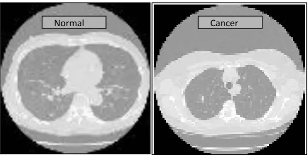
Fig. 1. Typical normal and cancerous LDCT image samples acquired from I-ELCAP
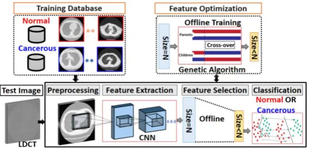
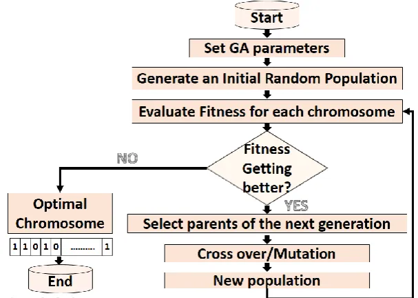
The proposed CADe system is composed from four steps (see Figure. 1): i. Initially preprocess the raw LDCT data in order to improve its contrast, ii. Extract compact deep learning features from the LDCT image,
iii. Optimize the extracted features in order to improve the detection accuracy, iv. Classify the LDCT as normal or cancerous, based on the optimized feature vector.
The main contributions of the proposed CADe system are two-folds:
Analysis of extracting different deep learning features from different architectures, including Alex [9] and VGG16 and VGG19 [10] networks
Optimization of the extracted deep learning features, using a smart genetic algo-rithm, running during the training phase of the CADe system, which does not only improve the accuracy of detection, but also reduces the feature dimensions, making the classification faster.
Fig. 2. Proposed CADe system for nodule detection composed of four steps: Preprocessing, feature extraction using CNN, feature selection using offline genetic algorithm training,
and classification, the output is either normal or cancerous
The current paper is an extension of our published paper in [11], with the following contributions:
i. Investigating more architectures rather than Alex network, including VGG16 and VGG19,
ii. Investigating more classifiers rather than the supported vector machine (SVM) used in [11], including K-nearest neighbor (KNN), and decision trees, and
iii. Adding more experimentations and discussions in order to precisely quantify the advantages and limitations of the proposed CADe system.
The rest of the paper is organized as following. Section 2 outlines the related work for lung nodule detection. Section 3 details the proposed CADe system. Section 4 demonstrates the experimental results and related discussions. Finally, Section 5 con-cludes the paper and outlines the future work.
2
Literature Review
In the literature, several research groups have developed different CADe systems for cancerous lung nodule detection. These systems can be categorized as traditional and deep Convolutional Neural Network (CNN) systems. In this section, each category will be overviewed, as well as its strength and limitations.
2.1 Traditional CADe systems
et al. [12] applied a 3D lung segmentation technique to identify the lung regions from the CT images. A feature vector, composed of 24 intensity-based, blobness, shape, and texture features were extracted from the segmented lung regions in order to detect lung nodules. A radial basis SVM was further used for the detection of large nodules. Li et al. [13] segmented the lung regions using a threshold-based technique. A set of inten-sity-based and texture features, including contrast, energy, correlation, homogeneity, and grey level concurrence matrix (GLCM), were extracted from the segmented lungs. Then a SVM was used for classification. Amer et al. [14] segmented the lung regions using bi-thresholding and morphological operations. A set of statistical, texture, histo-gram-based, wavelet features were fused using a genetic algorithm, to perform early lung nodule detection. Although traditional CADe system has achieved a considerable success in the detection of lung nodules, more sophisticated features are still need to be investigated in order to afford more accurate detection, especially for early cases.
2.2 Deep CNN CADe systems
Recently, deep learning CNNs have shown a remarkable success for lung nodule detection [3-4]. In CNN, medical images are directly processed, most often without segmenting the lung fields, throughout several convolutional layers that work as spa-tially localized filters and fully connected layers. These CNN architectures involve a very large number of parameters or weights in order to encode the images into a com-pact high-level feature space. To adjust the CNN parameters, training should be per-formed using a very large number of data images to avoid under-fitting. The extracted deep learned feature vector has shown a significant capability to describe precisely the training data and distinguish between normal and cancerous lung nodules [15]. For ex-ample, Jin et al. [16] trained a 3D CNN architecture, composed of eleven layers, using the segmented lung regions for the task of lung nodule detection. Their method achieved a detection accuracy of 87.5%. Shen et al. [17] achieved a detection accuracy of 87.14% using a CNN architecture that involved a multi-crop pooling of convolu-tional layers. Wang et al. [18] applied a multi-level feature pyramid network followed by a 3D non-maximum suppression and a 3D CNN to achieve a sensitivity of 95.8% over the LUng Nodule Analysis (LUNA16) dataset [18]. More recently, Winkels et al. [19] showed that using 3D CNN with group convolutions was able to reduce the number of false positives for lung nodules detection. The promising results of using deep learn-ing CADe systems throughout the literature encourage us to adopt uslearn-ing this approach to extract the LDCT image features.
3
Methods
relevant features are determined offline using a smart genetic algorithm (GA), which significantly reduces the feature space dimensions and improves the detection accuracy. Finally, a classification stage is applied to determine whether the test LDCT image contains pulmonary nodules or not. In this section, we will detail the procedures of each of these stages.
3.1 Database description
The LDCT images are collected from an online publically available database, i.e., the Early Lung Cancer Action Project (ELCAP) database [8]. In order to test the pro-posed CADe system, 320 LDCT images from 40 different subjects are selected ran-domly, including 160 normal cross-sections, and 160 cancerous ones, to avoid the bias during experimentations. In order to account for early lung nodule detection, nodules are in the range between 3 mm to 30 mm. Images are of resolution of 0.76×0.76×1.25 mm. Typical normal and cancerous samples of LDCT images, from the I-ELCAP pro-ject, are exemplified in Figure. 2.
3.2 Preprocessing of LDCT images
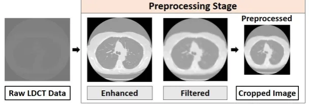
Preprocessing stage is implemented in three steps, as shown in Figure 3. First, the contrast of the raw image is enhanced using the histogram stretching technique [19]. Second, a smoothing Wiener filter is applied in order to remove the scanner noise. Fi-nally, the image is cropped to the standard size of the CNN model that is used for feature extraction (i.e., 227 × 227 for Alex architecture, and 224 × 224 for VGG16 and VGG19 architectures, see Table. 1).
Fig. 3. Preprocessing raw LDCT data in three steps: histogram stretching to improve image contrast, image smoothing using Wiener filer, and cropping to the standard input size of
the CNN model used for feature extraction
3.3 Feature extraction using (CNN)
under-fitting, these architectures were originally trained over a large number of images (1.2 million for Alex network [9], and 1.3 Million for VGG16 and VGG19 [10], in order to adjust their huge number of parameters (~60, 138, and 144 million for Alex, VGG16, and VGG19, respectively). In our experiments, we investigate using these dif-ferent CNN models to provide a reliable descriptor for LDCT images. Since the col-lected data size is small (320 LDCT images), the proposed CADe system applies trans-fer learning, where the parameters of the convolutional layers are kept unchanged (trained using the original data in [9,10]) and only the fully connected (FC) layers are trained and customized using the new database of interest. Transfer leaning is repeat-edly applied by different research teams, showing a remarkable success [22]. To apply the transfer learning, the last fully connected (FC) layer (output layer) is customized based on the number of classes, i.e., two neurons in our experiments (normal or cancer-ous). In the proposed CADe system, the FC layer, just before the output layer, is used as a compact high-level feature descriptor (of length 4096 for all the three investigated models, as shown in Table 1 to precisely describe the LDCT image.
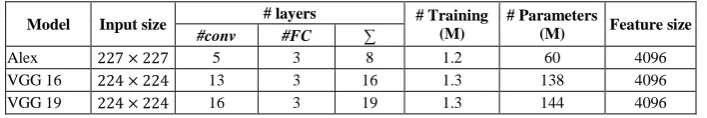
Table 1. Comparison between the different CNN models that are used for feature extraction (Alex, VGG16, and VGG19) with respect to the input space dimention, the number of layers (convolutional (conv), fully connected (FC), and total number of layers (∑)), the
number of the training images originally used, in millions (M), the number of network parameters, in milions (M), and the extracted feature size used in our experiments (i.e.,
the size of the FC layer just before the output layer)
Model Input size # layers # Training
(M)
# Parameters
(M) Feature size
#conv #FC ∑
Alex 227 × 227 5 3 8 1.2 60 4096
VGG 16 224 × 224 13 3 16 1.3 138 4096
VGG 19 224 × 224 16 3 19 1.3 144 4096
3.4 Genetic Algorithm (GA) feature optimization
Since transfer learning keeps the convolutional layers’ parameters unchanged (are not trained with the database of interest), the extracted feature vector is generic and may involve redundant information. In order to select the most relevant features for lung nodule detection, a smart GA is designed. The output of the GA is a binary chromosome of length 4096 bits, evaluated offline at the training phase of the CADe system, in which a bit of logic ‘1’ indicates that this feature is relevant and a bit of logic ‘0’ indicates that this feature is irrelevant, so it is removed from the optimized feature vector used in the test phase. The detailed of GA design is illustrated in Figure. 4.
𝐴𝐷 = 𝑇𝑃+𝑇𝑁
𝑇𝑃+𝑇𝑁+𝐹𝑃+𝐹𝑁 (1)
Where 𝑇𝑃, 𝑇𝑁, 𝐹𝑃, and 𝐹𝑁 denotes the true positive, true negative, false positive, and false negative, respectively.
For feature selection, a subset of training images is iteratively processed by the GA (i.e., one half of the training images and the other half is used to train the classifier; the last step in the proposed CADe system). At each iteration, genetic operations (selection, cross over, and mutation) are applied in order to form a new population. The process is repeated until the detection accuracy (fitness, 𝐴𝐷) no longer improves. After GA ter-mination, the chromosome of the best fitness (maximum detection accuracy, 𝐴𝐷) rep-resents the optimal feature vector that should be used for nodule detection, where a bit of ‘1’ in the chromosome means that the feature is selected and a bit of ‘0’ means that the feature is not selected (see Figure 4). The proposed CADe system uses the following GA setting: tournament selection [24], with a tournament size of four, blending cross-over [25], and adaptive feasible mutation [26]. The number of generations is set to a maximum of 100 generations.
Fig. 4. Details of the Genetic Algorithm (GA) proposed for feature selection at the training phase. Binary chromosomes are selected, where a bit of ‘1’ in the chromosome means
that the feature is selected and a bit of ‘0’ means the feature is not selected
3.5 Classification
(the other half) is used to train the classifier. For this job, we investigate different num-ber of classifiers, including, K-Nearest Neighbor (KNN), decision trees, and supported vector machines (SVM) with binary linear kernel in order to detect the pulmonary lung nodules.
3.6 Performance metrics
To evaluate the efficiency of the proposed CADe system, standard detection metrics are used on the test data, namely, the accuracy of detection (𝐴𝐷) (see Equation (1)), the specificity (𝑆𝑝), and the sensitivity (𝑆), defined as follows [23]:
𝑆𝑃 = 𝑇𝑁
𝑇𝑁+𝐹𝑃 (2)
𝑆 = 𝑇𝑃
𝑇𝑃+𝐹𝑁 (3)
4
Experimental Results and Discussion
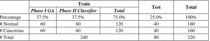
Collected data (320 image) is divided into 75% training and 25% test (see Table. 2). Training is carried throughout two phases, each with equal number of training images. The first phase is GA training, in order to select the most relevant features for the task of pulmonary nodule detection. The second phase is classifier training, based on the selected feature vector, in order to improve the detection accuracy. The details of the train and test datasets are illustrated in Table. 2. Note that all experiments select equal number of normal and cancerous images to avoid any bias.
Table 2. Data is divided into 75% for training and 25% for test. Training is performed in two phases. Training the GA to select the most releavant features and training the classifier based on the selected relevant features. Normal and cancerous images are selected
equally for each experiment to avoid the bias
Train
Test Total
Phase I GA Phase II Classifier Total
Percentage 37.5% 37.5% 75.0% 25.0% 100%
# Normal 60 60 120 40 160
# Cancerous 60 60 120 40 160
# Total 240 80 320
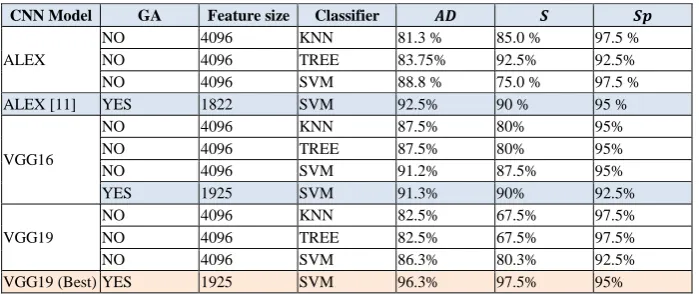
efficiently used for nodule detection. In addition, these results indicate the superior of using SVM over other classifiers due to its sophisticated nature.
To further optimize the feature descriptor, the proposed GA optimization is investi-gated, using the best type of classifier (SVM). As shown in Table. 3, the GA step can improve the detection accuracies to 92.5% ([11]), 91.3%, 96.3% using Alex, VGG16, and VGG19, respectively. In addition, the GA optimization was able to reduce the fea-ture dimensions from 4096 to 1822, 1925, and 1925, for Alex, VGG16, and VGG19, respectively. These results show the promise of using GA in the proposed CADe system to improve the overall system detection accuracy and to speed up the classifier, remov-ing the burden of irrelevant features. Figure 5 shows visual samples of the correctly classified LDCT images from different subjects using the proposed CADe system with GA optimization and SVM classifier using the three different CNN models.
Table 3. Experimental results of the proposed CADe system for early detection interms of three metrics (𝐴𝐷, 𝑆, 𝑆𝑝), with differnt architectures (Alex, VGG16, and VGG19) and
different settings: whether the proposed GA is used (YES) or not (NO), the classifer feature size, and the type of classifier used (KNN, decision tree (TREE), or SVM)
CNN Model GA Feature size Classifier 𝑨𝑫 𝑺 𝑺𝒑
ALEX
NO 4096 KNN 81.3 % 85.0 % 97.5 %
NO 4096 TREE 83.75% 92.5% 92.5%
NO 4096 SVM 88.8 % 75.0 % 97.5 %
ALEX [11] YES 1822 SVM 92.5% 90 % 95 %
VGG16
NO 4096 KNN 87.5% 80% 95%
NO 4096 TREE 87.5% 80% 95%
NO 4096 SVM 91.2% 87.5% 95%
YES 1925 SVM 91.3% 90% 92.5%
VGG19
NO 4096 KNN 82.5% 67.5% 97.5%
NO 4096 TREE 82.5% 67.5% 97.5%
NO 4096 SVM 86.3% 80.3% 92.5%
Fig. 5. Samples of correctly classified normal (first two columns) and cancerous (last two col-umns) images using the proposed CADe system, with Alex (first row), VGG16 (second
row), and VGG 19 (third row) models
To discuss the limitations of the proposed CADe system, Figure 6 exemplified sam-ples of false positives and false negatives of our system. As shown in samsam-ples, the vas-cular structure is close to nodule shapes, leading to false positive and false negative cases. In the future, we will investigate the fusion of the different model features in order to improve the accuracy.
Fig. 6. Samples of false positives (misclassified normal images, first row) and false negatives (misclassified cancer images, second row) of the proposed CADe system using GA and
5
Conclusion
A CADe system for early pulmonary nodules is presented. The CADe system is based on transfer learning to get a generic feature descriptor and a smart genetic algo-rithm (GA) to select the most relevant features for early pulmonary cancer detection. The CADe system accuracy is tested on an online publically available database; the Early Lung Cancer Action Project (ELCAP). The proposed CADe system shows prom-ising results in early lung cancer detection with respect to the state-of-the-art methods. In the future, more databases will be investigated in order to stand on the robustness of the proposed system.
6
References
[1]Siegel, R. L., Miller, K. D., and Jemal, A. (2019). Cancer statistics, 2019. CA: a cancer jour-nal for clinicians, 69(1), 7-34. https://doi.org/10.3322/caac.21551
[2]Ardila, D., Kiraly, A. P., Bharadwaj, S., Choi, B., Reicher, J. J., Peng, L, Tse, D., Etemadi, M., Ye, W., Corrado, G. and Naidich, D. P. (2019). End-to-end lung cancer screening with three-dimensional deep learning on low-dose chest computed tomography. Nature medicine, 25(6), 954. https://doi.org/10.1038/s41591-019-0447-x
[3]El-Regaily, S. A., Salem, M. A., Abdel Aziz, M. H., and Roushdy, M. I. (2018). Survey of computer aided detection systems for lung cancer in computed tomography. Current Medical Imaging Reviews, 14(1):3-18. https://doi.org/10.2174/1573405613666170602123329 [4]Zhang, G., Jiang, S., Yang, Z., Gong, L., Ma, X., Zhou, Z., Bao, C. and Liu, Q. (2018).
Au-tomatic nodule detection for lung cancer in CT images: A review. Computers in biology and medicine, 103:287-300. https://doi.org/10.1016/j.compbiomed.2018.10.033
[5]El-Baz, A., Beache, G. M., Gimel'farb, G., Suzuki, K., Okada, K., Elnakib, A., Soliman, A., and Abdollahi, B. (2013). Computer-aided diagnosis systems for lung cancer: challenges and methodologies. International journal of biomedical imaging, 2013. https://doi.org/10.1155/2013/942353
[6]Jacobs, C., and van Ginneken, B. (2019). Google's lung cancer AI: a promising tool that needs further validation. Nature Reviews Clinical Oncology, 1. https://doi.org/10.1038 /s41571-019-0248-7
[7]Pompe, E., de Jong, P. A., and Hoesein, F. A. M. (2019). Unravelling complexities of the subsolid pulmonary nodule-detection, characterization, natural history, monitoring and (fu-ture) patient management. Journal of Thoracic Disease, 11(Suppl 9):S1402. https://doi.org/10.21037/jtd.2019.03.07
[8]Early Lung Cancer Action Program (ELCAP), available from: http://www.via. cornell.edu/lungdb.html
[9]Krizhevsky, A., Sutskever, I., & Hinton, G. E. (2012). Imagenet classification with deep convolutional neural networks. Proceedings of the Advances in neural information pro-cessing systems. pp. 1097-1105
[10]Simonyan, K., & Zisserman, A. (2014). Very deep convolutional networks for large-scale image recognition. arXiv preprint arXiv:1409.1556.
[12]Setio, A. A., Jacobs, C., Gelderblom, J., and van Ginneken, B. (2015). Automatic detection of large pulmonary solid nodules in thoracic CT images. Medical physics, 42(10): 5642-5653. https://doi.org/10.1118/1.4929562
[13]Li, L., Wu, Y., Yang, Y., Li, L., and Wu, B. (2018, June). A New Strategy to Detect Lung Cancer on CT Images. 2018 IEEE 3rd International Conference on Image, Vision and Com-puting (ICIVC). pp 716-722 https://doi.org/10.1109/ICIVC.2018.8492820
[14]Amer, H. M., Abou-Chadi, F. E., Kishk, S. S., and Obayya, M. I. (2018, May). A Comput-er-Aided Early Detection System of Pulmonary Nodules in CT Scan Images. 7th Internation-al Conference on Software and Information Engineering. pp. 81-86. https://doi.org/10.11 45/3220267.3220291
[15]Setio, A. A. A., Traverso, A., De Bel, T., Berens, M. S., van den Bogaard, C., Cerello, P., Chen, H., Dou, Q., Fantacci, M.E., Geurts, B. and van der Gugten, R. (2017). Validation, comparison, and combination of algorithms for automatic detection of pulmonary nodules in computed tomography images: the LUNA16 challenge. Medical image analysis, 42:1-13. https://doi.org/10.1016/j.media.2017.06.015
[16]Jin, T., Cui, H., Zeng, S., and Wang, X. (2017). Learning deep spatial lung features by 3D convolutional neural network for early cancer detection. International Conference on Digital Image Computing: Techniques and Applications (DICTA). pp. 1-6 https://doi.org/10.110 9/DICTA.2017.8227454
[17]Shen, W., Zhou, M., Yang, F., Yu, D., Dong, D., Yang, C., Zang, Y., and Tian, J. (2017). Multi-crop convolutional neural networks for lung nodule malignancy suspiciousness classi-fication. Pattern Recognition, 61:663-673. https://doi.org/10.1016/j.patcog.2016.05.029 [18]Wang, B., Qi, G., Tang, S., Zhang, L., Deng, L., and Zhang, Y. (2018). Automated
pulmo-nary nodule detection: High sensitivity with few candidates. In International Conference on Medical Image Computing and Computer-Assisted Intervention (pp. 759-767). Springer, Cham. https://doi.org/10.1007/978-3-030-00934-2_84
[19]Winkels, M., and Cohen, T. S. (2019). Pulmonary nodule detection in CT scans with equivariant CNNs. Medical image analysis, 55, 15-26. https://doi.org/10.1 016/j.media.2019.03.010
[20]Andrews, H. C. (1976). Monochrome digital image enhancement. Applied optics, 15(2):495-503. https://doi.org/10.1364/AO.15.000495
[21]Fu, J., and Rui, Y. (2017). Advances in deep learning approaches for image tagging. APSIPA Transactions on Signal and Information Processing, 6. https://doi.org/10.1 017/ATSIP.2017.12
[22]Litjens, G., Kooi, T., Bejnordi, B. E., Setio, A. A. A., Ciompi, F., Ghafoorian, M., Van Der Laak, J.A., Van Ginneken, B., and Sánchez, C. I. (2017). A survey on deep learning in med-ical image analysis. Medmed-ical image analysis, 42:60-88. https://doi.org/10.10 16/j.media.2017.07.005
[23]Yerushalmy, J. (1947). Statistical problems in assessing methods of medical diagnosis, with special reference to X-ray techniques. Public Health Reports (1896-1970): 1432-1449 https://doi.org/10.2307/4586294
[24]Miller, B. L., and Goldberg, D. E. (1995). Genetic algorithms, tournament selection, and the effects of noise. Complex systems, 9(3):193-212
[25]Eiben, A. E., and Schippers, C. A. (1998). On evolutionary exploration and exploitation. Fundamenta Informaticae, 35(1-4):35-50 https://doi.org/10.3233/FI-1998-35123403 [26]Thierens, D. (2002, May). Adaptive mutation rate control schemes in genetic algorithms.
7
Authors
Ahmed Elnakib is an assistant professor, Electronics and Communications Engi-neering Department, Faculty of EngiEngi-neering, Mansoura University, Mansoura, Egypt. He received his Ph.D. degree from university of Louisville, USA, in 2013. His current research includes developing computer aided diagnostic systems, machine learning, stochastic modeling, detection of brain disorders, and signal and image processing. He has authored or coauthored more than 50 technical articles, appeared in world-renown journals including IEEE TMI and Medical Physics. Mr. Elnakib is a regular reviewer for a number of technical journals including Neurocomputing, Medical Image Analysis, IEEE TMI and IEEE TIP. Email: [email protected]
Hanan M. Amer is an assistant professor, Electronics and Communications Engi-neering Department, Faculty of EngiEngi-neering, Mansoura University, Mansoura, Egypt. She received her Ph.D. degree from Mansoura University, Egypt, in 2018. Her field of experience includes signal and image processing, medical image analysis, and lung can-cer. She has authored or coauthored more than 6 technical articles.
Fatma E. Z. Abou-Chadi is a professor and head of Electrical Engineering Depart-ment, Faculty of Engineering, The British University of Egypt, Cairo, Egypt. She ceived her BSc. degree from Ain-Shams University, Egypt, in 1974. In 1978, she re-ceived her Master degree in Electrical Communications Engineering from Mansoura University. She awarded the PhD degree at the Imperial College of Science and Tech-nology, University of London, UK in 1986, specializing in Biomedical Signal Pro-cessing and Informatics. Prof. Abou-Chadi has authored and co-authored more than 90 scientific articles and two books. Her scientific interests and current research work in-clude biomedical signal processing, medical imaging, digital signal processing, time-series analysis, and information technology in biomedicine. Prof. Abou-Chadi is a Sen-ior Member at Institute of Electrical and Electronics Engineers – IEEE since 1995, and a Member of the Society of IEEE Women in Engineering since 2000.