University of South Carolina
Scholar Commons
Theses and Dissertations
2015
The Recovery of Gut Barrier Function With
Selenium Rich Diet in Acute DSS-Induced Colitis
Sarah Depaepe
University of South Carolina
Follow this and additional works at:https://scholarcommons.sc.edu/etd Part of theExercise Science Commons
This Open Access Thesis is brought to you by Scholar Commons. It has been accepted for inclusion in Theses and Dissertations by an authorized administrator of Scholar Commons. For more information, please [email protected].
Recommended Citation
T
HE RECOVERY OF GUT BARRIER FUNCTION WITH SELENIUM RICH DIET IN ACUTEDSS-
INDUCED COLITISby
Sarah Depaepe
Bachelor of Science
University of South Carolina, 2013
Submitted in Partial Fulfillment of the Requirements
For the Degree of Master of Science in
Exercise Science
The Norman J. Arnold School of Public Health
University of South Carolina
2015
Accepted by:
Raja Fayad, Director of Thesis
Jim Carson, Reader
Xuewen Wang, Reader
Ray Thompson, Reader
D
EDICATIONThis thesis is dedicated to my sister. For without her I would not have had the
courage to keep going. She is a strong and highly motivated individual that encourages
all to pursue their dreams no matter what problems lie in their wake. She gives me a
sense of passion for the work I do in the lab and elsewhere in my life. She is the example
person I wish to become in this world. For she is going for her dreams, conquering the
obstacles, and achieving what was thought to be impossible. She has gone above and
beyond the call of sisterhood to help me prepare my thesis. She has taught me to
incorporate a higher level of thinking and given me the ability to do what I thought I
couldn’t.
I would also like to dedicate this thesis to Dr. Raja Fayad and my lab mates, Dr.
Arpit Saxena, Kamaljeet Kaur, and Alex Sougiannis. Dr. Fayad was a great teacher and
mentor. I will never forget his kind words and helpful guidance in my undergraduate and
masters degrees. He taught me to keep going, to never give up, and to push forward even
when times are rough. He will always be remembered. To my lab mates: I will never
forget all of your guidance. You have helped me in so many ways that I cannot express
my thanks enough. You have listened to all my questions and answered all my phone
calls when experiments go wrong. You have been there for me every step of the way. I
A
BSTRACTBackground: Acute Dextran Sodium Sulfate (DSS)-induced colitis is an
inflammatory ailment limited to the colon. It works to destroy the morphology and gut
barrier goblet and epithelial cells that aid in providing homeostasis. Selenium (Se) is an
essential micronutrient that has anti-inflammatory and antioxidant properties and is
known to play a role in reducing inflammation in areas elsewhere in the body. The
current study is focused on how Se alters gut barrier permeability and functionality
related to the recovery of tight junction regulation and mucin secretion. Methods:
C57BL/6 mice were randomly placed into control (normal water) and 2% DSS water
receiving groups and within these groups they were randomly given either a Se rich diet
or a control diet ad libidum. Hemotoxylin-Eosin and Alcian Blue staining was used to
study the colon morphology and to quantify the goblet to epithelial cell ratio. Western
Blot was used to analyze protein expression levels for MUC-2 and ZO-1. Gut barrier
permeability was assessed by administering FD4 and determining its plasma
concentration by spectrofluorescence. ELISA was used to study the colon-secreted
cytokine levels of TNF-α and IL-1β. Results: DSS + Se mice showed significantly lower
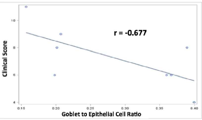
clinical scores, histopathology, and higher goblet to epithelial cell ratios compared to
DSS mice given a control diet. It is interesting to note that there was a main effect of diet
and DSS treatment with ZO-1 expression. We found no significant difference between
secretion. Conclusion: The data suggests that Se works to reduce the severity of colitis by
increasing ZO-1 expression and goblet cell content.
T
ABLE OFC
ONTENTSDEDICATION ... iii
ABSTRACT ... iv
LIST OF FIGURES ... vii
CHAPTER 1:PROPOSAL ... 1
INTRODUCTION ... 1
PURPOSE AND AIMS ... 4
WORKING MODEL ... 6
METHODS ... 7
REVIEW OF LITERATURE ... 10
REFERENCES... 25
CHAPTER 2:THE RECOVERY OF GUT BARRIER FUNCTION WITH SELENIUM RICH DIET IN ACUTE DSS-INDUCED COLITIS ... 31
ABSTRACT ... 32
INTRODUCTION ... 33
METHODS ... 36
RESULTS ... 40
DISCUSSION ... 43
FUTURE DIRECTIONS ... 48
FIGURE LEGENDS ... 50
FIGURES ... 51
L
IST OFF
IGURESFigure 1.1 Working model ...6
Figure 2.1 Effect of Se rich diet on clinical score...52
Figure 2.2 Effect of Se rich diet on the morphology of the colon ...53
Figure 2.3 Effect of Se rich diet on goblet cell content ...54
Figure 2.4 Correlation between clinical score and goblet cell content ...55
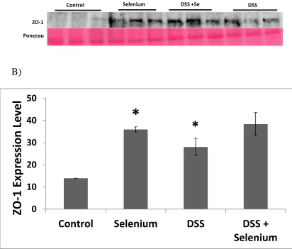
Figure 2.5 Effect of Se rich diet on tight junction protein ZO-1 ...56
Figure 2.6 Effect of Se rich diet on colon tissue secreted cytokines ...57
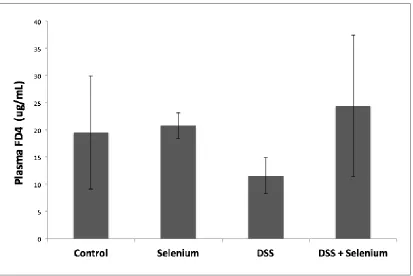
Figure 2.7 Effect of Se rich diet on gut barrier permeability ...58
CHAPTER
1:
PROPOSAL
I
NTRODUCTIONInflammatory bowel disease (IBD) has been a growing concern in the United
States and all over the world. IBD can be classified as either Ulcerative Colitis (UC) or
Crohn’s Disease (CD) and has been characterized by chronic uncontrolled inflammation
that results in damage to the lining of the gastrointestinal tract, blood in stools, diarrhea,
and weight loss (Abraham, 2009; Matter, 2011). It is estimated that as many as 1.4
million American’s, or 1 in every 200 people, suffer from IBD and as many as 70,000
new cases appear each year (Hanauer, 2006). The peak onset of the disease has been
shown to be from 15 to 30 years of age with most recent data suggesting a higher
incidence before the age of 20 (Abraham, 2009; M’Koma 2013). Studies have shown that
individuals suffering from IBD for at least 6-8 years are six times more likely to develop
colorectal cancer (M’Koma 2013; Mattar, 2011). Acute colitis is a single inflammatory
flare-up that presents the same symptoms of IBD except on a much lower scale
(Keshavarzian, 2003). These acute flare-ups have been attributed to genetic and
non-genetic factors. However, non-genetics have only been able to account for 20-25% of
susceptibility, while environmental (diet, exercise, drugs, smoking, and social stress),
immunological, and microbial factors play a larger role (Keshavarzian, 2003). Acute
colonic flare-ups that are frequent and reoccurring are more indicative of chronic
The GI tract must prevent leakage of gut bacteria into the abdominal cavity for if
it does not, acute inflammatory immune responses may occur (Matricon, 2008;
McGuckin, 2009). The intestines are home to trillions of commensal bacteria that make
up a microbiome. This population of bacteria is tightly regulated and the immune system
is highly responsive in distinguishing harmful bacteria from commensal (Johannson,
2013). Studies examining IBD development have found concomitant intestinal barrier
dysfunction and increased intestinal permeability allowing bacteria to leak outside of the
intestines (Matricon, 2010). The gut barrier is made of an outer mucus layer and an inner
single layer of epithelial cells that are held together by tight junctions (TJ) (Antoni,
2014). TJ are composed of zona occludens (ZO) that are located at the apical surface of
the epithelial cells. These cell junctions are the rate-limiting step in paracellular
permeability (Clayburgh, 2004). Inflammation can downregulate their overall expression
and translocate them to the inside of the cell away from the surface causing increased
paracellular leakage of noxious bacteria (Ma, 2004). Inflammatory cytokines tumor
necrosis factor alpha (TNF-α) and interleukin factor 1 beta (IL-1β have been shown to be
able to alter intestinal TJ permeability (Ma, 2004; Wang, 2005). Furthermore, reactive
oxygen species (ROS) may also cause oxidative stress-induced inflammation and lead to
decreased intestinal barrier function by downregulation of TJ proteins (Keshavarzian,
2003). TJs are the main regulatory site for paracellular permeability and are highly
investigated in inflammatory diseases (Clayburgh, 2004). However, further research is
needed in examining nutritional effects on TJ expression as well as how these effects may
In addition to tight junction regulation, the secretive functions of gut epithelial
cells can have a role in intestinal permeability. The mucus layer is composed of a
two-layered system that regulates the luminal bacterial environment and is important for the
protection of the barrier epithelial cells (Johansson, 2013). The inner layer is formed by
mucins (Muc 2, 3, and 4) that are secreted by goblet cells. In this layer there is relatively
no bacteria, which provides the protective function to the epithelial cells. On the other
hand, the outer layer has the same mucins as the inner layer, but here is where the
bacteria in the gut thrive (Johansson, 2013). UC cases show diminished outer layer of
mucus causing the bacteria and other noxious agents to move to the inside layer putting
them in direct contact with apical epithelial surface. The diminished outer layer has been
correlated with a loss of goblet cells and their secreted mucins (Dorofeyev, 2013). While
mucous production is important for gut protection, further work is needed to determine
how nutrition impacts the secretory function of gut epithelial cells.
Selenium (Se) is an essential micronutrient that exerts its anti-inflammatory and
antioxidant effects through many families of selenoproteins. Dietary supplementation of
Se has been shown to play a role in thyroid hormone metabolism, cardiovascular health,
prevention and reduction of cancer, and immune function (Huang, 2012). A deficiency in
Se has been negatively correlated with IBD, which suggests a crucial role of Se in
inflammatory pathology (Barrett, 2013). Research has shown that Se can shift
macrophage polarization from an M1 pro-inflammatory state to an M2 anti-inflammatory
state after an insult of injury (Nelson, 2011). Additionally, glutathione peroxidases (Gpx),
a major family of selenoproteins, have been found to decrease inflammation, reduce
carcinogenesis (Krehl, 2012). Deficiency in subtypes of Gpx, Gpx-1, Gpx-2, or both,
have shown development of spontaneous intestinal inflammation and increased apoptosis
of intestinal epithelial cells (Edelblum, 2006; Krehl, 2012). While Se has shown
beneficial effects in various disease states the effects on gut barrier dysfunction,
especially tight junction regulation and mucus production, is not very well understood.
Se has demonstrated positive effects in reducing inflammation and preserving
epithelial cells. In cell models of human breast cancer, Se has enhanced the function of
TJs by relocation of ZO-1 proteins to the apical surface, thus decreasing permeability
(Martin, 2007). Additionally, rat models examining stress and chemically induced gastric
ulcers have found Se to prevent gastric wall mucus depletion. (al-Moutairy, 1996). The
current study is focused on how Se alters gut barrier functionality related to the recovery
of tight junction regulation and mucin secretion. However, there are currently gaps in our
understanding or how Se can impact intestinal barrier function in a mouse model of acute
colitis. Thus, we examined Se function with a widely used mouse model of intestinal
inflammation, dextran sodium sulfate (DSS) - induced colitis (Perse, 2012). This model
has demonstrated acute, chronic, and relapsing experimental inflammation and has been
shown to closely resemble human IBD (Okayasu, 1990; Perse, 2012).
P
URPOSE ANDA
IMSThe purpose of this study is to examine the preventive and restorative effect of Se
on acute DSS-induced colitis in C57BL/6 mice. The severity of colitis in the mice will be
observed through assessment of clinical score and histopathology. Inflammatory
mediators that will be studied include expression of inflammatory cytokines TNF-α and
barrier function that will be studied include ZO-1 expression and localization, gut
permeability, mucus protein content, and goblet cell content. The overall hypothesis is
that a Se rich diet will decrease the severity of colitis by decreasing inflammation,
decreasing gut permeability, and increasing mucus protein content.
Specific Aim #1 will determine the effect of Se rich diet on the severity of colitis in mice
with DSS-induced colitis. The primary outcomes measured will be clinical score and
histopathology to indicate severity of colitis. The score consists of weight loss, diarrhea,
and hemoccult. We hypothesize that a Se rich diet will decrease the severity of colitis by
decreasing clinical score and histopathology as compared to control diet.
Specific Aim #2 will determine the effect of Se rich diet on gut barrier function in mice
with DSS-induced colitis. Primary outcomes measured will be gut barrier permeability.
Secondary outcomes include mucus protein expression, goblet cell content, tight junction
expression, and secreted tissue expression of inflammatory cytokines. We hypothesize
that a Se rich diet will improve gut barrier function by decreasing gut permeability as
compared to control diet. We also hypothesize that these positive changes will be
associated with increased mucus protein expression, goblet cell content, and tight
W
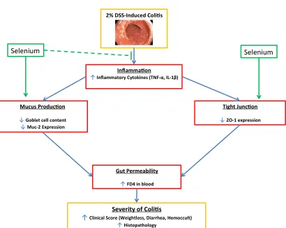
ORKINGM
ODELFigure 1.1 Working Model: This study is aimed to uncover the proposed model of the protective effect of Se on gut barrier function in C57BL/6 mice with acute DSS-induced colitis. The overall hypothesis is that a Se rich diet will decrease the severity
of colitis by decreasing inflammation, decreasing gut permeability, and increasing mucus protein content.
2% DSS-Induced Coli s
Inflamma on
↑Inflammatory Cytokines (TNF-α, IL-1β)
Tight Junc on
↓ZO-1 expression Mucus Produc on
↓Goblet cell content
↓Muc-2 Expression
Gut Permeability
↑FD4 in blood
Severity of Coli s
↑Clinical Score (Weightloss, Diarrhea, Hemoccult)
↑Histopathology
M
ETHODSAnimals and Housing
Four to five week old male and female C57BL/6 mice (n=33) were bred and
maintained in the animal resource facility at the University of South Carolina. They were
housed three-five per cage and maintained on a 12:12 light-dark cycle in a low stress
environment (22°, 50% humidity, low noise). Mice were split into two main groups:
control and experimental. The control group consisted of a mixed population, while only
males were placed in the experimental groups. Each main group was subdivided into
mice receiving either a Control diet (0.02ppm Se) or a Se rich diet (0.75ppm Se). After 1
week of either diet, experimental mice were given 2% Dextran Sodium Sulfate (DSS)
(MP Biochemicals, MW 36,000 – 50,000) dissolved in their drinking water for 5 days
followed by 5 days of normal drinking water to induce acute colitis. Control mice were
given normal drinking water ad libitum throughout the duration of the study. All animal
experiments were approved by the University of South Carolina’s Institutional Animal
Care and Use Committee.
Monitoring Animal Health
Food and water intake, as well as body weight, were measured every alternate day
for all mice throughout the length of the study. During and following DSS treatment in
experimental groups, mice were observed every alternate day for clinical signs of disease,
ranked by a point system as follows: 0= 0-5% weight loss; 1=6-10% weight loss;
2=11-15% weight loss; 3=16-20% weight loss; and 4=>20% weight loss. The appearance of
diarrhea was ranked as 0= well-formed pellets, 2= pasty and semi-formed stools that do
not adhere to the anus, 4= liquid stools with no form that do not adhere to the anus.
Positive hemoccult was scored as follows: 0= no blood or negative hemoccult, 2= some
blood (<50%) or positive hemoccult, and 4= gross bleeding (>50%) using hemoccult kit
(Beckman Coulter). The clinical score was then determined by adding and totaling the
scores of weight loss, diarrhea, and hemoccult.
Tissue Collection
All mice were sacrificed 17 days after initial induction of Se rich or Control diet.
The mice were sacrificed within 2 hours by cervical dislocation and tissue collection was
performed as a non-survival surgery. The mouse colon was excised and flushed with PBS
(EMD Chemicals) and three 1cm sections of each were cut. The first section was stored
at -80°C for protein expression studies. The second section was fixed in formalin (Fisher
Scientific) and stored in 70% ethanol, which was later cut into 5-6μm thin sections for
use in Hemotoxylin and Eosin (H&E) and Alcian Blue and Nuclear Fast Red staining.
The last section was placed in 12-well plates containing 1ml of RPMI 1640 media that
included 1% Penicillin-streptomycin (Mediatech, Inc) per well and was incubated for 24
hours at 37°C and 5% CO2. The RPMI media containing tissue cytokines was
centrifuged at 10,000g for 10 minutes at 15°C and the supernatant was collected and
Histology
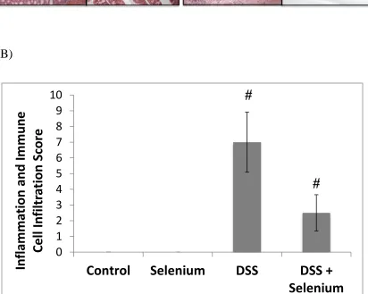
The histopathology of the colon was observed through H&E staining. A standard
protocol for H&E staining was used. The severity of colitis was quantified by a scale of 0
to 4, where 0= no infiltration and inflammation; 2= moderate infiltration and
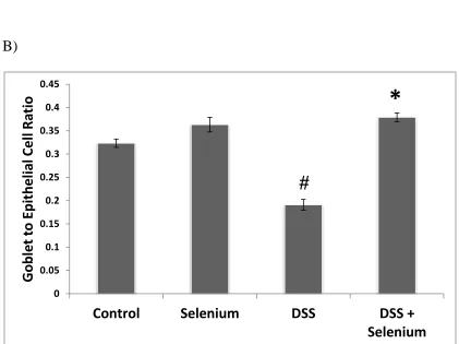
inflammation; and 4= severe infiltration and inflammation with distorted crypts. Alcian
Blue and Neutral Fast Red staining was used to detect goblet and epithelial cells in the
colon. Goblet cells were stained blue with Alcian Blue and epithelial cells were stained
pink with Neutral Fast Red. They were quantified by goblet to epithelial ratio using 6
crypts per colon section from each mouse for all treatment groups.
Gut Permeability
Gut barrier integrity was assessed in all mice by permeability to FITC-dextran
(MWav= 4000; FD4) (Sigma Aldrich). The FD4 was administered by gavage based on
the animal’s body weight in grams and diluted with 125mg/ml of PBS to five hour fasted
mice. Plasma was sampled before and 1 h after FD4 administration and measured for
florescence as previously described by Yang et al., 2003.
Enzyme-linked Immunosorbant Assay
The RPMI medium supplemented with secreted colon cytokines was used to
quantify the local concentrations of TNFα and IL-1β (BD biosciences) using
commercially available BD OptEIA enzyme-linked immunoabsorbant assay (ELISA)
kits, according to the manufacturer’s instructions. The local cytokine concentrations were
normalized by the estimated protein content in the colon supernatant by a Bradford
Western Blotting
Colon tissue samples frozen at -80°C were homogenized in RIPA buffer that was
supplemented with protease and phosphatase inhibitors (SIGMA). The samples were then
centrifuged at 10,000 rpm for 15 minutes and the supernatant was collected for protein
analysis by a standard Bradford assay. Protein homogenate from all groups was
electrophoresed on 7% SDS-PAGE gels and transferred to a nitrocellulose membrane for
3 hours at 4°C. The membrane was blocked by 1X PBS and 0.1% Tween 20 for 1 hour
and subsequently probed for ZO-1 (Abcam) and MUC-2 (Abcam) overnight. The bands
were detected using chemiluminescence and normalized relative to GAPDH expression
(Genetex). The bands were quantified by densitometry and expressed as mean area
density by using Image J software (Image J).
Statistical Analysis
XLStat for Windows (verson 2009.4.07) statistical software was used to perform all
statistical analysis. Independent two-tailed t-tests were used to determine significance for
all single variables. Statistical significance was considered at P < 0.05 level of confidence
R
EVIEW OFL
ITERATUREThe literature review for this study is divided into three main sections with further
subcategories to explore knowledge more in depth on specific topics. The first section
divulges inflammation and its role within injury and the immune response. The second
section draws attention to the gut barrier, its components, and how it relates to
inflammatory events. The final section sheds knowledge on the dietary nutrient called
selenium. This section will explore in detail the functions and roles of selenium in the
knowledge of links between gut barrier dysfunction, inflammation, and selenium will be
reviewed in this section.
I.
InflammationInjury or infection of tissues sets off chemical and physical signals that allow
infiltration of blood cells and fluid in order to promote healing. These events are
called an inflammatory response, which is a type of defense mechanism housed
within the immune system of the body. Its primary purpose is to contain, neutralize,
dilute, or wall off deleterious agents and has been characterized by distinct cardinal
signs, such as heat, swelling, redness, pain, and sometimes loss of function
(Anderson, 2001; Medzhitov, 2008). Inflammation may seem to be more
degenerative than reparative, but if closely regulated it plays a pivotal role in the
wound healing process. An acute response has been characterized by immediate and
nonspecific events. The immediate inflammatory action calls for the activation of
local macrophages and mast cells that release inflammatory mediators, such as
chemokines and cytokines, in order to attract white blood cells and clotting agents to
divest infectious agents for wound repair (Medzhitov, 2008). The white blood cells,
mainly neutrophils, and plasma proteins are normally housed within the
cardiovascular system and may venture into local tissue if the endothelial membrane
becomes leaky or slightly more permeable than normal. This infiltration of
inflammatory agents is necessary for repairing tissue damage, but it must be tightly
regulated and resolved quickly in order to decrease the risk for increasing mortality
and morbidity for diseases such as rheumatoid arthritis, diabetes, Crohn’s disease,
delayed and highly specific events, which is more of an unregulated inflammatory
response with an abundance of inflammatory mediators and white blood cells as
compared to acute inflammation (Eming, 2007; Ryan, 1977). Moreover, chronic
inflammation has been widely studied as a mechanism of the development of
modern human diseases, such as cancer (Shacter, 2002), Inflammatory Bowel
Disease (Zhang, 2012), Alzheimers (Bibi, 2014), and Chronic Obstructive
Pulmonary Disease (Maclay, 2013).
A. The Pathway of Inflammation
Injury promotes the immediate activation of an immune mediated response
that allows infiltration of polymorphonuclear (PMN) leukocytes, mainly
neutrophils, through the endothelial membrane. This process occurs via the
activation of the membrane by local proinflammatory cytokines: Tumor
Necrosis Factor-α (TNF-α), Interleukin -1β (IL-1β), and Interferon-γ (IFN-γ)
(Eming, 2007). After several days, the neutrophils are joined by larger
populations of activated macrophages via attraction by monocyte
chemoattractant protein -1 (MCP-1). Macrophages are widely studied
phagocytes that are thought to have primarily deleterious effects on tissues.
However, not only do they encompass a proinflammatory phenotype (M1),
but they also present an antagonist side, the anti-inflammatory phenotype
(M2). Studies have shown that M1 is the predominant phenotype during the
initial phase of acute inflammation due to its high T helper cell-1 (Th-1)
cytokine response (Mantovani, 2013; Romagnani, 2000). The Th-1 response is
located on the plasma membrane of macrophages. The binding stimulates a
cascade affect that activates protein kinase C (a major regulatory
Phosphorylation protein). PKC may phosphorylate Iκβ, which is bound to
nuclear factor kappa beta (Nfκβ) (Aveleira, 2010). Phosphorylation of this
complex allows Nf-κβ to dislocate into the nucleus, which promotes the
transcription of proinflammatory mediators, such as TNF-α, IL-1β, IL-6,
reactive oxygen species (ROS), inducible Nitric Oxide Synthase (iNOS)
CXCL9 and CXCL10 (Chazaud, 2014; Dohi, 2014; Lawrence, 2009;
Mantovani, 2013). After the wound has been cleansed of debris, further
leakage of cell contents has ceased, and neutrophils are phagocytized, the
macrophage will undergo polarization to an M2 phenotype to initiate the
second phase – healing (Ramaiah, 2007). The M2 response is of Th-2 type
and is activated by transforming growth factor-β (TGF-β), 1, 4, and
IL-10 and its primary role is to promote tissue repair and regeneration in order to
recover functionality of the damaged tissue (Chazaud, 2014). These activating
factors bind to the cell surface receptors on the macrophage and illicit
transcription and release of anti-inflammatory mediators (IL-4, IL-13, IL-10,
arginase, proline, CCL17, CCL22, and CCL24, VEGF, and MMPs), which
dampen inflammation and promote tissue repair (Mantovani, 2013; Martinez,
II.
Inflammation and the GutA. Epithelial Barrier
The gastrointestinal tract involves a large tube-like structure that runs from the
mouth to the anus. Consumed nutrients travel on the luminal side and remain
there unless excreted or selectively absorbed by the single layer of epithelial
cells that line this tract. The intestines are the primary areas in the
gastrointestinal tract that allow absorption of nutrients that are vital for
homeostatic functionality. Moreover, selective permeability is one of the key
protective functions of the intestinal epithelial cells. This feature allows the
uptake of nutrients and minimal exposure to various toxins, antigens, and
microorganisms (Lennernas, 1998). Molecules and ions may be selectively
taken into the epithelial cells through two types of transport. The first is
transcellular – the intake of substances through the apical membrane on the
extracellular surface or the basolateral membrane on the luminal side.
However, substances taking this route of transport require either a lipophilic
composition or a specific mechanism of ATP-dependent transport across the
membrane. Substances that do not have access to either of these requirements
may take the second route of transport – paracellular. This is the route, by
which the substances may travel into the intercellular space between adjacent
epithelial cells. However, this pathway is tightly regulated by cell junctions
1. Cell Junctions
i. Tight junctions – A type of epithelial and endothelial cell junction
located on the apical surface of the membrane. Tight junctions play a
vital role in paracellular transport by tightly regulating permeability of
essential molecules and ions and keeping out noxious agents (Gu,
2011). The degree of permeability may fluctuate due to the local
mucosal or luminal environmental stimuli as well as various
physiological and pathological conditions. Tight junctions are
integrated via an array of proteins as well as with an association of
scaffolding proteins. Occludins are transmembrane proteins that have a
PDZ domain that directly regulates paracellular trafficking or
permeability (Hwang, 2013). Zona Occludins (ZO) are cytoplasmic
scaffolding proteins that hold occludins in proper orientation through
interaction with the PDZ domain and binding to actin located in the
cell cytoskeleton. These PDZ domains allow scaffolding and structural
interaction of ZO and occludins by spacing these proteins in close
proximity (Gonzalez-Mariscal, 2000). The final proteins of interest
that play a pivotal role in permeability are the claudin family. These
proteins vary in their degree of leakiness and this function allows
permeability of certain types of ions (i.e claudin-8 reduces Na2+
permeability) (Gonzalez-Mariscal, 2008).
ii.Other Cell Junctions – Tight junctions may be the most apical junction
junctions to consider in regard to cell-cell environmental homeostasis.
The adherens junctions are located directly below the tight junctions.
The main structural protein components in these junctions consist of
cadherins (Dejana, 1995). They come in a variety of types, the most
important being E-cadherin. It comprises specific roles of cell-cell
adhesion and interaction with the actin cytoskeleton components. In
order for E-cadherin to exhibit these roles, it must be joined in a
complex with catenins (alpha, beta, or gamma)(Dejana, 1995). Gap
junctions reside below adherens junctions and are involved in cell-cell
exchange of ions and low molecular weight molecules. The passage of
molecules and ions is conducted through hemichannels made of
proteins called connexins. In the gastrointestinal tract, they are found
in abundance in the inner smooth muscle and studies have shown that
they may influence the contractile activity of the gut (Nielsen, 2012).
B. Mucus Layer
The GI lining in the mouth and the esophagus have multiple layers of
squamous epithelial cells. However, the stomach, small intestine, and large
intestine have a single, thin layer of these cells causing increased vulnerability
to damage and pathology. Aiding in protective defenses to these epithelial
cells in the lower part of the GI tract is the mucus layer. Mucus is a
transparent liquid layer that covers epithelial cells lining the GI tract and it is
made of proteins known as mucins. There are two different types of mucins:
transmembrane (MUC1, MUC3, MUC4, MUC12, MUC13, MUC16, and
MUC17) (Johansson, 2011). The small intestine has a single mucus layer,
while the large intestine is composed of 2 distinct layers with separate
functionality. The single layer of mucus in the small intestine is composed of
the mucus protein MUC2, as well as antibacterial peptides, which allows this
layer to be unattached to the epithelial cells and easily removable (Johansson,
2011). The function of the mucus in this region is to keep the surface of the
epithelia as well as within the crypts where stem cells lie as sterile as possible.
The colon is doubly lined with an outer mucus layer that is composed of
MUC2 that resembles the single mucus layer of the small intestine, while the
inner mucus layer is composed of MUC2 that is bound to the surface of the
epithelia by attachment of goblet cells (Johansson, 2013). The MUC2 from
the inner layer gradually becomes the outer layer in order to renew damaged
mucin proteins from noxious agents. The functionality of the two layers
remains separate. The inner layer protects the single layer of epithelial cells
from consumed deleterious agents and is considered a sterile environment,
while the outer layer houses the large populations of bacteria that make up the
microbiome of the large intestine. The bacteria found here is ultimately
essential for normal functionality of the colon and as long as it is regulated
and within the outer layer of mucus, the colon may function without
developed pathologies (Johansson, 2013). Like the epithelial cells, the mucus
layer is constantly renewed in order to keep up proper functionality. The notch
(epithelial cells) via hairy and enhancer of split-1 (HES-1) and when this
pathway is inhibited the secretory linage of cells or goblet cells are
differentiated by MATH-1 (Jeon, 2013).
C. Barrier Dysfunction via Inflammation
Inflammation is not only a process, by which healing occurs, but it has also
been studied as a mechanism for the development of disease. Studies have
shown that low grade inflammation involving infiltration of activated T cells
that release TNF-α and other noxious agents may be a primary contributing
factor in disease development, especially in the colon (Piche, 2014). Not only
does TNF-α stimulate the activation of Nfκβ to promote further inflammation,
but it also stimulates the disassembly of tight junction proteins (Aveleira,
2010). Studies have elucidated that PKC is an important regulator of tight
junction permeability. Activation of PKC may target downstream proteins,
such as Nfκβ or it may circle back and target the tight junctions themselves
(Aveleira, 2010; Gonzalez-Mariscal, 2008). PKC is not the only protein that
regulates tight junctions; myosin light chains (MLC) may alter their
permeability as well. TNF-α and IL-1 stimulate the phosphorylation of MLCs by activating myosin light chain kinases (MLCK). This promotes the
rearrangement of actin filaments in the cytoskeleton and loss of structural
support for the tight junctions, thus allowing increased permeability (Turner,
2009; Wang, 2005). Beta-Catenin (-Catenin) is a cytoplasmic protein that allows adherens junctions to be connected to the actin cytoskeleton for
proximity and aiding in tight regulation of permeability (Hurst, 1999).
Stimulation of the MLCK causes a contraction of the actin filaments in the
cytoskeleton, which results in rearrangement of not only ZO-1, but also -catenin (Hurst, 1999). These rearrangements lead to increased permeability
and allow infiltration of immune cells into the lumen of the GI tract and
subsequent inflammation. The goblet cell proteins HES-1 and MATH-1 are
important in UC and CD disease development (Zheng, 2011). Studies have
found that the expression of the transcription factor Hath-1 is essential for
goblet cell differentiation and that HES-1 suppresses Hath-1 leading to
suppression of goblet cell formation, which results in decreased mucus layers.
In UC there is an abnormal expression of HES-1, which promotes further
suppression of goblet cells (Zheng, 2011).
III.
Selenium, Inflammation, and the GutA. Selenium
Selenium (Se) is an essential trace element apart of the semimetals that is
incorporated into proteins – selenoproteins – via co-translational modification
by selenocysteine (SeCys). It is found within many food sources such as egg
noodles (34.7ug), beef liver (57.0ug), brazil nuts (839.2ug), canned tuna
(80.4ug), plain yogurt (8.1ug), and mushrooms (4.3ug) as well as a variety of
others (Holben, 1999). Intake of Se is an average of 40ug per day in Europe
and between 93ug (women) and 134ug (men) in the USA (Rayman, 2012).
Selenoproteins are a growing area of research and have so far been found to
infections, regulate thyroid hormones and function, improve mental health and
reproductive performance, and act as a potent antioxidant. Recently, multiple
studies have elucidated the role of Se on reducing the mortality of a variety of
cancers as well as possible metastasis (Clark, 1996; Harris, 2012; Nagy, 2013;
Rayman, 2012; Wrobel, 2013). The mechanism of action has been found to
be through sufficient activation of c-Jun NH2-terminal kinase 1 (JNK1)
leading to decreased -catenin signaling with subsequent decreased cell proliferation (Fang, 2010).
Dietary or supplemental Se-species are absorbed in the GI tract and are routed
to the liver where it is sorted into either the pathway for excretion by the
kidneys or for synthesis into other Se metabolites. Regulation of these
distribution pathways as been postulated as both active and passive; however,
it has not been elucidated as to which has primary control. Most Se is
transported out of the liver to other various locations as selenoprotein P (SeP),
where it may be metabolized into other families of selenoproteins. The main
family of importance is the glutathione peroxidases (GPxs) whose primary
properties include antioxidant functions. There are four GPx members: Gpx1
(cytosolic) – functions to reduce retroviral virulence, GPx2 (gastrointestinal) –
shows anti-apoptotic functions in colon crypts and maintains intestinal
mucosal integrity, GPx3 (plasma) – antioxidant within extracellular fluids,
and finally GPx4 (phospholipid) – present in high concentrations in the testis
B. Selenium and inflammation
The immune system relies on many defense mechanisms, such as generation
of reactive oxygen species (ROS) and inflammatory responses to protect the
body from pathogens and noxious agents. During an immune reaction ROS
may rise above normal concentrations, which may damage cell membranes,
proteins, or DNA causing mutations and possibly dysfunction (Huang, 2012).
H2O2 is a species of ROS that acts as a signaling molecule in the activation of
cysteine residues. GPxs target the H2O2 species and metabolizes them into
non-harmful agents. Preventative studies in humans have shown TNF-α
stimulated immune cells having markedly higher expression of GPxs and are
dependent on ROS concentration (Defi, 2011; Huang, 2012). Additionally, Se
may downregulate cytokine and adhesion molecule expression that are
released by macrophages (Roman, 2014). Studies looking at Se deficiency
and/or GPx Knockout (KO) mice show higher incidence for cancer as well as
spontaneous development of intestinal inflammation (Krehl, 2012; Roman,
2014). Inflammatory responses may not be a single event, but may be termed
as a flare-up and occur frequently or more gradually over a period of time.
During these flare-ups the actively inflamed mucosa shows reduced GPx
activity and longer duration of inflammation, especially in Crohn’s disease
(Pinto, 2013). Se supplementation studies suggest use as a therapeutic target
C. Selenium and Barrier Dysfunction
Cell-cell junction dysfunction and delocalization associated with
inflammatory events, especially in the gut, have been widely researched
(Al-Sadi, 2007; Groschwitz, 2009; Vaziri, 2012). Tight junctions are thought to be
the main target for therapy, since they play a major role in sealing the
membrane to prevent leakage of harmful agents into surrounding tissues.
Acute inflammation creates cracks in these tight seals to allow immune
defenses to clean the area of potential threats. However, if these junctions are
not resealed, the inflammation may not fully resolve and may develop into
chronic inflammation and/or cancer. Multiple therapies have been studied to
directly and indirectly restore these junctions to proper functionality:
Berberine (Gu, 2011), Moxibustion (Bao, 2011), Carbachol (Zhang, 2014),
and Vasoactive intestinal peptides (Conlin, 2009). Se is a dietary nutrient that
has shown plentiful benefits in healthcare and has been considered as a
therapeutic strategy in a colitis model as well as in a cancer model (Martin,
2007). However, research on tight junction restoration after an inflammatory
event with the use of Se is minimal. Studies suggest that Se may be used to
decrease ROS, which helps to eliminate a potential threat for tight junction
dysfunction (Keshavarzian, 2003). Furthermore, one study has observed the
effects of a Se, gamma linolenic acid, and iodine on tight junctions in human
breast cancer cells. This study revealed that supplementation with any of these
alone or in combination enhanced tight junction function by relocation of
(Martin, 2007). There are currently few to no studies looking at Se and mucus
production. There is one study that shows Se protects the mucosal layer by an
unknown mechanism in rats given diaspirin cross-linked hemoglobin
(DBBF-Hb) (Baldwin, 2002). More research is needed to elucidate the role of Se in
restoring barrier function and possible prevention of prolonged acute
inflammation in the gut.
D. DSS-Induced Colitis Mouse Model
Dextran sodium sulfate (DSS) is a negatively charged sulfated polysaccharide
that when ingested may induce damage. The DSS model of intestinal
inflammation in mice was developed by Okayasyu and collegues (1990) and
has been a widely used representative and reproducible model for IBD and
acute colitis for many years (Okayasu, 1990). This model promotes epithelial
damage by way of the toxic sulfate groups, which creates a large
inflammatory response in the intestines, especially the colon, for several days
after administration. The advantages to this model are that the dosages may be
varied in order to bring about acute injury, chronic injury, or a specific time
course of injury by dissolving DSS in drinking water. Another advantage to
this model is that the development of disease is slow and steady, which is
optimal for studying the different stages of disease pathogenesis. Furthermore,
it is known that DSS-induced colitis brings about disruption and changes the
expression of tight junction proteins as well as disrupts the mucosal layer of
the intestines by depletion of goblet cells (Chassaing, 2014). The typical
DSS (2-3%), which brings about weight loss, immune cell infiltration, and
barrier dysfunction. Studies looking into barrier restoration allowed mice to
have 2-3 days of regular drinking water following DSS treatment (Chassaing,
R
EFERENCESAbraham C, and Cho JH. Inflammatory bowel disease. The New England journal of
medicine 361: 2066-2078, 2009.
al-Moutairy AR, and Tariq M. Effect of vitamin E and selenium on hypothermic
restraint stress and chemically-induced ulcers. Digestive diseases and sciences 41: 1165-1171, 1996.
Al-Sadi R, Ye D, Said HM, and Ma TY. IL-1beta-induced increase in intestinal
epithelial tight junction permeability is mediated by MEKK-1 activation of canonical NF-kappaB pathway. The American journal of pathology 177: 2310-2322, 2010.
Al-Sadi RM, and Ma TY. IL-1beta causes an increase in intestinal epithelial tight
junction permeability. Journal of immunology 178: 4641-4649, 2007.
Anderson JM. Biological responses to materials. Ann Rev Mater Res 31: 81-110, 2001.
Antoni L, Nuding S, Wehkamp J, and Stange EF. Intestinal barrier in inflammatory
bowel disease. World journal of gastroenterology : WJG 20: 1165-1179, 2014.
Aveleira CA, Lin CM, Abcouwer SF, Ambrosio AF, and Antonetti DA. TNF-alpha
signals through PKCzeta/NF-kappaB to alter the tight junction complex and increase retinal endothelial cell permeability. Diabetes 59: 2872-2882, 2010.
Baldwin AL, and Wiley EB. Selenium reduces hemoglobin-induced epithelial damage
to intestinal mucosa. Artificial cells, blood substitutes, and immobilization biotechnology 30: 1-22, 2002.
Bao CH, Wu LY, Shi Y, Wu HG, Liu HR, Zhang R, Yu LQ, and Wang JH.
Moxibustion down-regulates colonic epithelial cell apoptosis and repairs tight junctions in rats with Crohn's disease. World journal of gastroenterology : WJG 17: 4960-4970, 2011.
Barrett CW, Singh K, Motley AK, Lintel MK, Matafonova E, Bradley AM, Ning W, Poindexter SV, Parang B, Reddy VK, Chaturvedi R, Fingleton BM, Washington
MK, Wilson KT, Davies SS, Hill KE, Burk RF, and Williams CS. Dietary selenium
deficiency exacerbates DSS-induced epithelial injury and AOM/DSS-induced tumorigenesis. PloS one 8: e67845, 2013.
Bibi F, Yasir M, Sohrab SS, Azhar EI, Al-Qahtani MH, Abuzenadah AM, Kamal
MA, and Naseer MI. Link between chronic bacterial inflammation and Alzheimer
Chassaing B, Aitken JD, Malleshappa M, and Vijay-Kumar M. Dextran sulfate sodium (DSS)-induced colitis in mice. Current protocols in immunology / edited by John E Coligan [et al] 104: Unit 15 25, 2014.
Chazaud B. Macrophages: supportive cells for tissue repair and regeneration.
Immunobiology 219: 172-178, 2014.
Clark LC, Combs GF, Jr., Turnbull BW, Slate EH, Chalker DK, Chow J, Davis LS, Glover RA, Graham GF, Gross EG, Krongrad A, Lesher JL, Jr., Park HK,
Sanders BB, Jr., Smith CL, and Taylor JR. Effects of selenium supplementation for
cancer prevention in patients with carcinoma of the skin. A randomized controlled trial. Nutritional Prevention of Cancer Study Group. Jama 276: 1957-1963, 1996.
Clayburgh DR, Shen L, and Turner JR. A porous defense: the leaky epithelial barrier
in intestinal disease. Laboratory investigation; a journal of technical methods and pathology 84: 282-291, 2004.
Conlin VS, Wu X, Nguyen C, Dai C, Vallance BA, Buchan AM, Boyer L, and
Jacobson K. Vasoactive intestinal peptide ameliorates intestinal barrier disruption
associated with Citrobacter rodentium-induced colitis. American journal of physiology Gastrointestinal and liver physiology 297: G735-750, 2009.
Defi IR, Yamazaki C, Kameo S, Kobayashi K, Nakazawa M, Shinya Y, Sato N,
Wada N, Shirakura K, and Koyama H. Acute phase response of selenium status and
glutathione peroxidase activity in blood plasma before and after total knee arthroplasty surgery. Biological trace element research 144: 388-395, 2011.
Dejana E, Corada M, and Lampugnani MG. Endothelial cell-to-cell junctions. FASEB
journal : official publication of the Federation of American Societies for Experimental Biology 9: 910-918, 1995.
Dohi T, Kawashima R, Kawamura YI, Otsubo T, Hagiwara T, Amatucci A,
Michaelson J, and Burkly LC. Pathological activation of canonical nuclear-factor
kappaB by synergy of tumor necrosis factor alpha and TNF-like weak inducer of apoptosis in mouse acute colitis. Cytokine 69: 14-21, 2014.
Dorofeyev AE, Vasilenko IV, Rassokhina OA, and Kondratiuk RB. Mucosal barrier
in ulcerative colitis and Crohn's disease. Gastroenterology research and practice 2013: 431231, 2013.
Edelblum KL, Yan F, Yamaoka T, and Polk DB. Regulation of apoptosis during
homeostasis and disease in the intestinal epithelium. Inflammatory bowel diseases 12: 413-424, 2006.
Eming SA, Krieg T, and Davidson JM. Inflammation in wound repair: molecular and
cellular mechanisms. The Journal of investigative dermatology 127: 514-525, 2007.
Fang W, Han A, Bi X, Xiong B, and Yang W. Tumor inhibition by sodium selenite is
Gonzalez-Mariscal L, Betanzos A, and Avila-Flores A. MAGUK proteins: structure and role in the tight junction. Seminars in cell & developmental biology 11: 315-324, 2000.
Gonzalez-Mariscal L, Lechuga S, and Garay E. Role of tight junctions in cell
proliferation and cancer. Progress in histochemistry and cytochemistry 42: 1-57, 2007.
Gonzalez-Mariscal L, Tapia R, and Chamorro D. Crosstalk of tight junction
components with signaling pathways. Biochimica et biophysica acta 1778: 729-756, 2008.
Groschwitz KR, and Hogan SP. Intestinal barrier function: molecular regulation and
disease pathogenesis. The Journal of allergy and clinical immunology 124: 3-20; quiz 21-22, 2009.
Gu L, Li N, Gong J, Li Q, Zhu W, and Li J. Berberine ameliorates intestinal epithelial
tight-junction damage and down-regulates myosin light chain kinase pathways in a mouse model of endotoxinemia. The Journal of infectious diseases 203: 1602-1612, 2011.
Guntaka SR, Samak G, Seth A, LaRusso NF, and Rao R. Epidermal growth factor
protects the apical junctional complexes from hydrogen peroxide in bile duct
epithelium. Laboratory investigation; a journal of technical methods and pathology 91: 1396-1409, 2011.
Hanauer SB. Inflammatory bowel disease: epidemiology, pathogenesis, and therapeutic
opportunities. Inflammatory bowel diseases 12 Suppl 1: S3-9, 2006.
Harris HR, Bergkvist L, and Wolk A. Selenium intake and breast cancer mortality in a
cohort of Swedish women. Breast cancer research and treatment 134: 1269-1277, 2012.
Holben DH, and Smith AM. The diverse role of selenium within selenoproteins: a
review. Journal of the American Dietetic Association 99: 836-843, 1999.
Huang Z, Rose AH, and Hoffmann PR. The role of selenium in inflammation and
immunity: from molecular mechanisms to therapeutic opportunities. Antioxidants & redox signaling 16: 705-743, 2012.
34. Hurst VI, Goldberg PL, Minnear FL, Heimark RL, and Vincent PA.
Rearrangement of adherens junctions by transforming growth factor-beta1: role of contraction. The American journal of physiology 276: L582-595, 1999.
Hwang I, An BS, Yang H, Kang HS, Jung EM, and Jeung EB. Tissue-specific
expression of occludin, zona occludens-1, and junction adhesion molecule A in the duodenum, ileum, colon, kidney, liver, lung, brain, and skeletal muscle of C57BL mice.
Journal of physiology and pharmacology : an official journal of the Polish Physiological Society 64: 11-18, 2013.
Jeon MK, Klaus C, Kaemmerer E, and Gassler N. Intestinal barrier: Molecular
Johansson ME, Ambort D, Pelaseyed T, Schutte A, Gustafsson JK, Ermund A, Subramani DB, Holmen-Larsson JM, Thomsson KA, Bergstrom JH, van der Post
S, Rodriguez-Pineiro AM, Sjovall H, Backstrom M, and Hansson GC. Composition
and functional role of the mucus layers in the intestine. Cellular and molecular life sciences : CMLS 68: 3635-3641, 2011.
Johansson ME, and Hansson GC. Mucus and the goblet cell. Digestive diseases 31:
305-309, 2013.
Johansson ME, Sjovall H, and Hansson GC. The gastrointestinal mucus system in
health and disease. Nature reviews Gastroenterology & hepatology 10: 352-361, 2013.
Keshavarzian A, Banan A, Farhadi A, Komanduri S, Mutlu E, Zhang Y, and Fields JZ. Increases in free radicals and cytoskeletal protein oxidation and nitration in the colon of patients with inflammatory bowel disease. Gut 52: 720-728, 2003.
Kevil CG, Oshima T, Alexander B, Coe LL, and Alexander JS. H(2)O(2)-mediated
permeability: role of MAPK and occludin. American journal of physiology Cell physiology 279: C21-30, 2000.
Krehl S, Loewinger M, Florian S, Kipp AP, Banning A, Wessjohann LA, Brauer
MN, Iori R, Esworthy RS, Chu FF, and Brigelius-Flohe R. Glutathione peroxidase-2
and selenium decreased inflammation and tumors in a mouse model of inflammation-associated carcinogenesis whereas sulforaphane effects differed with selenium supply.
Carcinogenesis 33: 620-628, 2012.
Lawrence T. The nuclear factor NF-kappaB pathway in inflammation. Cold Spring
Harbor perspectives in biology 1: a001651, 2009.
Lennernas H. Human intestinal permeability. Journal of pharmaceutical sciences 87:
403-410, 1998.
M'Koma AE. Inflammatory bowel disease: an expanding global health problem. Clinical
medicine insights Gastroenterology 6: 33-47, 2013.
Ma TY, Iwamoto GK, Hoa NT, Akotia V, Pedram A, Boivin MA, and Said HM.
TNF-alpha-induced increase in intestinal epithelial tight junction permeability requires NF-kappa B activation. American journal of physiology Gastrointestinal and liver physiology 286: G367-376, 2004.
Maclay JD, and MacNee W. Cardiovascular disease in COPD: mechanisms. Chest 143:
798-807, 2013.
Mantovani A, Biswas SK, Galdiero MR, Sica A, and Locati M. Macrophage plasticity
and polarization in tissue repair and remodelling. The Journal of pathology 229: 176-185, 2013.
Martin TA, Das T, Mansel RE, and Jiang WG. Enhanced tight junction function in
human breast cancer cells by antioxidant, selenium and polyunsaturated lipid. Journal of cellular biochemistry 101: 155-166, 2007.
Martinez FO, Sica A, Mantovani A, and Locati M. Macrophage activation and
Matricon J, Barnich N, and Ardid D. Immunopathogenesis of inflammatory bowel disease. Self/nonself 1: 299-309, 2010.
Mattar MC, Lough D, Pishvaian MJ, and Charabaty A. Current management of
inflammatory bowel disease and colorectal cancer. Gastrointestinal cancer research : GCR 4: 53-61, 2011.
McGuckin MA, Eri R, Simms LA, Florin TH, and Radford-Smith G. Intestinal
barrier dysfunction in inflammatory bowel diseases. Inflammatory bowel diseases 15: 100-113, 2009.
Medzhitov R. Inflammation 2010: new adventures of an old flame. Cell 140: 771-776,
2010.
Medzhitov R. Origin and physiological roles of inflammation. Nature 454: 428-435,
2008.
Nagy DT, Fulesdi B, and Hallay J. [The relationship between selenium and
gastrointestinal inflammatory diseases]. Orvosi hetilap 154: 1636-1640, 2013.
Nelson SM, Lei X, and Prabhu KS. Selenium levels affect the IL-4-induced expression
of alternative activation markers in murine macrophages. The Journal of nutrition 141: 1754-1761, 2011.
Nielsen MS, Axelsen LN, Sorgen PL, Verma V, Delmar M, and Holstein-Rathlou NH. Gap junctions. Comprehensive Physiology 2: 1981-2035, 2012.
Okayasu I, Hatakeyama S, Yamada M, Ohkusa T, Inagaki Y, and Nakaya R. A
novel method in the induction of reliable experimental acute and chronic ulcerative colitis in mice. Gastroenterology 98: 694-702, 1990.
Perse M, and Cerar A. Dextran sodium sulphate colitis mouse model: traps and tricks.
Journal of biomedicine & biotechnology 2012: 718617, 2012.
Piche T. Tight junctions and IBS--the link between epithelial permeability, low-grade
inflammation, and symptom generation? Neurogastroenterology and motility : the official journal of the European Gastrointestinal Motility Society 26: 296-302, 2014.
Pinto MA, Lopes MS, Bastos ST, Reigada CL, Dantas RF, Neto JC, Luna AS, Madi
K, Nunes T, and Zaltman C. Does active Crohn's disease have decreased intestinal
antioxidant capacity? Journal of Crohn's & colitis 7: e358-366, 2013.
Ramaiah SK, and Jaeschke H. Role of neutrophils in the pathogenesis of acute
inflammatory liver injury. Toxicologic pathology 35: 757-766, 2007.
Rayman MP. Selenium and human health. Lancet 379: 1256-1268, 2012.
Romagnani S. T-cell subsets (Th1 versus Th2). Annals of allergy, asthma & immunology
: official publication of the American College of Allergy, Asthma, & Immunology 85: 9-18; quiz 18, 21, 2000.
Roman M, Jitaru P, and Barbante C. Selenium biochemistry and its role for human
health. Metallomics : integrated biometal science 6: 25-54, 2014.
Ryan GB, and Majno G. Acute inflammation. A review. The American journal of
Shacter E, and Weitzman SA. Chronic inflammation and cancer. Oncology 16: 217-226, 229; discussion 230-212, 2002.
Tracey KJ. The inflammatory reflex. Nature 420: 853-859, 2002.
Turner JR. Intestinal mucosal barrier function in health and disease. Nature reviews
Immunology 9: 799-809, 2009.
Vaziri ND, Yuan J, Rahimi A, Ni Z, Said H, and Subramanian VS. Disintegration of
colonic epithelial tight junction in uremia: a likely cause of CKD-associated inflammation. Nephrology, dialysis, transplantation : official publication of the
European Dialysis and Transplant Association - European Renal Association 27: 2686-2693, 2012.
Wang F, Graham WV, Wang Y, Witkowski ED, Schwarz BT, and Turner JR.
Interferon-gamma and tumor necrosis factor-alpha synergize to induce intestinal epithelial barrier dysfunction by up-regulating myosin light chain kinase expression.
The American journal of pathology 166: 409-419, 2005.
Wrobel JK, Seelbach MJ, Chen L, Power RF, and Toborek M. Supplementation with
selenium-enriched yeast attenuates brain metastatic growth. Nutrition and cancer 65: 563-570, 2013.
Yang R, Han X, Uchiyama T, Watkins SK, Yaguchi A, Delude RL, and Fink MP.
IL-6 is essential for development of gut barrier dysfunction after hemorrhagic shock and resuscitation in mice. American journal of physiology Gastrointestinal and liver physiology 285: G621-629, 2003.
Zhang Y, and Li J. Carbachol ameliorates lipopolysaccharide-induced intestinal
epithelial tight junction damage by down-regulating NF-kappabeta and myosin light-chain kinase pathways. Biochemical and biophysical research communications 428: 321-326, 2012.
Zhang YZ, and Li YY. Inflammatory bowel disease: pathogenesis. World journal of
gastroenterology : WJG 20: 91-99, 2014.
Zheng X, Tsuchiya K, Okamoto R, Iwasaki M, Kano Y, Sakamoto N, Nakamura T,
and Watanabe M. Suppression of hath1 gene expression directly regulated by hes1 via
notch signaling is associated with goblet cell depletion in ulcerative colitis.
CHAPTER
2
T
HE RECOVERY OF GUT BARRIER FUNCTION WITH SELENIUM RICH DIET IN ACUTEDSS-
INDUCED ACUTE COLITIS1
1
A
BSTRACTBackground: Acute Dextran Sodium Sulfate (DSS)-induced colitis is an inflammatory
ailment limited to the colon. It works to destroy the morphology and gut barrier goblet
and epithelial cells that aid in providing homeostasis. Selenium (Se) is an essential
micronutrient that has anti-inflammatory and antioxidant properties and is known to play
a role in reducing inflammation in areas elsewhere in the body. The current study is
focused on how Se alters gut barrier permeability and functionality related to the
recovery of tight junction regulation and mucin secretion. Methods: C57BL/6 mice were
randomly placed into control (normal water) and 2% DSS water receiving groups and
within these groups they were randomly given either a Se rich diet or a control diet ad
libidum. Hemotoxylin-Eosin and Alcian Blue staining was used to study the colon
morphology and to quantify the goblet to epithelial cell ratio. Western Blot was used to
analyze protein expression levels for MUC-2 and ZO-1. Gut barrier permeability was
assessed by administering FD4 and determining its plasma concentration by
spectrofluorescence. ELISA was used to study the colon-secreted cytokine levels of
TNF-α and IL-1β. Results: DSS + Se mice showed significantly lower clinical scores,
histopathology, higher goblet to epithelial cell ratios compared to DSS mice given a
control diet. It is interesting to note that there was a main effect of diet and DSS treatment
permeability as well as for MUC-2 expression or IL-1β and TNF-α secretion.
Conclusion: The data suggests that Se works to reduce the severity of colitis by
increasing ZO-1 expression and goblet cell content.
Keywords: Selenium; Acute Colitis; Inflammation; Gut Barrier; Mucin; Tight Junction
I
NTRODUCTIONInflammatory bowel disease (IBD) has been a growing concern in the United
States and all over the world. IBD can be classified as either Ulcerative Colitis (UC) or
Crohn’s Disease (CD) and has been characterized by chronic uncontrolled inflammation
that results in damage to the lining of the gastrointestinal tract, blood in stools, diarrhea,
and weight loss (Abraham, 2009; Matter, 2011). It is estimated that as many as 1.4
million American’s, or 1 in every 200 people, suffer from IBD and as many as 70,000
new cases appear each year (Hanauer, 2006). The peak onset of the disease has been
shown to be from 15 to 30 years of age with most recent data suggesting a higher
incidence before the age of 20 (Abraham, 2009; M’Koma 2013). Studies have shown that
individuals suffering from IBD for at least 6-8 years are six times more likely to develop
colorectal cancer (M’Koma 2013; Mattar, 2011). Acute colitis is a single inflammatory
flare-up that presents the same symptoms of IBD except on a much lower scale
(Keshavarzian, 2003). These acute flare-ups have been attributed to genetic and
non-genetic factors. However, non-genetics have only been able to account for 20-25% of
susceptibility, while environmental (diet, exercise, drugs, smoking, and social stress),
immunological, and microbial factors play a larger role (Keshavarzian, 2003). Acute
colonic flare-ups that are frequent and reoccurring are more indicative of chronic
The GI tract must prevent leakage of gut bacteria into the abdominal cavity for if
it does not, acute inflammatory immune responses may occur (Matricon, 2008;
McGuckin, 2009). The intestines are home to trillions of commensal bacteria that make
up a microbiome. This population of bacteria is tightly regulated and the immune system
is highly responsive in distinguishing harmful bacteria from commensal (Johannson,
2013). Studies examining IBD development have found concomitant intestinal barrier
dysfunction and increased intestinal permeability allowing bacteria to leak outside of the
intestines (Matricon, 2010). The gut barrier is made of an outer mucus layer and an inner
single layer of epithelial cells that are held together by tight junctions (TJ) (Antoni,
2014). TJ are composed of zona occludens (ZO) that are located at the apical surface of
the epithelial cells. These cell junctions are the rate-limiting step in paracellular
permeability (Clayburgh, 2004). Inflammation can downregulate their overall expression
and translocate them to the inside of the cell away from the surface causing increased
paracellular leakage of noxious bacteria (Ma, 2004). Inflammatory cytokines tumor
necrosis factor alpha (TNF-α) and interleukin factor 1 beta (IL-1β have been shown to be
able to alter intestinal TJ permeability (Ma, 2004; Wang, 2005). Furthermore, reactive
oxygen species (ROS) may also cause oxidative stress-induced inflammation and lead to
decreased intestinal barrier function by downregulation of TJ proteins (Keshavarzian,
2003). TJs are the main regulatory site for paracellular permeability and are highly
investigated in inflammatory diseases (Clayburgh, 2004). However, further research is
needed in examining nutritional effects on TJ expression as well as how these effects may
In addition to tight junction regulation, the secretive functions of gut epithelial
cells can have a role in intestinal permeability. The mucus layer is composed of a
two-layered system that regulates the luminal bacterial environment and is important for the
protection of the barrier epithelial cells (Johansson, 2013). The inner layer is formed by
mucins (Muc 2, 3, and 4) that are secreted by goblet cells. In this layer there is relatively
no bacteria, which provides the protective function to the epithelial cells. On the other
hand, the outer layer has the same mucins as the inner layer, but here is where the
bacteria in the gut thrive (Johansson, 2013). UC cases show diminished outer layer of
mucus causing the bacteria and other noxious agents to move to the inside layer putting
them in direct contact with apical epithelial surface. The diminished outer layer has been
correlated with a loss of goblet cells and their secreted mucins (Dorofeyev, 2013). While
mucous production is important for gut protection, further work is needed to determine
how nutrition impacts the secretory function of gut epithelial cells.
Selenium (Se) is an essential micronutrient that exerts its anti-inflammatory and
antioxidant effects through many families of selenoproteins. Dietary supplementation of
Se has been shown to play a role in thyroid hormone metabolism, cardiovascular health,
prevention and reduction of cancer, and immune function (Huang, 2012). A deficiency in
Se has been negatively correlated with IBD, which suggests a crucial role of Se in
inflammatory pathology (Barrett, 2013). Research has shown that Se can shift
macrophage polarization from an M1 pro-inflammatory state to an M2 anti-inflammatory
state after an insult of injury (Nelson, 2011). Additionally, glutathione peroxidases (Gpx),
a major family of selenoproteins, have been found to decrease inflammation, reduce
carcinogenesis (Krehl, 2012). Deficiency in subtypes of Gpx, Gpx-1, Gpx-2, or both,
have shown development of spontaneous intestinal inflammation and increased apoptosis
of intestinal epithelial cells (Edelblum, 2006; Krehl, 2012). While Se has shown
beneficial effects in various disease states the effects on gut barrier dysfunction,
especially tight junction regulation and mucus production, is not very well understood.
Se has demonstrated positive effects in reducing inflammation and preserving epithelial
cells. In cell models of human breast cancer, Se has enhanced the function of TJs by
relocation of ZO-1 proteins to the apical surface, thus decreasing permeability (Martin,
2007). Additionally, rat models examining stress and chemically induced gastric ulcers
have found Se to prevent gastric wall mucus depletion. (al-Moutairy, 1996). The current
study is focused on how Se alters gut barrier functionality related to the recovery of tight
junction regulation and mucin secretion. However, there are currently gaps in our
understanding or how Se can impact intestinal barrier function in a mouse model of acute
colitis. Thus, we examined Se function with a widely used mouse model of intestinal
inflammation, dextran sodium sulfate (DSS) - induced colitis (Perse, 2012). This model
has demonstrated acute, chronic, and relapsing experimental inflammation and has been
shown to closely resemble human IBD (Okayasu, 1990; Perse, 2012).
M
ETHODSAnimals and Housing
Four to five week old male and female C57BL/6 mice (n=33) were bred and maintained
in the animal resource facility at the University of South Carolina. They were housed
three-five per cage and maintained on a 12:12 light-dark cycle in a low stress
control (n=15) and experimental (n=18). The control group consisted of a mixed
population, while only males were placed in the experimental groups. Each main group
was subdivided into mice receiving either a Control diet (0.02ppm Se) (n=17) or a Se rich
diet (0.75ppm Se) (n=16). After 1 week of either diet, experimental mice were given 2%
Dextran Sodium Sulfate (DSS) (MP Biochemicals, MW 36,000 – 50,000) dissolved in
their drinking water for 5 days followed by 5 days of normal drinking water to induce
acute colitis. Control mice were given normal drinking water ad libitum throughout the
duration of the study. All animal experiments were approved by the University of South
Carolina’s Institutional Animal Care and Use Committee.
Monitoring Animal Health
Food and water intake, as well as body weight, were measured every alternate day
for all mice throughout the length of the study. During and following DSS treatment in
experimental groups, mice were observed every alternate day for clinical signs of disease,
which included weight loss, diarrhea, and positive fecal hemoccult. Weight loss was
ranked by a point system as follows: 0= 0-5% weight loss; 1=6-10% weight loss;
2=11-15% weight loss; 3=16-20% weight loss; and 4=>20% weight loss. The appearance of
diarrhea was ranked as 0= well-formed pellets, 2= pasty and semi-formed stools that do
not adhere to the anus, 4= liquid stools with no form that do not adhere to the anus.
Positive hemoccult was scored as follows: 0= no blood or negative hemoccult, 2= some
blood (<50%) or positive hemoccult, and 4= gross bleeding (>50%) using hemoccult kit
(Beckman Coulter). The clinical score was then determined by adding and totaling the