0095-1137/11/$12.00 doi:10.1128/JCM.00339-11
Copyright © 2011, American Society for Microbiology. All Rights Reserved.
Improved Identification of Yeast Species Directly from Positive Blood
Culture Media by Combining Sepsityper Specimen Processing and
Microflex Analysis with the Matrix-Assisted Laser Desorption
Ionization Biotyper System
䌤
Yingjun Yan,
1,3Ying He,
1† Thomas Maier,
4Criziel Quinn,
1Gongyi Shi,
4Haijing Li,
1Charles W. Stratton,
1,2Markus Kostrzewa,
4and Yi-Wei Tang
1,2*
Departments of Pathology1and Medicine,2Vanderbilt University Medical Center, Nashville, Tennessee 37232; Department of
Laboratory Medicine, Sichuan Medical Science Academy and Sichuan Provincial People’s Hospital, Chengdu 610072, China3; and Bruker Daltonics Inc., Billerica, Massachusetts4
Received 16 February 2011/Returned for modification 16 March 2011/Accepted 26 April 2011
Current methods for identification of yeast from blood cultures may take several days after these microor-ganisms have been observed by Gram stain smears from positive blood cultures. We explored the use of a matrix-assisted laser desorption ionization (MALDI) Biotyper system in combination with Sepsityper speci-men processing and Microflex analysis for improved detection and identification of yeast species directly from positive blood culture specimens demonstrating yeast-like organisms by Gram stain. The limit of detection of yeast species in blood culture medium was determined to be 5.9 ⴛ 105
CFU, with intra- and interstrain coefficients of variation of 1.8 to 3.6% and 2.9%, respectively. A total of 42 yeast-containing positive blood culture specimens were processed, and the identification results were compared to those obtained by routinely used phenotypic methods. Specimens with discrepant results between the Biotyper and phenotypic methods were identified on the basis of internal transcribed spacer region sequencing. The MALDI Biotyper system correctly identified the 42 specimens to species level, including 28 (66.7%)Candida albicans, 8 (19.0%)Candida
parapsilosis, and 5 (11.9%)Candida tropicalisisolates and 1 (2.4%)Cryptococcus neoformansisolate. The entire
procedure, from specimen extraction to final result reporting, can be completed within 1 h. Our data indicated that the Sepsityper specimen processing and Microflex analysis by the MALDI Biotyper system provide a rapid and reliable tool for yeast species identification directly from positive blood culture media.
Rapid detection and identification of yeast species in blood specimens have been advocated in order to shorten the turn-around time for appropriate management of patients suffering from fungemia (25, 31, 33). While the automated, continuously monitoring blood culture system has reduced the delay for detecting the presence of blood-borne fungi, identification of such organisms still requires microscopic examination of the organism morphology after Gram staining as well as further identification after subculturing the organism onto solid me-dium.
Further identification of yeast-like fungal pathogens to spe-cies level is clinically important since intrinsic antibiotic resis-tance profiles vary among different yeast specimens (20). Cur-rent phenotypic methods for yeast identification, including colony morphology, germ tube test, urease activity, and the API 20C AUX strip (bioMeriuex, Durham, NC), take an ad-ditional 48 to 72 h to complete after the yeast-like fungal pathogens are observed in the blood culture media (33). Var-ious molecular techniques, including real-time PCR, fluores-centin situhybridization, and pyrosequencing, have been
de-veloped to speed the identification of blood-borne fungi (14, 23, 25), but they have not been implemented as routine tech-niques in the clinical microbiology laboratory.
Matrix-assisted laser desorption ionization–time of flight mass spectrometry (MALDI-TOF MS) has emerged as a rapid and powerful tool for microbial species identification (1, 3, 6, 12, 24). One such system, the MALDI Biotyper system (Bruker Daltonics Inc., Billerica, MA), has been successfully used to rapidly identify yeast-like fungal pathogens after they grow on solid medium as pure colonies (2, 5, 17, 28, 32). Although direct identification from blood culture media has been suc-cessfully applied to bacterial pathogens (8, 9, 15, 22, 27), pub-lished studies are limited and have yielded variable results for direct identification of yeast-like fungal pathogens directly from positive blood culture medium (8, 9, 16).
Development and optimization of a protocol for specimen processing are critical for yeast-like fungal pathogen identifi-cation directly from positive blood culture medium specimens. Recently, a blood culture medium processing kit, the MALDI Sepsityper, has become commercially available (Bruker Dal-tonics). This kit contains all reagents and materials required for processing positive blood culture medium. This kit includes a dedicated lysis solution that disrupts blood cells but not bacteria and yeast cell walls, as well as a wash solution that conditions the sample for subsequent mass spectrometric anal-ysis. In this study, we adapted and optimized it for yeast-containing positive blood culture medium processing and ap-* Corresponding author. Mailing address: Molecular Infectious
Dis-ease Laboratory, Vanderbilt University Hospital, 4605 TVC, Nashville, TN 37232-5310. Phone: (615) 322-0126. Fax: (615) 343-8420. E-mail: [email protected].
† Present address: Futian Hospital, Guangdong Medical College, Shenzhen, China.
䌤Published ahead of print on 4 May 2011.
2528
on May 16, 2020 by guest
http://jcm.asm.org/
plied the extract for yeast identification by the Microflex instrument in the MALDI Biotyper system.
(This study was presented in part at the 3rd Mass Spectrom-etry Applications to the Clinical Laboratory Annual Confer-ence & Exhibits [MSACL 2011], San Diego, CA, 5 to 9 Feb-ruary 2011.)
MATERIALS AND METHODS
Clinical specimen collection and phenotypic identification. Positive blood culture media randomly collected from the Bactec FX blood culture system (Becton Dickinson Diagnostic Instrument Systems, Sparks, MD) which demon-strated yeast-like fungal pathogens by Gram stain in the Clinical Microbiology Laboratory at the Vanderbilt University Medical Center (VUMC) during the entire year of 2009 were included in the study. The research project was ap-proved by the VUMC Institutional Research Board. The positive blood culture contents were subcultured onto 5% sheep blood agar plates, and the plates were incubated in a 35°C atmosphere for 24 to 48 h. Phenotypic identification and differentiation were performed by routine phenotypic methods, including colony morphology, germ tube test, urease activity, and an API 20C AUX strip (bio-Me´rieux) (21, 26).
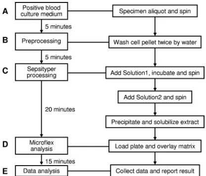
Specimen processing for MALDI-TOF MS analysis.Yeast-containing blood culture medium was processed using a modification of the product of the MALDI Sepsityper kit (270170; Bruker Daltoniks GmbH). Prior to the Sepsi-typer procedure, two brief washing/centrifugation steps were done to remove red blood cells and proteins from the blood culture broths. In brief, 1 ml of the blood culture fluid was centrifuged at 8,000⫻gfor 2 min, and the cell pellet was washed twice with 1 ml of water. The pellet was then resuspended in 1 ml water by vortexing, and 200l Sepsityper lysis solution (solution 1) was added. After incubation for 2 min at room temperature, the yeast cell pellet was obtained by centrifugation and resuspended into 1 ml Sepsityper washing solution (solution 2) by vortexing. The cell pellet was obtained by centrifugation and resuspended into 300l water by vortexing. An additional 900l absolute ethanol was added into the suspension, the contents were mixed, and a cell pellet was obtained by centrifugation. The initial supernatant was discarded completely, and the cell pellet was air dried and eluted into 50l of 70% formic acid and 50l of acetonitrile (Fig. 1). After a second centrifugation, the final supernatant was used for analysis.
MALDI-TOF mass spectrometry measurement.One microliter of extract was added to the target plate, and 1l of matrix solution (Bruker Daltonics) was then overlaid on the spot. The mixture was allowed to dry in air at room temperature and crystallized (Fig. 1). MALDI-TOF MS spectra measurement was performed on a Microflex LT instrument (Bruker Daltonics) according to the manufactur-er’s instructions. Spectra were collected using FlexControl (version 3.0) software in the linear positive mode in the mass range of 2,000 to 20,000m/z(laser frequency, 20 Hz; ion source 1 voltage, 20 kV; ion source 2 voltage, 18.4 kV; lens voltage, 9.1 kV). The standard Biotyper AutoX method was used for automated
measurement (12, 19). Before the specimens were processed, the Bruker Dal-tonics bacterial test standard (BTS; no. 255343) was measured for calibration of the instrument.
MALDI Biotyper identifications.Automated data analysis and identification of raw spectra were performed by the MALDI Biotyper (version 2.0) software (Bruker Daltonics) using a library of 3,476 entries and default settings (13, 18, 19). The current database covers 110 different yeast species, of which 66 are Candidaspecies. An identification log(score) value ranging from 0 to 3 was given for each specimen. The score indicated the pattern-matching extent, according to the specifications of the MALDI Biotyper system (12, 17, 28). Score values of 0 to 1.699 generally indicated no reliable identification; score values of 1.7 to 1.999 indicated probable genus identification; score values of 2.0 to 2.299 indicated secure genus identification, probable species identification; and score values of 2.300 to 3.000 indicated highly probable species identification.
Limit of detection (LOD) and reproducibility.A positive blood culture spec-imen (specspec-imen 2-4), which was vortexed vigorously until all yeast cells separated singly, as seen by Gram stain, was 10-fold serially diluted with a pooled negative blood culture specimen. Each dilution was performed in triplicate for MALDI Biotyper identification and CFU determination. For MALDI Biotyper identifi-cation, three scores for each sample were obtained for both intra- and inter-specimen reproducibility determination. The numbers of CFU were determined by a quantitative subculture of each diluted sample onto 5% sheep blood agar plates, which were incubated at 35°C for 48 h. Variations in identification log-(score) values between three measurement spots and between three preparations were used to determine the intra- and interspecimen reproducibilities, respec-tively.
28S rRNA gene sequencing.Specimens with discrepant identification results between the phenotypic and MALDI Biotyper methods were further identified by sequencing analysis. A primer set (28S-Haynes-40F [5⬘-GCA TAT CAA TAA GCG GAG GA-3⬘] and 28S-Haynes-654R [5⬘-GGT CCG TGT TTC AAG ACG-3⬘]) was used to target the conserved sequences in the V3 region of the 28S rRNA gene (7). A loopful of the purified yeast isolate was put into 1 ml of distilled water and heated at 95°C for 10 min, the suspension was centrifuged, and 1l of supernatant was used for PCR amplification. PCR amplification was carried out as previously described (11, 29). Nucleotide sequences were deter-mined bidirectionally and were analyzed with BLAST (Basic Local Alignment Search Tool) on the NCBI website.
RESULTS
Concentrations of yeast cells of five positive blood culture media ranged from 1.2⫻ 106 to 8.5 ⫻ 106 CFU/ml. Direct
application of the MALDI Sepsityper kit for specimen pro-cessing failed to yield reliable identification (data not shown). However, the two additional preprocessing washing steps added (Fig. 1, step B) prior to using the MALDI Sepsityper kit resulted in satisfactory extraction results. Sepsityper processing resulted in reliable satisfactory identification results for all 42 clinical positive blood culture medium specimens, with identi-fication log(score) values being ⬎1.9 (Tables 1 and 2). The LOD was determined with one positive clinical specimen (identified asCandida parapsilosis), which was serially diluted with pooled negative blood culture medium. The yeast load of this specimen was determined to be 5.9 ⫻ 106 CFU/ml by
quantitative subculture. The LOD of the MALDI Biotyper system for reliable yeast identification was 5.9⫻105CFU/ml
when it was used directly on positive blood culture medium. The intraspecimen coefficients of variation (CVs) of the iden-tification log(score) values were 1.83 to 3.55%, and the inter-specimen CV was 2.91% for the original, undiluted inter-specimen (Table 1).
[image:2.585.58.266.69.247.2]A total of 42 yeast-containing positive blood culture medium specimens were collected during the study period. Current phenotypic methods, including colony morphology, germ tube test, urease activity analysis, and an API 20C AUX strip, iden-tified these isolates to two genera and five species, including FIG. 1. Flowchart of the MALDI Biotyper system for yeast
iden-tification directly from positive blood culture media.
on May 16, 2020 by guest
http://jcm.asm.org/
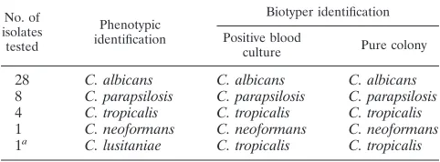
Candida albicans (n ⫽ 28, 66.7%), C. parapsilosis (n ⫽ 8, 19.0%),Candida tropicalis(n⫽4, 9.5%),Cryptococcus neofor-mans (n ⫽ 1, 2.4%), and Candida lusitaniae 1 (2.4%). The isolate identified asC. lusitaniae(isolate 43-154) had a poor match, with an identification ratio of 64% by the API 20C AUX strip. The MALDI Biotyper system was able to provide reliable identification to the species level for all 42 specimens (Table 2). The identification results were identical to those of phenotypic methods for all except one specimen, in whichC. tropicaliswas identified by the MALDI Biotyper system andC. lusitaniaewas identified by phenotypic methods. MALDI Bio-typer analysis on pure isolates obtained by subsequent subcul-ture gave results identical to those obtained when it was used directly on positive blood culture media (Table 2).
The specimen with discrepant identification results (speci-men 43-154) between the conventional methods and the MALDI Biotyper system was further analyzed by 28S rRNA gene sequencing. The nucleotide sequence of a partial 603-bp sequence of the 28S rRNA gene (GenBank accession number HQ214057) was identical to that of a clinicalC. tropicalis iso-late identified and reported previously (11).
The entire procedure, from specimen extraction to final re-sult reporting, was completed within 1 h.
DISCUSSION
In this pilot study, we explore the use of the MALDI Bio-typer system, which combines a blood culture medium speci-men processing kit (MALDI Sepsityper), a MALDI-TOF MS analyzer (Microflex), and dedicated software (MALDI Bio-typer, version 2.0), for rapid identification of yeast-like patho-gens directly from positive blood culture medium. With two additional preprocessing washing steps, the system reached a limit of detection of 105 CFU/ml with satisfactory intra- and
interspecimen reproducibilities. The entire procedure, from specimen extraction to final result reporting, can be completed within 1 h, providing another reliable tool for rapid yeast iden-tification directly from positive blood culture media.
Yeast-like fungal pathogens are among the commonest eti-ological agents of invasive fungal infections and are also seen in nosocomial bloodstream infections. Invasive fungal infec-tions and fungal nosocomial bloodstream infecinfec-tions are life-threatening, with overall and specific mortality rates of 60 and 49%, respectively (20). The selection of an effective treatment for candidemia depends on the infecting Candida species. There are more than 20Candida species, each with different antifungal susceptibility profiles (20, 30). At present, species determination requires from 2 to 5 days after collection of blood specimens for culture from patients. MALDI-TOF MS-based identification has the potential to shorten the time to results, thereby providing a substantial reduction in the delay before initiation of or adjustment to an effective therapy to improve therapeutic efficacy, minimize adverse effects, reduce costs, and lessen the risk of resistance development (6, 24).
[image:3.585.43.541.90.263.2]Previous published efforts on identification of yeast speci-mens by the MALDI-TOF MS-based procedures directly from yeast-containing positive blood culture specimens have yielded different findings (8, 9, 16). In this situation, the yeast-like fungi grew in liquid medium of complex composition due to the presence of both medium proteins and blood cells. In particular, the hemoglobin, proteins and peptides derived from leukocytes, and serum proteins must be discarded, as those yield strong signals in the mass spectra which hamper the TABLE 1. Limit of detection and reproducibility of MALDI Biotyper system for yeast identification directly from
a positive blood culture specimen
Sample 2-4
dilution No. of CFU/ml Identification
Score
% CV Specimen 1 Specimen 2 Specimen 3 Mean⫾SD
Undiluted 5.9⫻106 Reliable identification 2.230 2.112 2.211 2.184⫾0.063 2.90
2.234 2.245 2.170 2.216⫾0.041 1.83
2.204 2.354 2.229 2.262⫾0.080 3.55
1:10 5.9⫻105 Reliable identification 1.985 2.087 2.013 2.028⫾0.053 2.60
2.160 2.057 2.079 2.099⫾0.054 2.58
1.942 1.984 2.006 1.977⫾0.033 1.64
1:100 5.9⫻104 Not reliable identification 1.328 1.230 1.276 1.278⫾0.049 3.84
1.174 1.381 1.273 1.276⫾0.104 8.11
1.382 1.361 1.370 1.371⫾0.011 0.77
1:1,000 5.9⫻103 Not reliable identification 1.446 1.540 1.483 1.489⫾0.047 3.18
1.231 1.264 1.245 1.246⫾0.017 1.33
1.212 1.363 1.288 1.288⫾0.076 5.86
TABLE 2. Performance of MALDI Biotyper system for yeast identification on 42 yeast pathogens in blood
No. of isolates tested
Phenotypic identification
Biotyper identification
Positive blood
culture Pure colony
28 C. albicans C. albicans C. albicans 8 C. parapsilosis C. parapsilosis C. parapsilosis 4 C. tropicalis C. tropicalis C. tropicalis 1 C. neoformans C. neoformans C. neoformans 1a C. lusitaniae C. tropicalis C. tropicalis
a
The isolate was identified to beC. tropicalisby 28S rRNA gene sequencing.
on May 16, 2020 by guest
http://jcm.asm.org/
[image:3.585.42.283.628.717.2]interpretation of specific peaks. Development of a standard-ized and efficient specimen processing protocol to remove in-terference materials and yield clear microbial protein compo-nents is critical (10). In this study, we adapted and optimized a commercial kit, MALDI Sepsityper, for positive blood culture medium specimen processing. In our experience, the Sepsi-typer kit has the following characteristics: (i) identification of microorganisms directly from positive blood culture bottles is easy and rapid (less than 30 min); (ii) the sample preparation protocol is simple and uses only 1 ml sample material, few centrifugation steps, and only one tube for each analysis; (iii) identification of yeasts is reliable by the MALDI Biotyper system; and (iv) all reagents and consumables required for processing blood culture fluid are supplied in the kit ready to use. We added two washing steps to the specimen procedure prior to the MALDI Sepsityper analysis step to remove red blood cells and proteins. This modification made the MALDI Sepsityper an efficient tool for processing positive blood cul-ture media containing yeast-like pathogens.
In contrast to published protocols, which employed differ-ential centrifugation for the isolation of microorganisms in a positive blood culture and usually failed in identification of yeast, this method destroys the human cells (white and red blood cells) by lysis. The lysis solution used in this study is designed to lyse only human blood cells and not microorgan-isms. Therefore, in the high-speed centrifugation steps, only microorganisms, which are not affected by the lysis step be-cause of their robust cell walls, are precipitated. Thereby, in-terference of white blood cells, which are sedimented similarly to yeast cells and therefore cannot be separated from them by differential centrifugation, is prevented. We also applied two additional washing steps prior to the Sepsityper procedure, which resulted in more reproducible and satisfactory results on the basis of the identification of organisms in a total of 42 clinical specimens.
The sensitivity of MALDI-TOF MS to identify organisms in blood clearly depends on the inoculum (9, 10). Spiking exper-iments with Staphylococcus aureus and Escherichia coli indi-cated that organisms present at 107to 108CFU/ml were
cor-rectly identified, whereas 106CFU/ml yielded signals that were
indistinguishable from the background for the negative con-trols, indicating that it was necessary to deposit at least 104to
105 CFU on the MALDI target plate in order to obtain an
identifying spectrum (4). Additionally, the inoculum must con-tain sufficient microorganisms to overcome the background peaks derived from blood during measurement. In our exper-iment, we reached a limit of detection of 105 CFU/ml on a
clinical specimen withC. parapsilosis, which is more sensitive than that previously reported. This may be attributed to the adaptation and employment of the MALDI Sepsityper kit for processing of 1 ml of a positive blood specimen. The Sepsityper kit itself is much more effective at removing the interfering human cells (red and white blood cells) from the complex mixture and enriches microorganisms in a greater ratio than other methods. In addition, both intra- and interstrain identi-fication log(score) value CVs were small, suggesting that the Sepsityper kit for sample preparation possessed satisfactory reproducibility results similar to those achieved with bacterial and yeast pathogen identification methods (18, 28).
In summary, the direct MS fingerprinting method with
spec-imen processing by the modified MALDI Sepsityper kit offers a favorable combination of easy sample handling, accuracy, reproducibility, short turnaround time, and modest reagent costs. While our pilot study results are promising, more yeast-containing positive blood culture medium specimens covering a variety of genera and species need to be tested to fully evaluate this potentially revolutionary microbial identification tool.
ACKNOWLEDGMENTS
We thank Susan Sefers, Joni Williams, Bunny Ambrose, Jasper Benton, Rusty Bowden, Donna Brewer, Beth Brown, Sonia Cerruti, Emily Cauanaugh, Kathy Ewing, Pam Foster, Rene Gerald, Tonia Goodman, Mary Hedges, Monna Jedd, Lindsay Johnson, Kim Klocek, Sue May, Amy Montgomery, Kim Neville, Carla Nicholson, and Jen-nifer Steinhauer for helping collect clinical specimens.
This study was partially supported by Vanderbilt CTSA grant UL1 RR024975 from NCRR/NIH and an instrument lease agreement be-tween Vanderbilt University Medical Center and Bruker Daltonics.
REFERENCES
1.Alispahic, M., et al.2010. Species-specific identification and differentiation ofArcobacter,HelicobacterandCampylobacterby full-spectral matrix-asso-ciated laser desorption/ionization time of flight mass spectrometry analysis. J. Med. Microbiol.59:295–301.
2.Bader, O., et al.14 October 2010. Improved clinical laboratory identification of human pathogenic yeasts by matrix-assisted laser desorption ionization time-of-flight mass spectrometry. Clin. Microbiol. Infect. [Epub ahead of print.]
3.Cherkaoui, A., et al.2010. Comparison of two matrix-assisted laser desorp-tion ionizadesorp-tion–time of flight mass spectrometry methods with convendesorp-tional phenotypic identification for routine identification of bacteria to the species level. J. Clin. Microbiol.48:1169–1175.
4.Christner, M., et al.2010. Rapid identification of bacteria from positive blood culture bottles by use of matrix-assisted laser desorption–ionization time of flight mass spectrometry fingerprinting. J. Clin. Microbiol.48:1584– 1591.
5.Dhiman, N., L. Hall, S. L. Wohlfiel, S. P. Buckwalter, and N. L. Wengenack.
2011. Performance and cost analysis of matrix-assisted laser desorption ion-ization–time of flight mass spectrometry for routine identification of yeast. J. Clin. Microbiol.49:1614–1616.
6.Drancourt, M.2010. Detection of microorganisms in blood specimens using matrix-assisted laser desorption ionization time-of-flight mass spectrometry: a review. Clin. Microbiol. Infect.16:1620–1625.
7.Fell, J. W.1993. Rapid identification of yeast species using three primers in a polymerase chain reaction. Mol. Mar. Biol. Biotechnol.2:174–180. 8.Ferreira, L., et al.12 August 2010. Rapid method for direct identification of
bacteria in urine and blood culture samples by matrix-assisted laser desorp-tion ionizadesorp-tion time-of-flight mass spectrometry: intact cell vs. extracdesorp-tion method. Clin. Microbiol. Infect. [Epub ahead of print.]
9.Ferroni, A., et al.2010. Real-time identification of bacteria andCandida species in positive blood culture broths by matrix-assisted laser desorption ionization–time of flight mass spectrometry. J. Clin. Microbiol.48:1542– 1548.
10.Giebel, R., et al.2010. Microbial fingerprinting using matrix-assisted laser desorption ionization time-of-flight mass spectrometry (MALDI-TOF MS) applications and challenges. Adv. Appl. Microbiol.71:149–184.
11.Hall, L., S. Wohlfiel, and G. D. Roberts.2003. Experience with the MicroSeq D2 large-subunit ribosomal DNA sequencing kit for identification of com-monly encountered, clinically important yeast species. J. Clin. Microbiol.
41:5099–5102.
12.He, Y., H. Li, X. Lu, C. W. Stratton, and Y. W. Tang.2010. Mass spectrom-etry Biotyper system identifies enteric bacterial pathogens directly from colonies grown on selective stool culture media. J. Clin. Microbiol.48:3888– 3892.
13.Ilina, E. N., et al.2009. Direct bacterial profiling by matrix-assisted laser desorption-ionization time-of-flight mass spectrometry for identification of pathogenic Neisseria. J. Mol. Diagn.11:75–86.
14.Jordan, J. A., J. Jones-Laughner, and M. B. Durso.2009. Utility of pyrose-quencing in identifying bacteria directly from positive blood culture bottles. J. Clin. Microbiol.47:368–372.
15.La Scola, B., and D. Raoult.2009. Direct identification of bacteria in positive blood culture bottles by matrix-assisted laser desorption ionisation time-of-flight mass spectrometry. PLoS One4:e8041.
16.Marinach-Patrice, C., et al.2010. Rapid species diagnosis for invasive can-didiasis using mass spectrometry. PLoS One5:e8862.
on May 16, 2020 by guest
http://jcm.asm.org/
17.Marklein, G., et al.2009. Matrix-assisted laser desorption ionization–time of flight mass spectrometry for fast and reliable identification of clinical yeast isolates. J. Clin. Microbiol.47:2912–2917.
18.Mellmann, A., et al.2009. High interlaboratory reproducibility of matrix-assisted laser desorption ionization–time of flight mass spectrometry-based species identification of nonfermenting bacteria. J. Clin. Microbiol.47:3732– 3734.
19.Mellmann, A., et al.2008. Evaluation of matrix-assisted laser desorption ionization-time-of-flight mass spectrometry in comparison to 16S rRNA gene sequencing for species identification of nonfermenting bacteria. J. Clin. Microbiol.46:1946–1954.
20.Pappas, P. G., et al.2009. Clinical practice guidelines for the management of candidiasis: 2009 update by the Infectious Diseases Society of America. Clin. Infect. Dis.48:503–535.
21.Pincus, D. H., et al.1999. Rapid identification ofCandida dubliniensiswith commercial yeast identification systems. J. Clin. Microbiol.37:3533–3539. 22.Prod’hom, G., A. Bizzini, C. Durussel, J. Bille, and G. Greub.2010.
Matrix-assisted laser desorption ionization–time of flight mass spectrometry for direct bacterial identification from positive blood culture pellets. J. Clin. Microbiol.48:1481–1483.
23.Selvarangan, R., U. Bui, A. P. Limaye, and B. T. Cookson.2003. Rapid identification of commonly encounteredCandidaspecies directly from blood culture bottles. J. Clin. Microbiol.41:5660–5664.
24.Seng, P., et al.2009. Ongoing revolution in bacteriology: routine identifica-tion of bacteria by matrix-assisted laser desorpidentifica-tion ionizaidentifica-tion time-of-flight mass spectrometry. Clin. Infect. Dis.49:543–551.
25.Shepard, J. R., et al.2008. Multicenter evaluation of theCandida albicans/ Candida glabrata peptide nucleic acid fluorescent in situ hybridization method for simultaneous dual-color identification ofC. albicans andC. glabratadirectly from blood culture bottles. J. Clin. Microbiol.46:50–55.
26.Smith, M. B., D. Dunklee, H. Vu, and G. L. Woods.1999. Comparative performance of the RapID Yeast Plus System and the API 20C AUX clinical yeast system. J. Clin. Microbiol.37:2697–2698.
27.Stevenson, L. G., S. K. Drake, and P. R. Murray.2010. Rapid identification of bacteria in positive blood culture broths by matrix-assisted laser desorp-tion ionizadesorp-tion–time of flight mass spectrometry. J. Clin. Microbiol.48:444– 447.
28.Stevenson, L. G., S. K. Drake, Y. R. Shea, A. M. Zelazny, and P. R. Murray.
2010. Evaluation of matrix-assisted laser desorption ionization–time of flight mass spectrometry for identification of clinically important yeast species. J. Clin. Microbiol.48:3482–3486.
29.Tang, Y. W., et al.1998. Comparison of phenotypic and genotypic techniques for identification of unusual aerobic pathogenic gram-negative bacilli. J. Clin. Microbiol.36:3674–3679.
30.Tortorano, A. M., et al. 2004. Epidemiology of candidaemia in Europe: results of 28-month European Confederation of Medical Mycology (ECMM) hospital-based surveillance study. Eur. J. Clin. Microbiol. Infect. Dis.23:317–322.
31.Trenholme, G. M., et al.1989. Clinical impact of rapid identification and susceptibility testing of bacterial blood culture isolates. J. Clin. Microbiol.
27:1342–1345.
32.van Veen, S. Q., E. C. Claas, and E. J. Kuijper.2010. High-throughput identification of bacteria and yeast by matrix-assisted laser desorption ion-ization-time of flight mass spectrometry in conventional medical microbiol-ogy laboratories. J. Clin. Microbiol.48:900–907.
33.Weinstein, M. P., et al.1997. The clinical significance of positive blood cultures in the 1990s: a prospective comprehensive evaluation of the micro-biology, epidemiology, and outcome of bacteremia and fungemia in adults. Clin. Infect. Dis.24:584–602.