ISSN Online: 2158-2882 ISSN Print: 2158-284X
DOI: 10.4236/ijcm.2018.99055 Sep. 14, 2018 660 International Journal of Clinical Medicine
Effect of Specific Nutrients on Ovulation,
Oocytes Development, Gene Expression and
Coupling Success in Mice
P. Vitullo, C. Cossetti, M. A. Virmani
*Research, Innovation and Development, Sigma-Tau Health Science International BV, Utrecht, The Netherland
Abstract
Introduction: The roles of genetic, epigenetic, metabolic and other environ-mental factors such as nutrition and stress, are becoming evident for a suc-cessful and healthy pregnancy. This raises the possibility and question, if and how, we improve the probability of pregnancy and of a healthy fetus? The present study examined the role of metabolic, antioxidant and minerals and the results suggest that these factors may positively influence the oocyte qual-ity and the pregnancy rate. Methods: CD1 female mice aged 15 and 5 weeks were divided into four groups of ten each and treated by intragastric gavage daily for 3 weeks. G1: Vehicle; G2: Carnitines (L-carnitine 0.4 mg and acetyl-L-carnitine 0.12 mg/mouse); G3: Microelements (Zinc 4 ng, Copper 0.8 ng, Iron 7 ng/mouse); G4: G3+G2. At the end of the treatment period su-perovulation was induced and oocytes were collected to assess their quantity and quality. Further, in vitro fertilization (IVF) experiments were performed to assess the preimplantation embryo development. The birth success rate was also analyzed in old and young female. The mice were in vivo fertilized. qRT-PCR were performed to analyze a possible modulation in key genes of the reproductive process. Results: The number of oocytes was significantly higher in groups 2 and 4 compared to the control group. The oocyte number in group 3 was not affected. The level of degraded oocytes was 29.1% and 19.3% (group 2 and 4) versus 34.3% (control). Concomitantly, the numbers of embryos arriving to successful birth were also increased in G4, both in the old and young group of mice. Preliminary analysis of genes affected evi-denced that AMH was up regulated in the ovary and KITL in the uterus in group 2. Conclusion: Results showed that L-carnitine, acetyl-L-carnitine and micronutrients were able to improve both oocytes quality and success rate of pregnancy. Further studies are planned to further examine ways to improve pregnancy and fetal health.
How to cite this paper: Vitullo, P., Cosset-ti, C. and Virmani, M.A. (2018) Effect of Specific Nutrients on Ovulation, Oocytes Development, Gene Expression and Coupl-ing Success in Mice. International Journal of Clinical Medicine, 9, 660-674.
https://doi.org/10.4236/ijcm.2018.99055
Received: August 14, 2018 Accepted: September 11, 2018 Published: September 14, 2018
Copyright © 2018 by authors and Scientific Research Publishing Inc. This work is licensed under the Creative Commons Attribution International License (CC BY 4.0).
DOI: 10.4236/ijcm.2018.99055 661 International Journal of Clinical Medicine
Keywords
Pregnancy, L-Carnitine, Acetyl-L-Carnitine, Micronutrients, Oocytes, Birth, Fetal Health, Embryos, Key Genes, Fertilization, Reproductive
1. Introduction
Genes and cellular metabolism are intrinsically linked to fertility and successful pregnancy. It is estimated that 10 to 15 percent of couples have trouble getting pregnant or getting to a successful delivery, in the USA according to the Centers for Disease Control and Prevention (CDC). The infertility results due to issues with the female about one-third of the time and due to male infertility factors about one-third of the time. In the rest, the cause is either unknown or a combi-nation of male and female factors. As stated by the National Center for Health Statistics (NCHS), the fertility rates in the US have declined over the last few decades and many couples have trouble conceiving. The reasons for the declines are not completely understood but are thought to be related to a number of fac-tors such as nutrition, pollution, stress, and general lifestyle issues. Women who experience problems achieving a recognized conception also have elevated rates of early unrecognized pregnancy loss [1] and elevated rates of clinical sponta-neous abortion [2]. Indeed, the recent review by Gray and Becker [2] suggests that delayed conception and the early pregnancy loss may share a common eti-ology, possibly through events or exposures prior to or during implantation and embryogenesis. The decline in fertility could in part be explained by a decrease in the ovum quality, as well as the physiological processes involved in female fer-tility. At birth, there are approximately one million eggs; and by the time of pu-berty, only about 300,000 remain. Of these only about 300 to 400 will be ovu-lated during a woman’s reproductive lifetime, the question of how to improve egg quality is more recent. There is growing consensus that egg quality declines with age. This could be connected to the accumulation of cellular damage with time, a reduction in cellular metabolic processes, which in turn leads to a reduc-tion in energy producreduc-tion and in increase in cellular oxidative stress, which can trigger abnormal physiological processes especially immune and inflammation processes.
DOI: 10.4236/ijcm.2018.99055 662 International Journal of Clinical Medicine mitochondria results in aberrant functionality of the respiratory chain and oxid-ative phosphorylation resulting in reduced energy and increased cytotoxicity. Free radical production also induces inflammatory processes due to the mis-folding of proteins and other mechanisms. The use of antioxidant substances able to reduce or quench ROS may thus counteract cellular damage. Research into mitochondriotropic agents such as carnitines, coenzyme Q10 and nicotina-mide have shown potential to improve various factors associated with body energy, hormonal regulation, uterus weight as well as possible stabilization of cellular membranes and the genome [3]. The role of these compounds in epige-netics and effects upon genomic stability per se is also becoming important in fertility [4]. L-Carnitine, which is essential in fatty acid metabolism, has been shown to prevent mitochondrial damage induced in the rat choroid plexus by medium chain fatty acids [5] or by mitochondrial toxins [6].
1.2. The Emerging Role of Nutrigenomics, Metabolomics and
Oxidative Stress in Fertility
Many recent studies have shown that some biochemical and genetic alterations occur in the oocyte and take part in the natural history of female infertility [7] [8] [9]. Any dysfunction in the genes and therefore in the related protein, for example an enzyme protecting against reactive oxygen species (ROS) called su-peroxide dismutase I (SOD) is known to lead to female folliculogenesis defects and cause a failure in maintaining pregnancy.
Studies suggest that oxidative stress is associated with decreased female fertil-ity in animals and in-vitro models; epidemiological evidences strongly suggest that exposure to oxidative stress can influence the timing and maintenance of a viable pregnancy (e.g. preeclampsia).
The identification and evaluation of appropriate genetic, metabolic and bio-chemical markers correlating with early steps of disease onset and progression can be used as a key in preventing this type of dysfunction.
1.3. Carnitines and Female Fertility
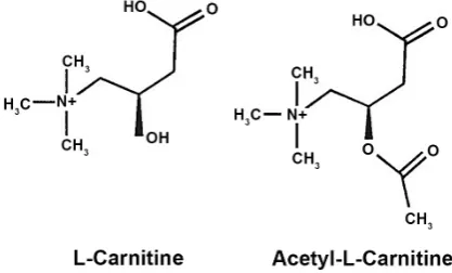
Carnitine is a natural product synthesized in mammals from the essential amino acids lysine and methionine or obtained from dietary sources (Figure 1).
DOI: 10.4236/ijcm.2018.99055 663 International Journal of Clinical Medicine
Figure 1. Chemical structures of L-carnitine and
acetyl-L-carnitine.
play a central role in fatty acid beta oxidation, that is involved in the acquisition of oocyte competence during which specific enzymes are expressed [12]. Studies show a correlation between the levels of CPT2 expression and embryo develop-mental competence [13]. Indeed, the inhibition of beta-oxidation during oocyte maturation or zygote division impairs subsequent blastocyst development. In contrast, L-carnitine supplementation during oocyte maturation significantly increases beta-oxidation and improves developmental competence [14]. In addi-tion, its acetylated form, acetyl-L-carnitine (ALC), protects cells exposed to neurotoxins and mitochondrial inhibitors [6]. Thus, together with antioxidant compounds like Q10 a protective action may be hypothesized on ovum and oth-er tissues vital for successful foth-ertility.
However, the quality of the ovum and the state of the uterus is also very im-portant for a successful pregnancy. There is growing consensus that egg quality declines with age and in conditions of abnormal energy balance, such as in po-lycystic ovary syndrome (PCOS). Indeed, what remain to be elucidated is the critical period between ovulation and implantation. During this period the suc-cessfully fertilized egg has to rely exclusively on its own energy reserves to start growing form the zygote to the blastocysts stage. The energy during this period comes from the mitochondria of the oocyte; if this energy runs out the egg stops dividing and implantation is not achieved. This would result in pregnancy loss. In the 70% of PCOS affected women (coupled or not with insulin resistance) in-hibition of ovulation, impaired of maturation of viable egg, and reduced egg quality and implantation rate, can occur. It is possible that improving the energy balance before the ovulation process would improve the success of pregnancy. Further the oocyte quality could be improved by substances such as carnitines and antioxidant compounds, increasing the chances of a successful pregnancy. All these factors are becoming important in the treatment of female infertility.
2. Material and Methods
2.1. Ethics Statement
DOI: 10.4236/ijcm.2018.99055 664 International Journal of Clinical Medicine Institute of Health. Animal care was conformed to the European Council Direc-tive 86/609/EEC and all experiments including animals were approved by the re-view board of the Italian National Institute of Health (Istituto Superiore di Sani-ta’, ISS) and authorized by the Italian Ministry of Health. Animals were sacri-ficed by cervical dislocation. All efforts were made to minimize suffering.
2.2. The Experiments Were Conducted in Three Steps
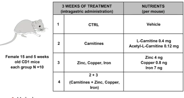
- First step: Female 8 weeks old CD1 mice were divided into four groups of ten each and treated daily for 3 weeks by intragastrical gavage. G1: Vehicle; G2: Carnitines (L-carnitine 0.4 mg and acetyl-L-carnitine 0.12 mg/mouse); G3: microelements (Zinc 4 ng, Copper 0.8 ng, Iron 7 ng/mouse); G4: G3+G2. The treatment dose for the mice was calculated approximately based on the rec-ommended healthy dietary allowances for human consumption. After treat-ment superovulation was induced, oocyte collected to assess quality and quantity. Moreover, in vitro fertilization (IVF) experiments were performed to evaluate the preimplantation embryos development [15]. Details for supe-rovulation, oocyte collection and IVF are given below (Figure 2).
- Second step: The ovary of the mice were surgically explanted and the RNA extracted, and expression of AMH, FSHB, CEPBP, LHR, and CDX2 genes was tested, being them related to ovary aging and minor production of cytes (CEPBP, LHR and CDX2), to ovarian reserve and stimulation of oo-cytes production (AMH and FSHB). RNA was extracted and gene expression evaluated by Reverse transcriptase PCR (RT-PCR) and quantitative real-time PCR as described below.
[image:5.595.214.538.543.716.2]- Third step: Female 15 and 5 weeks old CD1 mice were divided into four groups of ten each and treated daily for 3 weeks by intragastric gavage. G1: Vehicle; G2: Carnitines (L-carnitine 0.4 mg and acetyl-L-carnitine 0.12 mg/mouse); G3: microelements (Zinc 4 ng, Copper 0.8 ng, Iron 7 ng/mouse); G4: G3+G2. To assess the successful rate of birth in old and young female, mice were in vivo fertilized, as described below.
DOI: 10.4236/ijcm.2018.99055 665 International Journal of Clinical Medicine
2.3. Collection of Organs, Spermatozoa and Oocytes
Germ free CD1 female and male mice were purchased from Charles River (Cal-co, Italy). Viable somatic cell-free spermatozoa were obtained by puncturing cauda epididymis with a needle and collected after a 30 - 60 min “swim-up” se-lection step in FM medium [16] to perform IVF experiments. Oocytes for IVF experiment were obtained by surgical resection of ovaries as well as preimplan-tation embryos, obtained by natural breeding [16]. Surgically uterus and ovaries obtained were also stored in Trizol reagent (Invitrogen) for subsequent RNA ex-traction.
2.4. RNA Extraction
Organs from mice were lysed in Trizol reagent (Invitrogen) and total RNA was extracted according to manufacturer’s protocol, without further modification, except for the final step including two EtOH 75% washes rather than one.
2.5. Reverse Transcriptase PCR (RT-PCR) and Quantitative
Real-Time PCR
2 μg of total RNA extracted was reverse transcribed into cDNA using the iScript Advanced cDNA Synthesis kit for RT-qPCR (Bio-Rad). 200 ng of cDNA were qPCR amplified with specific primer pairs listed below:
AMH: For 5’-gtgagaggagaggggaacac-3’, Rev: 5’-gttctccagtctcccctagc-3’; CDX2: For 5’-ctgtcccttccctcgtcttt-3’, Rev: 5’-aactgtgttcggatcccctt-3’ CEBPB: For 5’-tgcggggttgttgatgtttt-3’, Rev: 5’-tgctcgaaacggaaaaggtt-3’; FSHb: For 5’-tcgtctgccttttagagcca-3’, Rev: 5’-ttcctcagccagcttcatca-3’ LHR: For 5’-acccggtgcttttacaaacc-3’, Rev: 5’-cgtcgtcccattgaatgcat-3’ GAPDH: For 5’-accacagtccatgccatcac-3’, Rev: 5-tccaccaccctgttgctgta-3’ Quantitative real time PCR (qPCR) was performed using SsoAdvanced Uni-versal SYBR Green Supermix (Bio-Rad) following the manufacturer’s instruc-tions in a 7500 Fast Real-Time PCR System (Applied Biosystems) under follow-ing conditions: one cycle of 95˚C for 30 s, 40 cycles of 98˚C for 15 s, 60˚C for 1 min and the instrument default settings for the Melt-Curve analysis. The an-nealing of the primers was at 60˚C. RNA levels were normalized to the level of GAPDH and calculated as delta-delta threshold cycle (ΔΔCT) [17]. All PCR ex-periments were performed in triplicate and the results were analyzed with qPCR 7500 Software Download v. 2.0.6.
2.6. Treatment and Induction of Superovulation in Mice
DOI: 10.4236/ijcm.2018.99055 666 International Journal of Clinical Medicine
2.7.
In Vitro
Fertilization
12 - 13 hours after hCG injection, female 8 weeks old CD1 mice were sacrificed; oviducts were removed and squeezed in 1 ml of FM medium. Aliquots of (1 – 2) × 106 spermatozoa were withdrawn and added to the egg-containing dishes.
Dishes containing both sperm cells and eggs were incubated for 6 hours at 37˚C with 5.3% CO2. Non-degraded eggs were then transferred to dishes containing 1
ml of fresh M-16 medium (Sigma) supplemented with BSA (4 mg/ml), overlaid with mineral oil and further cultured for 7 hours at 37˚C with 5.3% CO2. After
24 hr fresh medium was replaced. Embryos were allowed to grow for 5 days up to the blastocyst stage and, on the basis of morphology, the number of degraded and normal developing embryos was assessed under an inverted photomicros-cope stage by stage.
2.8.
In Vivo
Fertilization
12 - 13 hours after hCG injection, female 12 and 5 weeks old CD1 mice were mated with males of the same strain. The presence of a vaginal plug on the fol-lowing morning indicated successful mating. Developmental progression was followed up till the naturally birth of pups.
2.9. Statistical Analysis
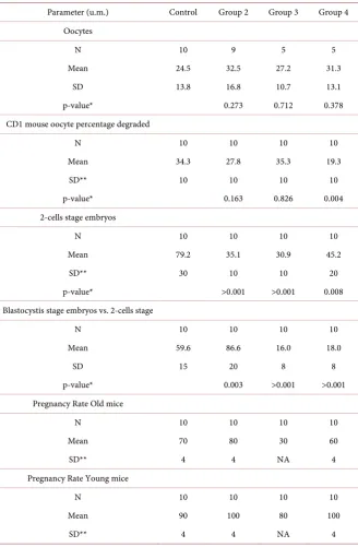
All the data were descriptively analyzed and presented as n, mean and standard deviation in Table 1 and as mean in Table 2. The figures were created using the data included in the tables. The p-values were derived from the t-test comparing each group with the control one.
3. Results
3.1. Carnitine Treatment Improve Quality and Quantity of Oocyte
The results of the study that were in part already published [14] and show that the mean number of oocytes/mouse was higherin the groups 2 (carnitines) and 4 (microelements plus carnitines): 32.5 (p = 0.273) and 31.3 (p = 0 .378) respec-tively, versus control group: 24.5 (Figure 3 and Table 1).The number of oocytes in group 3 was not significantly affected (27.2). Fur-thermore, the number of degraded oocytes in these groups was positively regu-lated: 27.8% (p = 0.163) and 19.3% (p = 0.004) (group 2 and 4 respectively) ver-sus 34.3% (control group) (Figure 4 and Table 1).
DOI: 10.4236/ijcm.2018.99055 667 International Journal of Clinical Medicine
Table 1. Summary of relevant results.
Parameter (u.m.) Control Group 2 Group 3 Group 4 Oocytes
N 10 9 5 5
Mean 24.5 32.5 27.2 31.3
SD 13.8 16.8 10.7 13.1
p-value* 0.273 0.712 0.378
CD1 mouse oocyte percentage degraded
N 10 10 10 10
Mean 34.3 27.8 35.3 19.3
SD** 10 10 10 10
p-value* 0.163 0.826 0.004
2-cells stage embryos
N 10 10 10 10
Mean 79.2 35.1 30.9 45.2
SD** 30 10 10 20
p-value* >0.001 >0.001 0.008
Blastocystis stage embryos vs. 2-cells stage
N 10 10 10 10
Mean 59.6 86.6 16.0 18.0
SD 15 20 8 8
p-value* 0.003 >0.001 >0.001
Pregnancy Rate Old mice
N 10 10 10 10
Mean 70 80 30 60
SD** 4 4 NA 4
Pregnancy Rate Young mice
N 10 10 10 10
Mean 90 100 80 100
SD** 4 4 NA 4
*p-values were derived from t-test on each group vs. control group; **estimated.
3.2. Carnitine Treatment Positively Affect Preimplantation
Development
DOI: 10.4236/ijcm.2018.99055 668 International Journal of Clinical Medicine
Table 2. Summary of CEBP, LHR and CDX2 gene expression by group.
Parameters Control Group 2 Group 3 Group 4
Stimulation of oocytes production
AMH 1 2.278
Stimulation of oocytes production
FSHB 1 1.264
Gene Analysis
CEBPB 1 0.586 0.7804 0.7006
Gene Analysis
LHR 1 0.7766 1.6521 0.0156
Gene Analysis
[image:9.595.208.539.468.660.2]CDX2 1 0.8876 0.5211 0.4408
Figure 3. Mean of oocytes.
Figure 4. CD1 mouse oocyte percentage degraded.
DOI: 10.4236/ijcm.2018.99055 669 International Journal of Clinical Medicine
Figure 5. % 2-cells stage embryos.
Figure 6. % Blastocysts stage embryos vs. 2-cells stage.
3.3. Gene Expression Analysis after Carnitine Treatment
After treatment, we decided to explore the possible effect of the different sup-plementations on the gene expression in ovary. Particularly we focused on CEPBP, LHR and CDX2 whose expression is related to ovary aging and minor production of oocytes, AMH and FSHB, influencing the ovarian reserve and stimulation of oocytes production.
Preliminary analysis of genes affected in these processes showed some inter-esting trends. The expression levels of AMH and FSHB was upregulated in group 2, the expression of AMH was more than 2 times higher than the control group, FSHB was slightly over expressed (Figure 7 and Table 2). In group 3 and 4 no significant effect on AMH or FSHB expression was evidenced.
[image:10.595.206.538.269.449.2]DOI: 10.4236/ijcm.2018.99055 670 International Journal of Clinical Medicine
Figure 7. Stimulation of oocytes production.
Figure 8. Gene analyses.
LHR expression shows a peculiar pattern; it was slightly down regulated in group 2 (0.8 times compared to the control), completely switched off in group 4, and upregulated in group 3 (1.6 times the control). The expression of CDX2 was down regulated in all the three groups, to a minor extent in group 2, about 0.9 times the control, and to a maximum extent in group 4, where it was reduced to about 0.4 times that of the control.
3.4.
In Vivo
Evaluation of Pregnancy Rate
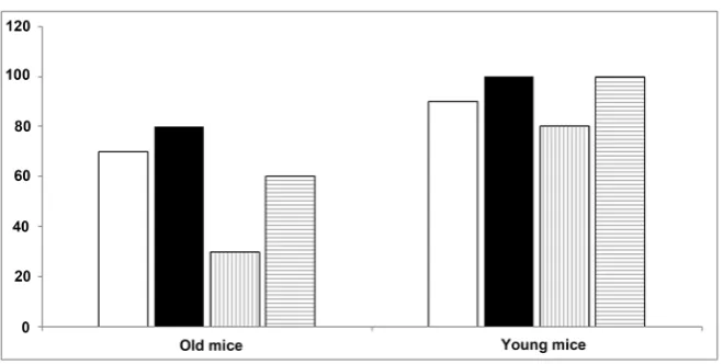
We verified if the above described genes expression regulation had some effect on the pregnancy rate in young and old mice (Figure 9 and Table 1).
The Group 2 old mice showed a significantly higher rate of pregnancy pared to control (80% vs 70%), Group 2 and group 4 young mice show a com-parable higher rate of pregnancy compared to control (100% compared to 90%). Group 3 of both young and old mice showed a lower rate of pregnancy com-pared to control group, in particular 30% vs 70% in old mice.
4. Discussion
DOI: 10.4236/ijcm.2018.99055 671 International Journal of Clinical Medicine
Figure 9. % Pregnant.
In particular, we targeted the oocytes, evaluating the effect of supplementation on their quantity and quality, which resulted significantly improved in the groups assuming carnitines alone ad carnitine plus microelements. These results, prompted us to investigate both the causes and the consequences of the im-provement in terms of quality and quantity of oocytes. In order to start to eluci-date the molecular mechanism underling the oocytes status, we performed a pre-liminary evaluation of the expression of genes that are known to be in some way related to the female fertility. In particular: AMH (anti mullerian hormone) is produced by pre-antral follicles and is a biomarker of granulosa cell mass, and thus indirectly, of ovarian reserve [18]; FSHB (Follicle stimulating hormone Beta subunit) is part of FSH, hormone inducing the oocytes production; the expres-sion of these two genes is thus directly related to an improvement of ovarian re-serve and stimulation of oocytes production. CEBPB (CCAAT/enhancer binding protein beta) encodes a transcription factor important for the transcription of genes involved in immune and inflammatory responses, among other processes; LHR (Luteinizing hormone receptor) is the receptor of Luteinizing hormone (LH) by gonadotropic cells in the anterior pituitary gland and triggers ovulation and development of the corpus luteum [19]; CDX2 is a member of the caud-al-related homeobox gene family, is related to placenta development and impor-tant in early embryogenesis [20][21].
The expression of these last three genes is correlated to ovary aging and minor production of oocytes. Taking into account that this was a preliminary evalua-tion of the expression of the above menevalua-tioned genes, requiring following inves-tigation, it seems quite clear a general positive effect of carnitine supplementa-tion, mainly alone, in the direction of up regulation of the genes that favorite the fertility (AMH, FSH, FSHB), and down regulation of the genes that impair it, as also related with aging (CEBPB, LHR, CDX2).
ef-DOI: 10.4236/ijcm.2018.99055 672 International Journal of Clinical Medicine fect we administered the different supplementation protocols to two groups of mice differing in age young mice 5 weeks old, and old mice 8 weeks old. As ex-pected, the global pregnancy rate was higher in all the four supplementation groups for young mice than old, but, among the groups of same age, again the groups receiving carnitine, alone or with macronutrients, showed a pregnancy rate significantly higher. Taking all together these results, we accumulated a strong evidence that the supplementation of carnitine has a beneficial effect on the female fertility, with an improvement of number and quality of oocytes, which is probably due or at least strongly connected with the up regulation of genes improving fertility and down regulation of genes which impair it, and with a final outcome in an increase of pregnancy rate.
An explanation could be that L-carnitine, which is essential in fatty acid me-tabolism, has been shown to prevent mitochondrial damage induced in the rat choroid plexus by medium chain fatty acids or by mitochondrial toxins. Key factors in the process of conception are the production of the ovum, the quality of the ovum and the state of the uterus. There is growing consensus that egg quality declines with age and in conditions of abnormal energy balance such as in polycystic ovary syndrome (PCOS). There is a critical period between ovula-tion and implantaovula-tion when the egg has to rely exclusively on its own energy re-serves contained within the zona pellucida. This energy comes from the fixed number of mitochondria present at moment of ovulation. If energy runs out the egg stops dividing and implantation is not achieved. In PCOS insulin resistance inhibits ovulation, impairs maturation of viable eggs, reduces egg quality and implantation. Restoring energy balance and providing adequate energy stores to the egg prior to ovulation would improve success of pregnancy. Further the oo-cyte genome quality could be improved by substances such as carnitines which improve genomic stability and therefore reduce chances of aneuploidy.
5. Conclusion
Environmental and stressful conditions can cause damage to the reproductive organs, eggs and general physiological processes of the body involved in the re-productive processes. The normal gene response may also be affected by meta-bolic dysfunctions. The metameta-bolic compounds, such as carnitines, antioxidants and micronutrients may play an important role in fertility by effects on energy and free radical formation during cellular metabolic processes. Further im-proved mitochondrial function could provide adequate energy for successful conception as well as the period up to implantation in the uterus. Targeting ovum, tubal and uterus function may provide new approaches to improve fertil-ity diseases however further experimental studies are needed in this field.
Conflicts of Interest
DOI: 10.4236/ijcm.2018.99055 673 International Journal of Clinical Medicine
References
[1] Hakim, R.B., Gray, R.H. and Zacur, H. (1998) Alcohol and Caffeine Consumption and Decrease Fertility. Fertility and Sterility, 70, 632-637.
https://doi.org/10.1016/S0015-0282(98)00257-X
[2] Gray, R.H. and Becker, S. (2000) Selected Topics in the Epidemiology of Reproduc-tive Outcomes. Epidemiologic Reviews, 22, 71-75.
https://doi.org/10.1093/oxfordjournals.epirev.a018027
[3] Virmani, M.A., et al. (2013) Food, Nutrigenomics and Neurodegeneration—Neuro- protection by What You Eat! Molecular Neurobiology, 48, 353-362.
https://doi.org/10.1007/s12035-013-8498-3
[4] Virmani, M.A. and Angeli, A. (2013) Role of Nutrigenomics and Metabolic Markers in Evaluating Oxidative Stress and Inflammation Dysfunctions in Female Fertility. Abstract and Posters, Vitafoods, Geneva.
[5] Kim, C.S., et al. (1990) L-Carnitine Prevents Mitochondrial Damage Induced by Octanoid Acid in the Rat Choroid Plexus. Brain Research, 536, 335-338.
https://doi.org/10.1016/0006-8993(90)90046-E
[6] Virmani, M.A., et al. (1995) Protective Actions of L-Carnitine and Acetyl-L-Carnitine on the Neurotoxicity Evoked by Mitochondrial Uncoupling or Inhibitors. Pharma-cological Research, 32, 383-389. https://doi.org/10.1016/S1043-6618(05)80044-1
[7] Binienda, Z., et al. (1999) Protective Effect of L-Carnitine in the Neurotoxicity In-duced by Mitochondrial Inhibitor 3-Nitrorpopionic Acid (3-NPA). Annals of the New York Academy of Sciences, 890, 173-178.
https://doi.org/10.1111/j.1749-6632.1999.tb07992.x
[8] Matzuk, M.M. and Lamb, D.J. (2002) Genetic Dissection of Mammalian Fertility Pathways. Nature Cell Biology, 4, s41-49.
https://doi.org/10.1038/ncb-nm-fertilityS41
[9] Virant-Klun, I., Knez, K., Tomazevic, T. and Skutella, T. (2013) Gene Expression Profiling of Human Oocytes Developed and Matured In Vivo or In Vitro. BioMed Research International, 2013, Article ID: 879489.
https://doi.org/10.1155/2013/879489
[10] Mansour, G., Abdelrazik, H., Sharma, R.K., Radwan, E., Falcone, T. and Agarwal, A. (2009) L-Carnitine Supplementation Reduces Oocyte Cytoskeleton Damage and Embryo Apoptosis Induced by Incubation in Peritoneal Fluid from Patients with Endometriosis.Fertility and Sterility, 91, 2079-2086.
https://doi.org/10.1016/j.fertnstert.2008.02.097
[11] Virmani, M.A., Krismanovic, L.Z., Stojilkovic, S.S. and Catt, K.J. (1991) Stimulatory Effects of L-Acetylcarnitine on the Pituitary-Gonadal Axis in Female Rats. In: Ada-shi, E.Y. and Mancus, S., Eds., Major Advances in Human Female Reproduction, Raven Press, 73, 291-296.
[12] Debbie, M., Frida, E., Isabelle, L., Stephanie, B., Timur, G. and Yves, M. (2012) Car-nitine Content in the Follicular Fluid and Expression of the Enzymes Involved in Beta-Oxidation in Oocyte and Cumulus Cells. Journal of Assisted Reproduction and Genetics, 29, 1221-1225. https://doi.org/10.1007/s10815-012-9855-2
[13] Dunning, K.R., Cashman, K., Russel, D.L., Thompson, J.G., Norman, R.J. and Robker, R.L. (2010) Beta-Oxidation Is Essential for Mouse Oocyte Development Competence and Early Embryo Development. Biology of Reproduction, 83, 909-918. https://doi.org/10.1095/biolreprod.110.084145
DOI: 10.4236/ijcm.2018.99055 674 International Journal of Clinical Medicine
Ovulation and Oocytes Quality in Mice. Giornale Italiano di Ostetricia e Ginecolo-gia, 37, 212-214.
[15] Virmani, A., Diedenhofen, A. and Zerelli, S. (2014) Mitochondriotropic Com-pounds in Energy, Oxidative Stress and Inflammation: Role in Reproductive Health, Fertility and Successful Pregnancy. Giornale Italiano di Ostetricia e Ginecologia, 36, 293-296.
[16] Vitullo, P., Sciamanna, I., Baiocchi, M., Sinibaldi-Vallebona, P. and Spadafora, C. (2012) LINE-1 Retrotransposon Copies Are Amplified during Murine Early Emb-ryo Development. Molecular Reproduction and Development, 79, 118-127. https://doi.org/10.1002/mrd.22003
[17] Meng, R. (2018) Relative Quantification: Data Management and Analysis Settings. Bio-Rad Laboratories Gene Expression Division.
http://www.bio-rad.com/genomics/pcrsupport
[18] Greene, A.D., Patounakis, G. and Segars, J.H. (2014) Genetic Associations with Di-minished Ovarian Reserve: A Systematic Review of the Literature. Journal of As-sisted Reproduction and Genetics, 31, 935-946.
https://doi.org/10.1007/s10815-014-0257-5
[19] Yding Andersen, C. (2017) Inhibin-B Secretion and FSH Isoform Distribution May Play an Integral Part of Follicular Selection in the Natural Menstrual Cycle. Mole-cular Human Reproduction, 23, 16-24.https://doi.org/10.1093/molehr/gaw070
[20] Beck, F., Erler, T., Russell, A. and James, R. (1995) Expression of Cdx-2 in the Mouse Embryo and Placenta: Possible Role in Patterning of the Extra-Embryonic Membranes. Developmental Dynamics, 204, 219-227.
https://doi.org/10.1002/aja.1002040302