Embolization Treatment of High-Flow Priapism
Kyung Rae Kim, MD
11Department of Radiology, University of North Carolina at Chapel Hill, Chapel Hill, North Carolina
Semin Intervent Radiol 2016;33:177–181
Address for correspondence Kyung Rae Kim, MD, Department of Radiology, University of North Carolina at Chapel Hill, 2019 Old Clinic, Campus Box 7510, Chapel Hill, NC 27599-7510
(e-mail: [email protected]).
Objectives: Upon completion of this article, the reader will be able to describe the pathophysiology, diagnosis, and treat-ment options of high-flow priapism.
Accreditation: This activity has been planned and imple-mented in accordance with the Essential Areas and Policies of the Accreditation Council for Continuing Medical Education (ACCME) through the joint providership of Tufts University School of Medicine (TUSM) and Thieme Medical Publishers, New York. TUSM is accredited by the ACCME to provide continuing medical education for physicians.
Credit: Tufts University School of Medicine designates this journal-based CME activity for a maximum of 1 AMA PRA Category 1 Credit™. Physicians should claim only the credit commensurate with the extent of their participation in the activity.
Priapism is defined as complete or partial penile tumescence that continues for more than 4 hours beyond sexual stimulation or is entirely unrelated to sexual stimulation.1–3It is a complex medical emergency with varied pathophysiology, usually requir-ing prompt medical, and sometimes surgical, management to prevent complications such as irreversible erectile dysfunction.2 The name priapism is derived from Priapus, the ancient Greek god of fertility. Mythological tales describe the cursing of the unborn Priapus while in Aphrodite’s womb, an act of vengeful spitefulness by the goddess Hera, which left the unfortunate child with enormous over-sized genitalia and an outcast from Mount Olympus. His image was adopted as a symbol of fertility.4
Thefirst reported case of priapism in the English medical literature was published in the Lancet by Tripe in 1845.5 Subsequently in 1914, Frank Hinman Sr. published a land-mark article describing the natural history of priapism.6Later in 1960, Frank Hinman Jr. proposed that venous stasis, increased blood viscosity, and ischemia were responsible for priapism, and he emphasized that failure to correct these abnormalities in the penile environment was the primary cause for treatment failures.7
Currently, priapism remains a common reason for many men to be seen in the emergency department. In fact, between 2006 and 2009, it is estimated that there were 32,462 visits made to emergency departments in the United States for priapism. This represents a national incidence of 5.34 per 100,000 men per year.2For clinical management, priapism can be stratified into three groups: low-flow pria-pism (ischemic, veno-occlusive), stuttering priapria-pism (inter-mittent), and high-flow priapism (nonischemic, arterial).1,2
Anatomy and Physiology of Normal Penile
Erection
The penis consists of three longitudinally oriented bodies or corpora. The dorsolaterally located paired corpora cavernosa are surrounded by the thick tunica albuginea and consist of multiple smooth muscle and endothelial-lined sinusoids, which are capable of significant volume expansion with consequential penile tumescence. The single ventral corpus Keywords
►
priapism
►
embolization
►
erectile dysfunction
►
ischemic
►
nonischemic
►
interventional
radiology
Abstract
Priapism is prolonged erection that persists beyond or is unrelated to sexual stimulation.
There are two main types of priapism: high
flow and low
flow. The treatment of priapism
will differ depending on the diagnosis of these two different types. In particular,
interventional radiology plays a key role in treating patients with high-
fl
ow priapism.
This article will review the diagnosis and treatment of the high-
fl
ow priapism.
Issue ThemeMen’s Health; Guest Editor, Charles Burke, MD
Copyright © 2016 by Thieme Medical Publishers, Inc., 333 Seventh Avenue, New York, NY 10001, USA.
Tel: +1(212) 584-4662.
DOI http://dx.doi.org/ 10.1055/s-0036-1586152. ISSN 0739-9529.
spongiosum is enveloped by a thinner layer of tunica albu-ginea and surrounds the penile urethra, extending distally to form the glans penis. The penis is supplied by the penile artery, which is a branch of the internal pudendal artery, which in turn arises from the anterior division of the internal iliac artery. The penile artery divides into three branches: the bulbar artery (to the corpus spongiosum), the dorsal artery, and the cavernosal artery (main blood supply to the erectile tissue of the corpus cavernosum).4
In theflaccid penis, the high resting tone of the cavernosal arterioles and sinusoids results in low blood volume inflow and outflow. The onset of an erection is allowed by smooth muscle relaxation and consequently a significant increase in cavernosal arterial bloodflow. The enlarging sinusoids com-press the exiting venules and emissary veins, thereby pas-sively restricting venous outflow. This situation of high inflow with restricted outflow leads to an elevated intracorporal pressure, eventually causing reduced inflow when systolic pressure is exceeded.4
Low-Flow Priapism (Ischemic,
Veno-occlusive)
Low-flow priapism, which accounts for more than 95% of all priapic episodes, is a compartment syndrome at the level of the corpora cavernosa and is characterized by a persistent painful erection with little or absent inflow through the cavernosal arteries.1 In the majority of cases, priapism is idiopathic; blood dyscrasias, the use of recreational drugs and alcohol abuse, malignancy, neurological diseases, and intracavernosal injection of vasoactive drugs account for the remaining 30 to 40% of episodes. As arterial inflow is reduced, there are time-dependent changes in the corporal metabolic environment that lead to progressive hypoxia, hypercarbia, acidosis, and glucopenia. This causes a progressive damage to the corporeal smooth muscle that becomes progressively irreversible as ischemic time continues. Although it is still a matter of debate as to the precise time when this process becomes irreversible, intervention after 49 to 72 hours may help relieve the pain and reverse the erection but will not be able to reverse the necrosis of the smooth muscle. The necrotic smooth muscle will be progressively substituted by scar tissue, with consequent penile shortening and onset of refractory erectile dysfunction. Therefore, low-flow priapism is an emergency that requires immediate treatment, as any delay in the management results in an increased risk of onset of severe erectile dysfunction and penile shortening.8
Stuttering Priapism (Intermittent)
Stuttering priapism is a variant of low-flow priapism and characterized by a pattern of recurrence. The term has historically described recurrent, unwanted, and painful erec-tions in men with sickle-cell disease. Patients typically are awakened by erections that persist for less than 3 hours but become progressively painful due to the onset of ischemia. Unfortunately, males with sickle-cell disease may experience stuttering priapism from childhood; in these patients, the
pattern of stuttering may increase in frequency and duration, leading up to a full episode of unrelenting ischemic priapism. Any patient who has experienced an episode of ischemic priapism is also at risk for stuttering priapism.1
High-Flow Priapism (Nonischemic, Arterial)
High-flow priapism is a persistent erection caused by unregu-lated cavernous arterial inflow. It wasfirst described by Burt et al in 1960 when a man developed a persistent erection following traumatic coitus.9The high-flow etiology of pria-pism is much less common than the low-flow condition, and the etiology is largely attributed to trauma. It is typically the result of injuries to the crura or corpora resulting in laceration of the cavernous artery or one of its branches within the corpora. This can be the result of either blunt or penetrating injuries and include such mechanisms as straddle injury, coital trauma, kicks to the penis or perineum, pelvic fractures, birth canal trauma to the newborn male, needle lacerations, com-plications of penile diagnostics, vascular erosions complicating metastatic infiltration of the corpora, and following surgical interventions.10–12In addition, high-flow priapism may arise as a delayed complication of genital or perineal trauma secondary to the development of an arteriocavernosalfistulas. Any mechanism that lacerates a cavernous artery or arteriole can produce unregulated pooling of blood in the sinusoidal space with consequence erection, but the cavern-ous environment does not become ischemic secondary to the continuous influx of arterial blood.13The corpora are tumes-cent but not rigid, and patients do not complain of pain with erection.1In contrast to the low-flow condition, high-flow priapism is not considered an emergent situation and does not require immediate intervention.
Evaluation and Diagnosis of Priapism
History
High-flow priapism should be suspected when there is no pain associated with the erection, the erection duration has not been accompanied by progressive discomfort, and the patient has a history as described earlier (►Table 1). The onset of posttraumatic high-flow priapism may be delayed by hours to days following the initial injury.1
Physical Exam
In low-flow priapism, the corpora cavernosa are rigid and very tender to palpation.14In patients with high-flow pria-pism, there is minimal to no pain, and on exam the penis is engorged or only partially erect.15Depending on the location
Table 1 Clinical characteristics of priapism
High-flow priapism Low-flow priapism
Trauma history Yes No
Pain No Yes
Duration >4 h Any
of the trauma and the elapsed time since the traumatic event, there may be residual bruising and some tenderness on palpation.
Laboratory Testing
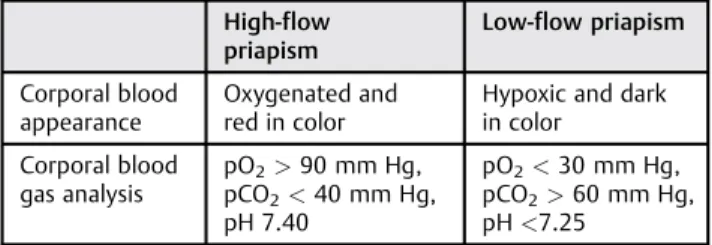
Laboratory evaluation should include a complete blood count, white blood cell with blood cell differential, platelet count, and coagulation profile to assess for anemia, to rule out infection and to detect hematologic abnormalities. Urine and plasma toxicology should be performed if recreational drugs are suspected from the psychosocial history. A corporal blood gas analysis is recommended in the evaluation of priapism in order to differentiate between low-flow and high-flow priapism (►Table 2).
Color Doppler Ultrasonography
Color Doppler ultrasonography (CDU) is an adjunct to the corporal blood aspirate in differentiating low-flow from high-flow priapism. CDU is performed using a high-frequency transducer (7 MHz or higher). With the patient in the frog-leg position and the scrotum elevated, the cavernosal arteries are traced from their origin in the perineum along the ventral aspect of the penile shaft. In low-flow priapism, cavernosal arterialflow typically demonstrates a“high-resistance, low-velocity” waveform and arterial flow is usually absent. In high-flow priapism, CDU demonstrates a “low-resistance, high-velocity” arterial waveform.4,16The sensitivity of CDU in localizing an arteriocavernosalfistula is nearly 100%.17On gray-scale ultrasonography, the arteriocavernosalfistula is a hypoechoic area surrounded by echogenic tissue.
Magnetic Resonance Imaging
The use of magnetic resonance imaging (MRI) in the diagnosis of priapism has not been well established, and MRI has a limited role in the initial diagnosis given the emergency situation of priapism. There has been at least one report of MRIfindings in four cases of high-flow priapism.18In this study, MRI demonstrated the arteriocavernosalfistula in all patients. The authors of that article pointed out that while CDU demonstrates characteristicflow patterns of priapism, MRI has better potential in tissue characterization and dem-onstration of a thrombus or arteriocavernosalfistula.18 Addi-tional benefits of MRI include its ability to reliably predict nonviable smooth muscle within the corpora after episodes of priapism, as well as detecting unusual conditions such as malignant infiltration.14
Treatment of High-Flow Priapism
Patients with high-flow priapism do not need to be managed urgently because this does not result in ischemia within the corpora cavernosa. Conservative management should be the first-line treatment as there is a 60% chance of spontaneous resolution.19However, there is increasing clinical evidence that long-standing, high-flow priapism could eventually lead to cavernosal fibrotic changes and erectile dysfunction; therefore, patients who do not respond to conservative management may require more aggressive treatment.20,21
Conservative management consists of compression and application of ice, with the aim of inducing vasospasm of the cavernosal artery to facilitate clot formation and oblit-eration of the fistula. If a cavernosal pseudoaneurysm is identified on CDU, ultrasound-guided compression of the pseudoaneurysm can be attempted.22 Intracavernosal injection ofα-adrenergic agonists (useful for the treatment of the low-flow type) is not recommended for high-flow priapism because of the different pathophysiology and substantial risk of cardiovascular adverse effects.23
If conservative management is not successful, selective embolization of the cavernosal artery can be attempted. This method, first described by Wear et al in 1977, allows for temporary occlusion of the cavernosal artery, thus permitting cicatricial closure of thefistula and subsequent rechanneling of the embolized artery.24 Recurrent or residual erectile dysfunction after embolization treatment has been reported in approximately 15 to 22% of high-flow priapism patients.20,21,25,26
Several occlusive agents can be used for selective arterial embolization, including autologous blood clot, gelatin sponge, microcoils, polyvinyl alcohol, and glue. All of these occlusive agents achieve a similar 75% resolution rate.13
Autologous blood clot, a temporary occlusive agent, has been successfully used often in children in the embolization offistulas in the treatment of priapism.27,28However, recur-rence within a few hours has been reported, and the blood clot can be technically more difficult to manipulate.29Gelatin sponge is a temporary occlusive agent that lasts 2 to 6 weeks after being injected. Detumescence can be achieved with gelatin sponge in theory without permanent vascular abla-tion30(►Fig. 1).
Microcoils are permanent occlusive agents. Hence, there are theoretically increased risks of permanent vascular oc-clusion and subsequent erectile dysfunction with their use. Glue is an ionic permanent occlusive agent that polymerizes when in contact with blood, and rapidly adheres to adjacent tissues; extreme care must be taken during its use.29
In the literature, recurrence rates of 30 to 40% have been reported for high-flow priapism treated by selective embolization, explaining the frequent need for repeated embolization.20
Surgical management with transcorporalfistula ligation is reserved for cases where repeated embolization has failed, as it carries a higher risk of future erectile dysfunction. Compli-cations of surgical management include erectile dysfunction (as high as 50%), penile gangrene, gluteal ischemia, purulent cavernositis, and perineal abscess.31
Table 2 Corporal blood analysis
High-flow priapism
Low-flow priapism
Corporal blood appearance
Oxygenated and red in color
Hypoxic and dark in color
Corporal blood gas analysis
pO2>90 mm Hg,
pCO2<40 mm Hg,
pH 7.40
pO2<30 mm Hg,
pCO2>60 mm Hg,
pH<7.25
Conclusion
Priapism can be the result of a variety of etiologies and is often a treatable condition. During the initial evaluation, it is critical to differentiate high-flow and low-flow etiologies because the pathophysiology and treatment are different. Patients with low-flow priapism must be treated emergently because of the high risk of complications. Patients with high-flow priapism may be treated initially with conservative therapy, reserving more aggressive treatments for those patients who do not respond to conservative therapy. Selec-tive transcatheter embolization of the arteriocavernosal
fistula with absorbable/nonabsorbable agents has been shown to be effective, with the results of selective emboliza-tion superior to surgical ligaemboliza-tion with fewer complicaemboliza-tions.
Disclosure
The author has no financial or nonfinancial conflicts relevant to this article.
References
1 Broderick GA, Kadioglu A, Bivalacqua TJ, Ghanem H, Nehra A, Shamloul R. Priapism: pathogenesis, epidemiology, and manage-ment. J Sex Med 2010;7(1, Pt 2):476–500
2 Roghmann F, Becker A, Sammon JD, et al. Incidence of priapism in emergency departments in the United States. J Urol 2013;190; (4):1275–1280
3 Tay YK, Spernat D, Rzetelski-West K, Appu S, Love C. Acute man-agement of priapism in men. BJU Int 2012;109(Suppl 3):15–21 4 Halls JE, Patel DV, Walkden M, Patel U. Priapism: pathophysiology and
the role of the radiologist. Br J Radiol 2012;85(Spec No 1):S79–S85 5 Tripe J. Case of continued priapism. Lancet 1845;2:1274–1276 6 Hinman F. Priapism: report of cases and a clinical study of the
literature with reference to its pathogenesis and surgical treat-ment. Ann Surg 1914;60(6):689–716
7 Hinman F Jr. Priapism; reasons for failure of therapy. J Urol 1960; 83:420–428
8 Garaffa G. Priapism. London: Springer-Verlag; 2013:49–54 9 Burt FB, Schirmer HK, Scott WW. A new concept in the
manage-ment of priapism. J Urol 1960;83:60–61
10 Dubocq FM, Tefilli MV, Grignon DJ, Pontes JE, Dhabuwala CB. High flow malignant priapism with isolated metastasis to the corpora cavernosa. Urology 1998;51(2):324–326
11 Chan PT, Bégin LR, Arnold D, Jacobson SA, Corcos J, Brock GB. Priapism secondary to penile metastasis: a report of two cases and a review of the literature. J Surg Oncol 1998;68(1):51–59 12 Lutz A, Lacour S, Hellstrom W. Conversion of low-flow to
high-flow priapism: a case report and review (CME). J Sex Med 2012; 9(4):951–954, quiz 955
13 Montague DK, Jarow J, Broderick GA, et al; Members of the Erectile Dysfunction Guideline Update Panel; American Urological Associ-ation. American Urological Association guideline on the manage-ment of priapism. J Urol 2003;170(4, Pt 1):1318–1324
14 Levey HR, Segal RL, Bivalacqua TJ. Management of priapism: an update for clinicians. Ther Adv Urol 2014;6(6):230–244 15 Huang YC, Harraz AM, Shindel AW, Lue TF. Evaluation and
manage-ment of priapism: 2009 update. Nat Rev Urol 2009;6(5):262–271 16 Donaldson JF, Rees RW, Steinbrecher HA. Priapism in children: a
comprehensive review and clinical guideline. J Pediatr Urol 2014; 10(1):11–24
17 Hakim LS, Kulaksizoglu H, Mulligan R, Greenfield A, Goldstein I. Evolving concepts in the diagnosis and treatment of arterial high flow priapism. J Urol 1996;155(2):541–548
18 White C, Gulati M, Gomes A, Rajfer J, Raman S. Pre-embolization evaluation of high-flow priapism: magnetic resonance angiogra-phy of the penis. Abdom Imaging 2013;38(3):588–597
19 Keck B, Lotter G, Wieland WF, et al. Sonographic diagnosis of a posttraumatic arteriocavernosal fistula resulting in high-flow priapism. J Clin Ultrasound 2012;40(1):60–62
20 Ciampalini S, Savoca G, Buttazzi L, et al. High-flow priapism: treatment and long-term follow-up. Urology 2002;59(1):110–113 21 Savoca G, Pietropaolo F, Scieri F, Bertolotto M, Mucelli FP, Belgrano E. Sexual function after highly selective embolization of cavernous artery in patients with highflow priapism: long-term followup. J Urol 2004;172(2):644–647
Fig. 1 A 21-year-old male with high-flow priapism after blunt perineal trauma. (a) Selective left internal pudendal arteriogram demonstrates arteriocavernosalfistula in the cavernosal artery (arrow). (b) Arterio-gram after embolization with gelatin sponge demonstrates no further
22 Volgger H, Pfefferkorn S, Hobisch A. Posttraumatic high-flow priapism in children: noninvasive treatment by color Doppler ultrasound-guided perineal compression. Urology 2007;70(3): 590.e3–590.e5
23 Towbin R, Hurh P, Baskin K, et al. Priapism in children: treatment with embolotherapy. Pediatr Radiol 2007;37(5):483–487 24 Wear JB Jr, Crummy AB, Munson BO. A new approach to the
treatment of priapism. J Urol 1977;117(2):252–254
25 Kim KR, Shin JH, Song HY, et al. Treatment of high-flow priapism with superselective transcatheter embolization in 27 patients: a multicenter study. J Vasc Interv Radiol 2007;18(10):1222–1226 26 Langenhuijsen JF, Reisman Y, Reekers JA, de Reijke Th M. Highly
selective embolization of bilateral cavernous arteries for post-traumatic penile arterial priapism. Int J Impot Res 2001;13(6): 354–356
27 Kress O, Heidenreich A, Klose KJ, Wagner HJ, Alfke H. Super-selective embolization with coils in high-flow priapism. Cardio-vasc Intervent Radiol 2002;25(4):326–329
28 Yesilkaya Y, Peynircioglu B, Gulek B, Topcuoglu M, Inci K. Autologous blood-clot embolisation of cavernosal artery pseudoaneurysm causing delayed high-flow priapism. Pol J Radiol 2013;78(2):54–56 29 O’Sullivan P, Browne R, McEniff N, Lee MJ. Treatment of“ high-flow”priapism with superselective transcatheter embolization: a useful alternative to surgery. Cardiovasc Intervent Radiol 2006; 29(2):198–201
30 Cohen GS, Braunstein L, Ball DS, Roberto PJ, Reich J, Hanno P. Selective arterial embolization of idiopathic priapism. Cardiovasc Intervent Radiol 1996;19(1):47–49
31 Burnett AL, Sharlip ID. Standard operating procedures for pria-pism. J Sex Med 2013;10(1):180–194