Address for correspondence Dr. Xuefeng Wan
Associate Professor, Department of Dermatology, 1stAffiliated Hospital of Xinjiang Medical University,China.
Email: [email protected]
Original Article
A retrospective observational study on clinical and
histopathological correlational analysis of malignant
melanoma
Introduction
A clear regional and racial divergence with
increased incidence in western countries is
shown by malignant melanoma.
1,2Malignant
melanoma is derived from the neuroectodermal
mucosa.
3Melanoma accounts for 1-3 percent of
malignancies in children and adults.
4This age
group is < 2% of all patients with melanoma.
4,5However, melanoma incidents across all age
groups have increased in recent decades.
6,7Initial
Qazi Syed Irfanullah Shah, Shaista Rasheed*, Paride Abliz, Fan Jun Wei, Syed Ikram Ullah, Xue Feng Wan*
Department of Dermatology, 1st Affiliated Hospital of Xinjiang Medical University China.
* Department of Pediatrics, Northwest General Hospital & Research Center, Peshawar, KPK, Pakistan.
Abstract
Background Malignant melanoma derives from the neuroectodermal mucosa. Melanoma accounts for 1-3 percent of malignancies in children and adults. The incidence and clinical properties of the disease are multifactorial and have biological and environmental variables. Tumor thickness was correlated to age, blood supply, site and nerve supply separately.Objective This study was made to determine the clinical and histopathological correlation of malignant melanoma.
Material and Methods A retrospective observational study was conducted at a tertiary care hospital, Urumqi in China to study the clinical and histopathological correlation of malignant melanoma. A detailed history and thorough physical examination of patients, lab tests including primary or secondary tumor biopsies, complete blood count and PET scans help in diagnosis. Clinical staging was done on the basis of regional lymph node involvement and distant metastasis. Results The highest no. of patients (28.20%) was recorded for 60-69 years age group followed by (25.64%) in 70-79 years age group. The largest no. (30.76%) of cases comprised of acral lentiginous melanoma as well as nodular melanoma followed by lentigo maligna melanoma (20.51%) and superficial spreading melanoma (17.95%). The highest no. of patients (35.90%) had tumor thickness of 1-2 mm followed by (20.51%) cases had 2-4 mm, (17.95%) cases had <1 mm. However, not a single patient had tumor thickness of >4 mm in our study.
Conclusion This study investigated histopathological and clinical results. The main result we found was that the trunk was involved to major extent than the periphery. Frequently involved subtypes were nodular melanoma and acral melanoma. Female are more commonly affected in this study then male 2:1.
Key words
diagnosis is carried out via morphological,
clinical,
dermoscopic,
microscopic
or
immunohistochemical sizes: a strong tumor
forecast depth in mm.
8,9Melanoma is a type of
melanocyte
generated
skin
cancer.
The
incidence and clinical properties of the disease
are multifactorial and have biological and
environmental
variables.
10,13According
to
Ackerman's malignant melanoma study in the
Caucasians, approximately 80 percent are
novices, while only about 20 percent are linked
to existing nevi melanocytes mainly in the trunk
and proximity. Nevi is a combination of SSM
and nodular (NM) melanomas, whilst Nevi is a
result of Lentigo malignant (LMMs) and Acral
Lentiginous (ALM) melanoma. In Asia, over
50% of melanomas in ALM not exposed to the
sun as in the Caucasians are reported.
15,16In this
respect, ultraviolet radiation seems unconnected
to black and Asian melanoma.
17The lesion
thickness is usually the most accurate of all the
prime melanoma forecasts in Breslow.
18,20In
every patient: age, sex, anatomy, histogenetic
type, the thickness of Breslow, the presence of
ulceration, stage, and symptoms like bleeding,
color, and dimensional variations, alterations,
and previous trauma, all were chosen to
determine clinical or pathological properties.
Materials and Methods
Study population / Sample size / Clinical data
The present study includes 39 patients of
malignant melanoma of all different stages
including 13 male and 26 female patients of
melanoma.
The study was designed for current analysis after
the institutional review board's permission prior
to medical records and examination. The basic
statistic data also included a database, diseases
(anatomic sites, histologic subtypes, ulceration,
Breslow thickness, treatment methods, operating
methods, and system treatment) and published
predictive factors. The patient's characteristic
features like age, sex, and performance were
reported. The database was designed and
checked, re-examined and added to the database
medical documents of every patient submitted to
the
pathology
department
if
malignant
melanoma had been confirmed. In addition,
detailed history and physical examination of
patients was included in the institutional
protocol. Tests included tumor biopsy, complete
blood count, serum electrolytes, metabolic or
liver screen, chest CT, abdominal CT,
ultra-reactive and PET scans. The PET-CT reporting
was done to examine the foci on the skin or
other tissues and organs carefully. Testing of the
concentric tuft or other tissues was done by
positron emission tomography PET-CT. Cases
were treated with surgery and postoperative
samples were sent for pathological assessment.
In patients with skin melanoma in clinical stage
I, local excision is performed without a biopsy.
Further assessment tests were necessary for
patients who were entitled to our future
institutional trials. Either for clinical or
pathological stages, the American Joint Cancer
Committee (AJCC) stage-system (6
thedition)
was used. Clinical stages were based on
available categories, regional involvement of
lymph nodes and distant metastasis without
Breslow's pathological thickness analysis for
referred patients.
Data analysis
Table 1 Distribution of patients among stages on the bases of Age, Gender, and Anatomic location
Characteristics Stages Total
(n = 39)
0 1 2 3 4
Age group (in years)
< 39 0 2 0 1 0 3 (7.69%)
40 – 49 1 1 4 1 0 7 (17.94%)
50 – 59 3 0 4 0 0 7 (17.94%)
60 – 69 2 4 1 4 0 11 (28.20%)
70 – 79 3 2 3 2 0 10 (25.64%)
>80 0 0 0 1 0 1 (2.56%)
Total (n=39) 9 9 12 9 0 39 (100%)
Mean Age 59. 92 years
Median range 61 years
Range Min age: 33years Max age: 82 years Gender
Male 3 4 4 2 0 13 (33.33%)
Female 6 5 8 7 0 26 (66.66%)
Total (n=39) 9 9 12 9 0 39 (100%)
Anatomic Location
Head and Neck 0 1 0 1 0 2 (5.13%)
Face 1 1 0 0 0 2 (5.13%)
Trunk 1 2 1 2 0 6 (15.38%)
Upper Extremities
Thumb 0 2 2 0 0 4 (10.26%)
Palm 1 0 0 0 0 1 (2.56%)
Lower Extremities
Finger 1 0 4 0 0 5 (12.82%)
Heel 0 2 1 2 0 5 (12.82%)
Valve 0 0 1 0 0 1 (2.56%)
Waist 1 0 0 0 0 1 (2.56%)
Other 3 1 5 3 0 12 (30.76%)
Total (n=39) 10 7 14 8 0 39 (100%)
the final follow-up visit for melanoma. In each
case, the tumor lesions were taken to the safety
limits and sutured to the "per primam" and
subsequently
sent to
the
histopathologic
analytics laboratory. The excised fragments
were fixed to 10 percent of tamponed formalin
and processed using the standard paraffin
integration method. Paraffin blocks were first
cut into usual stained sections of
Hematoxylin-Eosin in the 3-4-micron thick segments for the
immunohistochemical analysis of
poly-L-lysine-coated glass slides the serial parts were then
carried out and highlighted.
Histology and immunohistochemistry
developed. HMB45 and Melan have a positive
expression in the cytoplasm and S-100 in the
nucleus. The cell membrane has a positive CK
expression and the coloring of the nucleus or
cell membrane of the brown tumor cell was
considered beneficial for any coloration.
Statistical Analysis
Data was compiled in Microsoft Excel and
statistical analysis was done using a statistical
software PASW SPSS version 20. Medium
range descriptive statistics, like mean ± standard
deviation
have
been
performed
with
demographic and epidemic characteristics at a
95% Confidence Interval. Hazard ratio was
carried out at a significant P-value of 0.005 for
inferential statistics, Chi-Square test,
Kaplan-Merrier test, Mann-Whitney U test.
Observations and Results
See
Table 1
Demographic and epidemic characteristics
In the current study 39 patients at various tumor
stages were included, out of which 13 were male
and 26 were female with male-female ratio 1:2.
The mean age was 59.92 years for the 39
patients diagnosed. (Median age; 56 years,
range; 17-79 years, 95% CI 49.37-59.14). The
highest no. of patients (28.20%) were recorded
for 60-69 years age group followed by (25.64%)
in the age group of 70-79 years.
Tumor anatomic location and staging
Tumor was mainly located in lower extremities
(61.52%), including finger (12.82%), heel
(12.82%), valve (2.56%), waist (2.56%) and
other extremes (30.76%), followed by upper
extremities (12.85%) including thumb (10.26%)
and palm (2.56%) while (5.13%) patients of
melanoma had tumor in head and neck,
(15.38%) patients of melanoma on the trunk
(Table 2)
.
Histopathological subtype & clinical diagnosis
Table 2 shows that the largest (30.76%) cases
consisting of acral lentiginous melanoma as well
as
nodular
melanoma
followed
by
LentigoMaligna melanoma (20.51%), superficial
spreading melanoma (17.95%). Most of the
patients 37 out of 39 (94.87%) had clinically
diagnosed malignant melanoma while only
(2.56%) diagnosed nevus and (2.56%) verrucous
nevus
(Table 3)
.
Table 2 Distribution of patients among stages according to histopathological subtype and clinical diagnosis
Characteristics Stages Total
(n = 39)
0 1 2 3 4
Histopathological subtype
Acral Lentiginous Melanoma 2 2 4 4 0 12 (30.76%)
LentigoMaligna Melanoma 5 2 0 1 0 8 (20.51%)
Nodular Melanoma 2 3 4 3 0 12 (30.76%)
Superficial Spreading Melanoma 0 2 4 1 0 7 (17.95%)
Total (n = 39) 9 9 12 9 0 39 (100%)
Clinical Diagnosis
Malignant Melanoma (MM) 9 8 12 8 0 37 (94.87%)
Nevus 0 1 0 0 0 1 (2.56%)
Verrucous nevus 0 0 0 1 0 1 (2.56%)
Table 3 Classification of patients by age (in years), tumor thickness, Clark Level
Tumor Thickness No. (%)
In Situ 10 (25.64%)
Less than 1 MM 7 (17.95%)
1 – 2 MM 14 (35.90%)
2 – 4 MM 8 (20.51%)
More than 4 MM 0 (0%)
Total 39 (100%)
Duration Age (Yr) No. (%)
0 – 1 10 (25.64%)
2 – 4 19 (48.72%)
5 – 9 6 (15.38%)
More than 10 4 (10.26%)
Total 39 (100%)
Clark level No. (%)
I epidermal layer 9 (23.08%)
II dermis layer 6 (15.38%)
III epidermis layer 12 (30.77%)
IV dermal layer 12 (30.77%)
V 0 (0%)
Total 39 (100%)
Tumor Thickness and Clark Level
Table 3
shows that according to the present
study, the highest no. of patients 14 out of 39
(35.90 %) had tumor thickness of 1-2 mm
followed by 10 out of 39 patients of melanoma
(25.64%) had tumor thickness in situ melanoma,
8 (20.51%) patients had 2 - 4 mm tumor
thickness, 7 (17.95%) had less than 1 mm tumor
thickness. However, not a single patient had
tumor thickness more than 4 mm in our study.
The study focused that (48.72%) patients of
melanoma had tumor for 2-4 years followed by
(25.64%) for 0-1 year, (15.38%) for 5-9 years
and only (10.26%) patients of melanoma had
tumor for more than 10 years. (30.77%) patients
had Clark level III (epidermis layer), (30.77%)
had Clark level IV (dermal layer), followed by
(23.08%) Clark level I (epidermal layer),
(15.38%) had Clark level II (dermis layer), while
no patients of melanoma were recorded for
Clark level V.
The correlation between demographic and
epidemic characteristics and staging
Figure 1 Classification of Patients on the basis of Tumor Thickness
Table 4 Relationship between histopathological subtypes with statistical analysis
Parameter
Mann-Whitney U (P-value)
Significance
ALM vs. LMM 0.378 NS
ALM vs. NMM 0.002* S
ALM vs. SSM 0.602 NS
LMM vs. NMM 0.057 NS
LMM vs. SSM 0.512 NS
NMM vs. SMM 0.004* S
ALM = Acral Lentiginous, LMM = LentigoMaligna, NMM= Nodular, SSM = Superficial Spreading S = statistically significant with P < 0.005, NS= Not Significant
Results indicated that there was no statistical
difference between sex and staging (P=0.352),
age and staging (P=0.389) and area and stage
(P=0.399). There was however a strong
association between histopathology subtype and
staging (P=0.03). The study also showed that
nodular melanoma was associated at higher
levels
(Table 4)
.
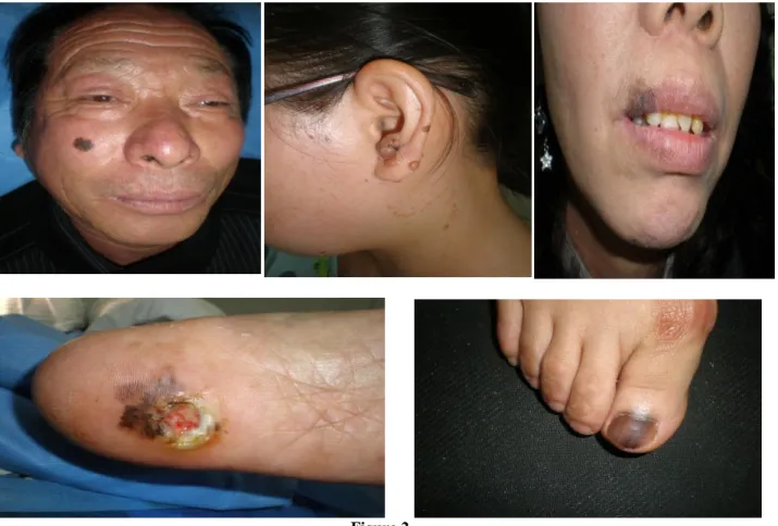
Gender and age
Figure 2
Table 5 Univariate analysis based on the model of Cox proportional hazard
Characteristics Mortality (%) P-Value Hazard Ratio
Gender
Male 3/13 (25%) 0.133 1
Female 4/26 (15.38%) 0.506
Age Group (in years)
≤ 50 2/10 (16.7%) 0.789 1
50-69 3/18 (18.3%) 0.797 0.945
≤ 69 2/11 (20.2%) 0.833 1.212
Location
Head and Neck 1/2 (50%) 0.259 1
Upper extremities 1/5 (9.7%) 0.323 0.356
Lower extremities 4/24 (15.7%) 0.100 0.387
Trunk 2/6 (30.6%) 0.513 0.599
Clark Level
I – III 3/27 (9%) 0.029* 1
IV –V 3/12 (26.95%) 3.561
Histopathological subtypes
Acral Lentiginous Melanoma 2/12 (17.4%) 0.780 1
LentigoMaligna Melanoma 0/8 (0%) 0.986 0.000
Nodular Melanoma 4/12 (29.9%) 0.223 1.999
Superficial Spreading Melanoma 2/7 (23.9%) 0.891 1.045
Location of primary tumors
According to the Kalpan - Merier analysis, we
Invasion level
The Clark IV-V death rate was about three times
that of a lower invasion rate, which was
statistically significant (P=0.029). The mortality
rate was three times higher. We were reported
similar survival differences based on the level of
invasion according to Kalpan-Merier analysis.
Tumor thickness
There was no big difference between the studies
< 2 mm thickness and > 2 mm tumor survival
rate (P=0.063). According to the analysis from
Kalpan-Merier, no significance was discovered
between the survival rate and the thickness of
the tumor.
Histopathological subtypes
Based
on
the
clinical
histopathological
classification, we found no distinction in the
survival rates, according to Kalpan- Merier
analysis.
Discussion
Studies on Melanoma were performed in the
Southern European regions such as Italy,
8,10,13,15some melanoma research studies were also
performed in Spain
11,12and have all been
published in Spanish Literature.
12Malignant
Melanoma is a tumor that arise from the
epithelium. (YU and LIU) Malignant melanoma
usually involves the skin, but not frequently the
mucosa. Rural communities also have high
re-occurrence and metastasis (YU and LIU) and
poor prognosis. However, cases of the malignant
melanoma have increased nowadays, but still not
sufficient to conclude about all the aspects of
primary and recurrent Malignant Melanoma. For
this reason, we took 39 patients (13 males and
26 females) of malignant melanoma of all
different stages. The present study shows the sex
ratio of the sample study population (1:2;
male-to-female) which is unlike the one observed in
Korean reports which were similarly designed.
11-13
Our results, however, are similar to the study
conducted (chi et al) as sex ratio 1:1.8
(male-to-female) The mean age of our sample study
population (39 cases) was 54.25 years, with a
mean age of 55 years and a range (17-79 years)
similar to the study given by Chi et.al. Also, it
was found approximately the same as the study
performed in South Korea having a mean age of
50s.
8,11,12Present study shows that most common location
of primary melanoma was in lower extremes
(61.52%), including finger (12.82%), heel
(12.82%), valva (2.56%), waist (2.56%) and
other extremities (30.76%), followed by upper
extremities (12.85%), including thumb (10.26%)
and palm (2.56%). (5.13%) cases had a tumor at
head and neck, (15.38%) in the trunk which was
near about the same to the study of South
Korea.
8,11,12In
the
current
study,
the
histopathological
subtypes
of
malignant
melanoma showed the largest (30.76%) cases of
acral lentiginous melanoma as well as nodular
melanoma followed by lentigo malignant
melanoma (20.51%), superficial spreading
melanoma (17.95%), which differs from the
results of Won et al
14, Lee et al
11and Chun et
al.
13In our study, 10 out of 39 cases (25.64%) had
tumor thickness in situ melanoma. The highest
no. of patients (35.90%) had tumor thickness 1-2
mm followed by (20.51%) cases had 1-2 mm,
(17.95%) cases had <1 mm however not a single
patient had tumor thickness >4 mm in our study
which differs with the study observed by
(Kyuangwook Nam et. al.) according to this,
other study.
and stages (
P
=0.389) and location and stage
(
P
=0.399) in relation to demographic and
epidemic properties. There was however a major
link between histopathology and staging
(
P
=0.03). Studies have also demonstrated that
nodular melanoma is seen more frequently than
acral lentiginous melanoma (
P
=0.002) and
superficial spreading melanoma (
P
=0.004).
Approximately the same results were observed
in the study (Kuang Wook Nam et. al.) In this
study, the Clark IV-V-level death rate was about
three times as high as the low invasive tumor
which was statistically significant (
P
=0,029).
According to the Kalpan-Merier analysis, we
demonstrated a significant difference in the
survival rate depending on the level of invasion
that the KuangWook Nam et al study assumes.
Conclusion
Trunk was the most common tumor location
followed by lower extremity. The most frequent
subtypes were nodular melanoma and Acral
melanoma. Females are more commonly
affected than males 2:1. A relationship was
found between stages and Clark level, especially
stage III-IV tumors with IV-V Clark level, and
correlated with high mortality rates.
Limitation
However, due to the small number of cases
future clinical studies with large sample sizes,
randomization,
grouping,
and
long-term
monitoring periods are required in order to
conclude these findings more precisely.
Conflict of interest
We have no conflict of interest to disclose that
the process and results of this study are not
affected by the relevant equipment, materials,
and pharmaceutical companies.
References
1. Norton SA. Betel: consumption and consequences. J Am Acad Dermatol 1998; 38(1): 8188.
2. Tilakaratne WM, Klinikowski MF, Saku T, Peters TJ, Warnakulasuriya S. Oral submucous fibrosis: a review on etiology and pathogenesis. Oral Oncol 2006; 42(6): 561-568.
3. Prasad ML, Busam KJ, Patel SG, Hoshaw Woodard S, Shah JP and Huvos AG: Clinicopathologic differences in malignant melanoma arising in oral squamous and sinonasal respiratory mucosa of the upper aerodigestive tract. Arch Pathol Lab Med 127: 997 1002, 2003.
4. Aldrink JH, Selim MA, Diesen DL, Johnson J, Pruitt SK, Tyler DS, et al. Pediatric melanoma: a single-institution experience of 150 patients. J Pediatr Surg 2009; 44: 1514– 1521.
5. Moore-Olufemi S, Herzog C, Warneke C, Gershenwald JE, Mansfield P, Ross M et al. Outcomes in pediatric melanoma: comparing prepubertal to adolescent pediatric patients. Ann Surg 2011; 253: 1211–1215.
6. Hamre MR, Chuba P, Bakhshi S, Thomas R, Severson RK. Cutaneous melanoma in childhood and adolescence. Pediatr Hematol Oncol 2002; 19: 309–317.
7. Strouse JJ, Fears TR, Tucker MA, Wayne AS. Pediatric melanoma: risk factor and survival analysis of the surveillance, epidemiology and end results database. J Clin Oncol 2005; 23: 4735–4741.
8. Bauer J, Buttner P, Murali R, et al. BRAF mutations in cutaneous melanoma are independently associated with age, anatomic site of the primary tumor and the degree of solar elastosis at the primary tumor site, Pigment Cell Melanoma Res 2011; 24:345-351.
9. Sekulic A, Haluska P Jr, Miller AJ, et al. Malign melanoma in the 21st century: the emerging molecular landscape, Mayo Clin Proc. 2008; 83(7): 825-46.
10. Reintgen DS, McCarty KM Jr, Cox E, et al. Malignant melanoma in black American and white American populations: a comparative review. JAMA 1982; 248:1856-9.
Japan. J Invest Dermatol 1989; 92: 210S-213S.
12. Collins RJ. Melanoma in the Chinese of Hong Kong: emphasis on volar and subungual sites. Cancer 1984; 54:1482-8. 13. Choi SJ, Bae YC, Moon JS, et al. An
analysis of the clinical and histopathological pattern of malignant melanoma. J Korean Soc Plast Reconstr Surg 2007; 34: 557-61. 14. Ackerman AB. What naevus are dysplastic,
a syndrome and the commonest precursor of malignant melanoma? A riddle and an answer. Histopathology. 1988; 13: 241-56. PMID:3056824
15. Togawa Y, Nakamura Y, Kamada N, Kambe N, Takahashi Y, Matsue H. Melanoma in association with an acquired melanocytic nevus in Japan: a review of cases in the literature. Int J Dermatol 2010; 49: 1362. PMID: 21155082
16. Sheen YS, Liao YH, Lin MH, Chiu HC, Jee SH, Liau JY, et al. Insulin-like growth factor II mRNA-binding protein 3 expression
correlates with poor prognosis in acrallentiginous melanoma. PLoS One. 2016; 11: e0147431.
17. Al-Jamal MS, Griffith JL, Lim HW. Photoprotection in ethnic skin. Dermatologica Sinica 2014; 32:217-24. 18. MacKie R, Hunter JA, Aitchison TC, Hole
D, McLaren K, Rankin R, et al. Cutaneous malignant melanoma, Scotland, 1979–89. The Scottish Melanoma Group. Lancet 1992; 339: 971–975.
19. Breslow A. Thickness, cross-sectional areas and depth of invasion in the prognosis of cutaneous melanoma. Ann Surg 1970; 172: 902–908.