Article
Inter-Molecular Electrostatic Interactions Stabilizing
the Structure of the PD-1/PD-L1 Axis: A Structural
Evolutionary Perspective
Wei Li1 *
1
2
3
4
5
6
7
8
1 InstituteofSpecialEnvironmentMedicine,NantongUniversity,No.9,SeyuanRoad,NantongCity,Jiangsu
Province,P.R.China;[email protected]
* Correspondence:[email protected]
Abstract: PD-1/PD-L1 axis is one key therapeutic target against tumor cell immune escape.
Structurally essential to the PD-1/PD-L1-linked immune escape is the binding interface of the
PD-1/PD-L1 complex structure. Incorporating currently available PD-1/PD-L1-related experimental
structures, this article unveils two sets of experimentally observed inter-molecular electrostatic
interactions which stabilize the binding interface of the PD-1/PD-L1 complex structure. For the first
time, this article proposes an evolutionary structural hypothesis that, as a result of natural selection,
PD-1 is able to genetically mutate itself to structurally disrupt the PD-1/PD-L1 axis towards the
restoration of T cell-mediated anti-tumor immunity.
Keywords: PD-1; PD-L1; Cancer-linked genetic mutation; Electrostatic repulsion; Structural evolution 9
1. Introduction 10
The PD-1/PD-L1 axis is a pivotal component of tumor cell immune escape mechanism, where 11
the engagement of PD-L1 (on tumor cells) to PD-1 (on T cells) exhausts T cell-mediated anti-tumor 12
immunity [1,2]. To counteract PD-1/PD-L1-linked tumor cell immune escape, one option is the
13
dissociation of the two cell membrane-bound immune checkpoints, i.e., the disruption of the structural 14
bond along the PD-1/PD-L1 axis, which is to be broken at the interface of tumor and T cells, such that 15
the immune system restores its capacity to recognize and kill tumor cells [3–5]. For example, to restore
16
T cell-mediated anti-tumor immunity, targeting the PD-1/PD-L1 axis using checkpoint inhibitors 17
(antibodies against PD-1 or PD-L1, or soluble PD-1 molecules with high affinity to PD-L1) allows 18
tumor cells to be detected and attacked by T cells, which has emerged as a potentially feasible strategy 19
for the treatment of tumors [6–10]. Nevertheless, clinical responses are mixed, highlighting the need
20
for further investigation into the structural aspects of the PD-1-PD-L1 axis in tumor cell immune escape 21
[11–14]. 22
Therefore, this article incorporates currently available PD-1/PD-L1-related experimental 23
structures (as of December 24, 2019) [15] and experimentally observed cancer-linked genetic mutation
24
of the two immune checkpoints, and puts forward a set of in silico electrostatic analysis of all 25
PD-1-PD-L1 complex structures (as of December 24, 2019), aiming at deciphering and visualizing 26
the inter-molecular electrostatic interactions that are instrumental in the structural stability of the 27
PD-1/PD-L1 complex. 28
2. Materials and Methods 29
2.1. Experimentally determined PD-1-PD-L1 structures 30
As of December 24, 2019, a total of 52 PD-1- and/or PD-L1-related structures (Supplementary file 31
supp.pdf) have been experimentally determined and deposited in PDB [15]. 32
PDB ID Chain ID Protein Name (Synonym)
3BIK A PD-L1 (CD274)
3BIK B PD-1 (CD279)
3BIK C PD-1 (CD279)
3SBW A PD-1 (CD279)
3SBW B PD-1 (CD279)
3SBW C PD-L1 (CD274)
4ZQK A PD-L1 (CD274)
4ZQK B PD-1 (CD279)
5IUS A PD-1 (CD279)
5IUS B PD-1 (CD279)
5IUS C PD-L1 (CD274)
5IUS D PD-L1 (CD274)
Table 1.Experimental structures of the PD-1-PD-L1 complex in PDB as of December 24, 2019, along with their respective PDB IDs, chain IDs and (alternative) protein names.
Among the 52 PD-1- and/or PD-L1-related structures, four represent the PD-1-PD-L1 complex 33
structure (Table1), which are to be analysed in detail and discussed further below.
34
2.2. Structural analysis of the PD-1-PD-L1 complex 35
First, the InterProSurf server (http://curie.utmb.edu/pdbcomplex.html) [16] was used to identify
36
interfacial amino acid residues of the PD-1-PD-L1 complex structures (Table1). Subsequent structural
37
analysis, including salt bridging and hydrogen bonding analysis [17], was performed as described
38
previously for all 52 experimentally determined PD-1-PD-L1 complex structures as of December 24, 39
2019. 40
3. Results 41
To investigate the structural stability of the binding interface of the PD-1/PD-L1 complex structure, 42
it is necessary to first chart out the entire interfacial salt bridging and hydrogen bonding networks of 43
the two immune checkpoints. Thus, an set of residue-specific structural analysis was carried out at the 44
binding interface of the PD-1-PD-L1 complex structure. Specifically, an amino acid residue is taken 45
into further consideration of subsequent structural analysis only when it fulfills no less than three of 46
the four criteria below, 47
1. it is associated with at least one cancer-linked mutation (BioMuta). 48
2. it is located at the interface of the PD-1-PD-L1 complex (InterProSurf). 49
3. it is involved in the salt bridging network [17] at the PD-1-PD-L1 binding interface.
50
4. it is involved in the hydrogen bonding network [17] at the PD-1-PD-L1 binding interface.
51
3.1. The cervical cancer-linked Glu136Gln mutation of PD-1 52
According to the InterProSurf interfacial residue analysis [16], Glu136 of PD-1 sits at the interface
53
of the four PD-1-PD-L1 complex structures (Table1). Further structural analysis reveals that Glu136
54
and Ser137 of PD-1 forms an intra-molecular hydrogen bond within PD-1 (PDB ID: 2M2D, Table2).
55
Moreover, Glu136 and Arg139 of PD-1 forms a stable intra-molecular salt bridge within PD-1 (PDB 56
IDs: 2M2D and 5GGR, supplementary fileGlu136.pdf).
PDB Acceptor (A) Donor (D) Hydrogen (H) D-A (Å) H-A (Å) ∠ADH(°)
2M2D_25 O, A_GLU_136 OG, A_SER_137 HG, A_SER_137 2.75 1.79 9.71
Table 2. The hydrogen bond formed between Glu136 and Ser 137 of PD-1. In this table, 2M2D_25 represents the 25thstructural model in the NMR ensemble (PDB ID: 2M2D), the residue naming scheme isChain ID_residue name_residue number,∠ADHrepresents the angle formed by acceptor (A), donor (D) and hydrogen (H)(∠ADH).
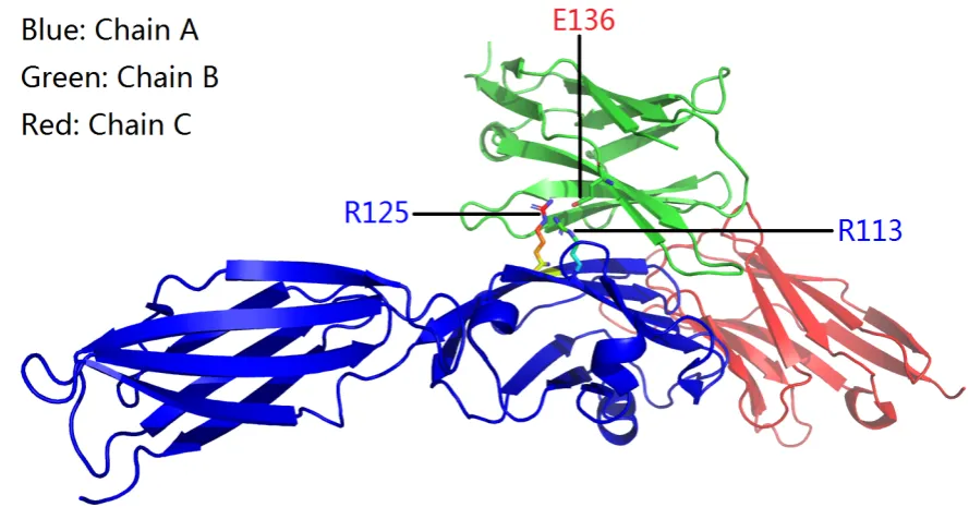
Figure 1.An overview of the structurally identified side chain salt bridges between an amino acid trio, i.e., Glu136 of PD-1 and Arg113 and Arg125 of PD-L1 for PDB ID 3BIK (Table1).
Interestingly, for the two PD-1-PD-L1 complex structures (3BIK and 4ZQK, Table1), Glu136 of
58
PD-1 forms salt bridges (Figures1and2) with Arg113 and Arg125 of PD-L1, which both sit at the
59
structural interface of the two immune checkpoints (PDB IDs: 3BIK, 4ZQK and 5IUS, and 3SBW, 60
Table1), although no cancer-linked mutation was clinically identified yet for Arg113 and Arg125 of
61
PD-L1 as of December 24, 2019. 62
SB1 Color =Green, Distance = 3.149 Å
Atomic coordinate ATOM 777 NH2 ARG A 113 12.637 -2.086 -32.968 1.00 70.85 N Atomic coordinate ATOM 2538 OE2 GLU B 136 11.442 -1.132 -30.215 1.00 72.18 O
SB2 Color =Blue, Distance= 3.033 Å
Atomic coordinate ATOM 870 NH2 ARG A 125 14.394 -1.252 -29.687 1.00 69.69 N Atomic coordinate ATOM 2537 OE1 GLU B 136 11.832 -1.667 -28.118 1.00 63.00 O
SB3 Color =Red, Distance = 3.001 Å
Atomic coordinate ATOM 870 NH2 ARG A 125 14.394 -1.252 -29.687 1.00 69.69 N Atomic coordinate ATOM 2538 OE2 GLU B 136 11.442 -1.132 -30.215 1.00 72.18 O
Table 3.The structurally identified inter-molecular side chain salt bridges between Glu136 of PD-1 and Arg113 and Arg125 of PD-L1 for PDB ID 3BIK (Table1). In this table, Color =Green, Color =Blueand Color =Redrepresent the three interfacial salt bridges as shown in Figure2with the corresponding colors, respectively.
Given that Glu136 of PD-1 is associated with the cervical cancer-linked Glu136Gln (E136Q) 63
Figure 2.The structurally identified inter-molecular side chain salt bridges between Glu136 of PD-1 and Arg113 and Arg125 of PD-L1 for PDB ID 3BIK). In this figure, the side chain salt bridges are shown as dotted short cylinders with different colors, the details of the salt bridges are included in Table3.
and installs the neutral side chain group of Gln136, along with the hydrogen with partial positive 65
charge in the side chain NH2group of Gln136. Similar to the spinal muscular atrophy-linked binding
66
interface-disrupting Gln136Glu (Q136E) mutation [17], the E136Q substitution here gives rise to a
67
reversal in the charge carried by the side chain of Glu136, shifting the local electrostatic attraction (the 68
side chain salt bridges and hydrogen bond of Gln136 of PD-1, Figures1and2) to local electrostatic
69
repulsion [18,19] between Gln136’s side chain (with partial positive charge) and the positively charged
70
side chains of Arg113 and Arg125. 71
Figure 3.To facilitate tumor cell immune escape, PD-L1 binds to PD-1 as a cat catches hold of a mouse, towards the exhaustion of T cell-mediated anti-tumor immunity [1,2,4,5]. Reproduced with permission from Everett Collection (https://www.everettcollection.com).
Overall, the net structural and functional consequence of this E136Q substitution is the local 72
electrostatic equilibrium perturbation [17] at the binding interface of the PD-1-PD-L1 complex, and
73
as shown in Figure1, it is likely that this cervical cancer-linked E136Q substitution disrupts the
binding interface of PD-1 and PD-L1, thereby allowing PD-1 to distance itself away from PD-L1, which 75
suppresses the PD-1/PD-L1 axis towards the restoration of T cell-mediated anti-tumor immunity. 76
This structural evolution hypothesis is likened to a situation where PD-L1 (the cat, Figure3) uses
77
its claws (Arg125 of PD-L1) to catch hold of PD-1 (the mouse, Figure3) via the Glu136 residue of
78
PD-1 (Figure2) to facilitate tumor cell immune escape, highlighting the functional importance of the
79
interacting interfacial residues of the PD-1-PD-L1 complex structure [20] On the other hand, however,
80
PD-1 is able to hide itself (via a Glu136Gln substitution) and escape from the recognition (the claw) and 81
binding of PD-L1 (the cat, Figure3), towards the restoration of T cell-mediated anti-tumor immunity
82
[1,2,4,5]. 83
3.2. The breast cancer-linked Met70Lys mutation of PD-1 84
As discussed above, Arg125 of PD-L1 is like one claw of the cat (PD-L1, Figure3), allowing
85
PD-L1 to bind to Glu136 of PD-1 (the mouse, Figures3,1and2) towards the establishment of the
86
PD-1-PD-L1 axis. After a set of comprehensive structural analysis for Arg125 of PD-L1 (supplementary 87
fileArg125.pdf), it turned out that Arg125 allowed PD-L1 to bind to Glu70 of a PD-1 mutant (PDB ID: 88
5IUS, Table1), too.
89
Here, PDB ID 5IUS corresponds to an crystal structure of human PD-L1 in complex with high 90
affinity PD-1 mutant (supplementary filesupp.pdf). Quite intriguingly, the 70thresidue of wild-type
91
PD-1 is methionine (Met, M), which is associated with the breast cancer-linked M70K mutation of PD-1. 92
Therefore, the M70E substitution (PDB ID: 5IUS, Table1) contributes to the local structural stability
93
and thus high affinity of the PD-1 mutant to PD-L1 as a total of five inter-molecular interfacial salt 94
bridges formed between Glu70 (of PD-1) and Arg125 (of PD-L1) at the binding interface of the two 95
immune checkpoints, as listed in the supplementary fileArg125.pdf.
96
In contrast, however, the breast cancer-linked M70K mutation of PD-1 does exactly the opposite 97
to the structural and functional consequence of the human-introduced M70E mutation, in the sense 98
that the M70K mutation establishes a local electric charge reversal for the side chain of the 70thresidue
99
of PD-1 and disrupts the inter-molecular salt bridges at the binding interface of the two immune 100
checkpoints, where the net structural and functional consequence is the local electrostatic energy 101
equilibrium perturbation (if not disruption) [17] at the binding interface of the PD-1-PD-L1 complex.
102
Overall, the human-introduced M70E and the breast cancer-linked M70K substitutions are like the 103
two sides of one coin, and serve as two excellent examples of how human intervention does exactly the 104
opposite to the effect of genetic mutation-driven natural selection in the structural evolution of PD-1, 105
where its M70K substitution removes the hydrophobic side chain of its Met70 and installs instead a 106
positively charged side chain of at its 70thresidue site (Lys70), and thereby establishes a structurally
107
destabilizing electrostatic repulsive force [17–19] between Lys70 (of PD-1) and Arg125 (of PD-L1) at
108
their binding interface. On the other side of the coin, while the M70E substitution of PD-1 removes 109
the hydrophobic side chain of its Met70 and installs instead a negatively charged side chain at its 70th
110
residue site (Glu70), and thereby establishes a structurally stabilizing electrostatic attractive force, i.e., 111
five inter-molecular interfacial salt bridges between Glu70 (of PD-1) and Arg125 (of PD-L1) to further 112
consolidate the PD-1-PD-L1 axis and consequently the tumor cell immune escape mechanism, too. 113
4. Conclusion 114
1. This article highlights an amino acid residue trio (Glu136 of PD1 and Arg125 and Arg113 of 115
PD-L1, Figures 1), which are inextricably linked to inter-molecular electrostatic interactions
116
towards the structural stabilization of the PD-1-PD-L1 axis in tumor cell immune escape. 117
2. This article highlights two cancer-linked genetic mutations of PD-1, i.e., the cervical cancer-linked 118
Glu136Gln(E136Q) mutation of PD-1 and the breast cancer-linked Met70Lys (M70K) mutation of 119
PD-1, which are at play in the inter-molecular electrostatic interactions towards the structural 120
3. With the two experimentally observed cancer-linked genetic mutations (E136Q and M70K) of 122
PD-1, this article puts forward an evolutionary structural hypothesis that, from a structural point 123
of view, PD-1 is able to genetically mutate itself (Figures3) to structurally disrupt the PD-1/PD-L1
124
axis towards the restoration of T cell-mediated anti-tumor immunity. Specifically, genetic 125
variation-induced electrostatic repulsion [17,18] is involved in the evolutionary suppression
126
of the tumor cell immune escape mechanism, where the electric charge reversal constitutes 127
a functional significant unfavourable electrostatic energy contribution towards the structural 128
perturbation (if not disruption) of the PD-1-PD-L1 complex structure [1,3,4].
129
4. With this evolutionary structural hypothesis, this article presents further discuss Tyr68 of PD-1 130
and Asp122 of PD-L1 at the structural interface of the two immune checkpoints, which is to be 131
described in detail below. 132
5. Discussion 133
5.1. Tyr68 of PD-1 and Asp122 of PD-L1: a structural evolutionary perspective 134
As of December 24, 2019, no cancer-linked mutation was clinically identified yet either for Tyr68 of 135
PD-1 or for Asp122 of PD-L1. Nonetheless, both Tyr68 of PD-1 and Asp122 of PD-L1 sit at the binding 136
interface of PD-1 and PD-L1, according to the three experimental structures of PD-1-PD-L1 complex 137
(PDB IDs: 3BIK, 4ZQK and 5IUS, Table1). In PDB ID 5IUS (Table1), a Tyr68His (Y68H) substitution
138
was introduced to PD-1 to replace the hydrophobic side chain (of Tyr68) with one positively charged 139
side chain of His68. After the Y68H substitution, three inter-molecular side chain salt bridges (Figures4
140
and5) were formed between His68 of the mutant PD-1 and Asp122 of PD-L1.
141
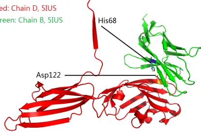
Figure 4.An overview of two salt-bridged amino acid residues at the binding interface of PD-1 (green) and PD-L1 (red) according to their complex structure (PDB ID: 5IUS, Table1).
From Figure4, it can be seen that His68 (of mutant PD-1) and Asp122 (of PD-L1) constitute two
142
structurally stabilizing electrostatic clips [17] at the binding interface of the two immune checkpoints.
143
Thus, the Y68H substitution contributes to the local structural stability and consequently high affinity 144
Asp122.pdf) were formed between the side chains (Figures4and5) of His70 (of PD-1) and Asp122 (of 146
PD-L1). 147
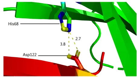
Figure 5.The structurally identified inter-molecular side chain salt bridges between His68 of PD-1 (mutant) and Asp122 of PD-L1 for PDB ID 5IUS). In this figure, the side chain salt bridges are shown as dotted yellow short cylinders, 3.8 represents the distance (3.8 Å) of the salt bridge formed between Nε2 (blue) of His68 of PD-1 (mutant) and Oδ2(yellow) of Asp122 of PD-L1, 2.7 represents the distance (2.7 Å) of the salt bridge formed between Nε2(blue) of His68 of PD-1 (mutant) and Oδ1(yellow) of Asp122 of PD-L1 (supplementary fileAsp122.pdf).
Thus, it is reasonable to not rule out the possibility that a Tyr68Asp (or Tyr68Glu) substitution 148
will take place in future in the scaffold of PD-1 and is clinically identified and linked to cancer 149
patient(s). This prediction stems from the same structural evolutionary hypothesis, where a Tyr68Asp 150
(or Tyr68Glu) substitution of PD-1 is able to establish an electrostatic repulsive force between Asp122 of 151
PD-L1, in addition to removing the three structurally stabilizing inter-molecular side chain salt bridges 152
(Figures4and5) formed between His68 of the mutant PD-1 and Asp122 of PD-L1. On the contrary,
153
when a Tyr68Asp (or Tyr68Glu) substitution of PD-1 does take place in future, then this structural 154
evolutionary hypothesis predicts again that an Asp122Arg (or Asp122Lys) substitution of PD-L1 155
will take place, too, towards the restoration of structurally stabilizing inter-molecular electrostatic 156
interactions between the two immune checkpoints. Similarly, this situation is likened to that in Figure3,
157
where the cat (PD-L1 of tumor cell) uses its claw to catch hold of the mouse, i.e., PD-1 of T cell, towards 158
the exhaustion of T cell-mediated anti-tumor immunity [1,2,4,5].
159
5.2. Application of gene-editing technologies to structurally disrupt the PD-1-PD-L1 axis 160
As discussed above, the PD-1-PD-L1 axis is but one of the many tumor immune escape 161
mechanisms, which makes tumor a difficult foe for the immune system, and explains at least partly 162
why PD-1/PD-L1 antibody-based clinical responses are mixed [11–13], highlighting the need to tackle
163
tumors from multiple angles, such that there is less chance for it to acquire resistance and avoid 164
elimination by the immune system. Similarly, if a tumor is targeted and treated precisely, there is less 165
chance for adverse effects to take place. Thus, against the PD-1/PD-L1-linked tumor cell immune 166
escape, this structural evolutionary mechanism provides a novel perspective for the application of 167
gene-editing [8,21–23] technologies in the treatment of tumors, since the engagement of PD-L1 (or even
168
PD-L2, supplementary filesupp.pdf) (on tumor cells) to PD-1 (on T cells) is central to the PD-1/PD-L1
axis, which causes T cell exhaustion, a hyporesponsive state of T cells in tumor microenvironment, 170
with increased inhibitory receptors, decreased effector cytokines and impaired cytotoxicity [24].
171
For instance, since Arg125 and Arg133 are like two claws of the cat (Figure3), site-specific
172
genetic mutations such as Arg125Glu (R125E) and Arg113 (R113E) for PD-L1 are able to establish two 173
structurally destabilizing electrostatic repulsive forces between them and Glu136 of PD-1, because 174
not only are the two claws removed to prevent PD-L1 from binding to PD-1 (Figure3), but also is the
175
cat (PD-L1) energetically reluctant to catch hold of the mouse (PD-1, Figure3) due to the electrostatic
176
repulsions between its Glu136 and Glu113 and Glu125 of PD-L1, and also are the approaching PD-1 177
molecules trying to stay further away from PD-L1 due to the electrostatic repulsions between Glu136 178
of PD-1 and Glu113 and Glu125 of PD-L1, too. 179
Author Contributions:Cconceptualization, W.L.; methodology, W.L.; software, W.L.; validation, W.L.; formal 180
analysis, W.L.; investigation, W.L.; resources, W.L.; data curation, W.L.; writing–original draft preparation, W.L.; 181
writing–review and editing, W.L.; visualization, W.L.; supervision, W.L.; project administration, W.L.; funding 182
acquisition, not applicable. 183
Funding:This research received no external funding. 184
Acknowledgments: I thank Dr. Yongdong Niu (Department of Pharmacology, Shantou University Medical 185
College) for his insights on this topic. 186
Conflicts of Interest:The author declares no conflict of interest. 187
188
1. Beatty, G.L.; Gladney, W.L. Immune Escape Mechanisms as a Guide for Cancer Immunotherapy. Clinical 189
Cancer Research2014,21, 687–692. doi:10.1158/1078-0432.ccr-14-1860. 190
2. Salmaninejad, A.; Valilou, S.F.; Shabgah, A.G.; Aslani, S.; Alimardani, M.; Pasdar, A.; Sahebkar, A. 191
PD-1/PD-L1 pathway: Basic biology and role in cancer immunotherapy. Journal of Cellular Physiology2019, 192
234, 16824–16837. doi:10.1002/jcp.28358. 193
3. Chen, L.; Han, X. Anti–PD-1/PD-L1 therapy of human cancer: past, present, and future. Journal of Clinical 194
Investigation2015,125, 3384–3391. doi:10.1172/jci80011. 195
4. Jiang, X.; Wang, J.; Deng, X.; Xiong, F.; Ge, J.; Xiang, B.; Wu, X.; Ma, J.; Zhou, M.; Li, X.; Li, Y.; Li, G.; Xiong, 196
W.; Guo, C.; Zeng, Z. Role of the tumor microenvironment in PD-L1/PD-1-mediated tumor immune 197
escape.Molecular Cancer2019,18. doi:10.1186/s12943-018-0928-4. 198
5. Constantinidou, A.; Alifieris, C.; Trafalis, D.T. Targeting Programmed Cell Death -1 (PD-1) and Ligand 199
(PD-L1): A new era in cancer active immunotherapy. Pharmacology & Therapeutics2019,194, 84–106. 200
doi:10.1016/j.pharmthera.2018.09.008. 201
6. Fusi, A.; Festino, L.; Botti, G.; Masucci, G.; Melero, I.; Lorigan, P.; Ascierto, P.A. PD-L1 202
expression as a potential predictive biomarker. The Lancet Oncology 2015, 16, 1285–1287. 203
doi:10.1016/s1470-2045(15)00307-1. 204
7. Cyranoski, D. CRISPR gene-editing tested in a person for the first time. Nature 2016,539, 479–479. 205
doi:10.1038/nature.2016.20988. 206
8. Zhan, T.; Rindtorff, N.; Betge, J.; Ebert, M.P.; Boutros, M. CRISPR/Cas9 for cancer research and therapy. 207
Seminars in Cancer Biology2018. doi:10.1016/j.semcancer.2018.04.001. 208
9. Liu, Y.; Wu, L.; Tong, R.; Yang, F.; Yin, L.; Li, M.; You, L.; Xue, J.; Lu, Y. PD-1/PD-L1 Inhibitors in Cervical 209
Cancer. Frontiers in Pharmacology2019,10. doi:10.3389/fphar.2019.00065. 210
10. Li, Y.; Liang, Z.; Tian, Y.; Cai, W.; Weng, Z.; Chen, L.; Zhang, H.; Bao, Y.; Zheng, H.; Zeng, S.; Bei, C.; Li, Y. 211
High-affinity PD-1 molecules deliver improved interaction with PD-L1 and PD-L2. Cancer Science2018, 212
109, 2435–2445. doi:10.1111/cas.13666. 213
11. Pauken, K.E.; Wherry, E.J. Overcoming T cell exhaustion in infection and cancer. Trends in Immunology 214
2015,36, 265–276. doi:10.1016/j.it.2015.02.008. 215
12. Liang, Z.; Tian, Y.; Cai, W.; Weng, Z.; Li, Y.; Zhang, H.; Bao, Y.; Li, Y. High-affinity human PD-L1 variants 216
attenuate the suppression of T cell activation.Oncotarget2017,8. doi:10.18632/oncotarget.21729. 217
13. Pawelec, G. Is There a Positive Side to T Cell Exhaustion? Frontiers in Immunology 2019, 10. 218
14. Wang, Y.; Zhou, S.; Yang, F.; Qi, X.; Wang, X.; Guan, X.; Shen, C.; Duma, N.; Aguilera, J.V.; Chintakuntlawar, 220
A.; Price, K.A.; Molina, J.R.; Pagliaro, L.C.; Halfdanarson, T.R.; Grothey, A.; Markovic, S.N.; Nowakowski, 221
G.S.; Ansell, S.M.; Wang, M.L. Treatment-Related Adverse Events of PD-1 and PD-L1 Inhibitors in Clinical 222
Trials.JAMA Oncology2019,5, 1008. doi:10.1001/jamaoncol.2019.0393. 223
15. Berman, H.; Henrick, K.; Nakamura, H. Announcing the worldwide Protein Data Bank. Nature Structural 224
& Molecular Biology2003,10, 980–980. doi:10.1038/nsb1203-980. 225
16. Negi, S.S.; Schein, C.H.; Oezguen, N.; Power, T.D.; Braun, W. InterProSurf: a web 226
server for predicting interacting sites on protein surfaces. Bioinformatics 2007, 23, 3397–3399. 227
doi:10.1093/bioinformatics/btm474. 228
17. Li, W. How do SMA-linked mutations ofSMN1lead to structural/functional deficiency of the SMA 229
protein?PLOS ONE2017,12, e0178519. doi:10.1371/journal.pone.0178519. 230
18. Li, W.; Shi, G. How CaV1.2-bound verapamil blocks Ca2+influx into cardiomyocyte: Atomic level views. 231
Pharmacological Research2019,139, 153–157. doi:10.1016/j.phrs.2018.11.017. 232
19. Li, W. Characterising the interaction between caenopore-5 and model membranes by NMR spectroscopy 233
and molecular dynamics simulations. PhD thesis, University of Auckland, 2016. 234
20. Magnez, R.; Thiroux, B.; Taront, S.; Segaoula, Z.; Quesnel, B.; Thuru, X. PD-1/PD-L1 binding studies using 235
microscale thermophoresis. Scientific Reports2017,7. doi:10.1038/s41598-017-17963-1. 236
21. Tsai, S.Q.; Nguyen, N.T.; Malagon-Lopez, J.; Topkar, V.V.; Aryee, M.J.; Joung, J.K. CIRCLE-seq: a 237
highly sensitive in vitro screen for genome-wide CRISPR–Cas9 nuclease off-targets. Nature Methods 238
2017,14, 607–614. doi:10.1038/nmeth.4278. 239
22. Zuo, E.; Sun, Y.; Wei, W.; Yuan, T.; Ying, W.; Sun, H.; Yuan, L.; Steinmetz, L.M.; Li, Y.; Yang, H. Cytosine 240
base editor generates substantial off-target single-nucleotide variants in mouse embryos. Science2019, p. 241
eaav9973. doi:10.1126/science.aav9973. 242
23. Yang, J.; Hu, L. Immunomodulators targeting the PD-1/PD-L1 protein-protein interaction: From antibodies 243
to small molecules.Medicinal Research Reviews2018,39, 265–301. doi:10.1002/med.21530. 244
24. Jiang, Y.; Li, Y.; Zhu, B. T-cell exhaustion in the tumor microenvironment. Cell Death & Disease2015, 245
![Figure 3. To facilitate tumor cell immune escape, PD-L1 binds to PD-1 as a cat catches hold of a mouse,towards the exhaustion of T cell-mediated anti-tumor immunity [1,2,4,5]](https://thumb-us.123doks.com/thumbv2/123dok_us/7997042.1327996/4.595.74.521.73.330/figure-facilitate-immune-escape-catches-exhaustion-mediated-immunity.webp)