Scholarship@Western
Scholarship@Western
Electronic Thesis and Dissertation Repository
4-3-2012 12:00 AM
Optically Driven PH Gradient Generator Based on Self-Assembled
Optically Driven PH Gradient Generator Based on Self-Assembled
Proton Pumps for Activating Hydrogel Microactuators
Proton Pumps for Activating Hydrogel Microactuators
Khaled M. Al-Aribe
The University of Western Ontario
Supervisor
Professor George Knopf
The University of Western Ontario
Graduate Program in Mechanical and Materials Engineering
A thesis submitted in partial fulfillment of the requirements for the degree in Doctor of Philosophy
© Khaled M. Al-Aribe 2012
Follow this and additional works at: https://ir.lib.uwo.ca/etd
Part of the Mechanical Engineering Commons
Recommended Citation Recommended Citation
Al-Aribe, Khaled M., "Optically Driven PH Gradient Generator Based on Self-Assembled Proton Pumps for Activating Hydrogel Microactuators" (2012). Electronic Thesis and Dissertation Repository. 415.
https://ir.lib.uwo.ca/etd/415
This Dissertation/Thesis is brought to you for free and open access by Scholarship@Western. It has been accepted for inclusion in Electronic Thesis and Dissertation Repository by an authorized administrator of
ASSEMBLED PROTON PUMPS FOR ACTIVATING HYDROGEL MICROACTUATORS
(Spine title: Optically driven pH gradient generator based on self-assembled proton pumps)
(Thesis format: Monograph)
by
Khaled M. Al-Aribe
Graduate Program in Engineering Science Department of Mechanical & Materials Engineering
A thesis submitted in partial fulfillment of the requirements for the degree of
Doctor of Philosophy
School of Graduate and Postdoctoral Studies
ii
THE UNIVERSITY OF WESTERN ONTARIO
SCHOOL OF GRADUATE AND POSTDOCTORAL STUDIES
CERTIFICATE OF EXAMINATION
Supervisor
______________________________ Dr. George K. Knopf
Examiners
______________________________ Dr. John Yeow
___________________________ Dr. Jun Yang
_______________________ Dr. Samuel Asokanthan
___________________________ Dr. Robert Sobot
The thesis by
Khaled M. Al-Aribe
entitled:
Optically driven pH gradient generator based on self-assembled proton
pumps for activating hydrogel microactuators
is accepted in partial fulfillment of the requirements for the degree of
Doctor of Philosophy
iii
ABSTRACT
This dissertation presents a new approach for developing a biologically inspired photo-electro-chemo- mechanical microactuator by exploiting the ion pumping characteristics of bacteriorhodopsin (bR) proton pumps and the pH sensitivity of smart hydrogels. The ultimate goal of this project is to prove the viability of integrating bR monolayer into novel actuation applications using molecular level architectures. To accomplish this, the bR proton pumps are molecularly labelled, organized, and directionally immobilized on Au-coated substrate, and then integrated with pH sensitive hydrogel. When responding to an incident light beams, the internal proton pumping mechanism is mathematically modeled for quantifying the processing of the photonic energy into electro-chemical potential. Experimental and theoretical findings indicate that the photo-electric response of the dry bR is attributed to charge displacement and recombination; whereas, the response of the aqueous bR measured is a real proton pumping mechanism. The photo-electric properties, light source conditions all have influence on the observed photo-electric response characteristics.
iv
leads to volume transition and associated mechanical work. The generated mechanical work is exploited in microactuation techniques with interest in microfluidic valves to control the flow in the microchannels.
Based on the presented work the bR monolayer shows great potential for becoming a viable biomaterial for use in optical sensing and actuation. Many industrial and biomedical applications may benefit from the presented advances in generating higher performance micro-systems.
v
ACKNOWLEDGEMENTS
I would like to acknowledge the many people who offered me help, guidance, and support throughout my academic career at the University of Western Ontario. Primary, among them was my thesis advisor, Dr. George Knopf. Dr. Knopf took me into his laboratory, had confidence on me to work on this project. It gives me a great honor to thank Dr. Knopf for his encouragement, guidance, support and patience. I would also like to thank Dr. Amrjeet Bassi for his guidance in the biochemical parts of the project.
I would also like to thank my mother Hamida. My mother was encouraging me to be successful in academia since I was in the elementary school. I would like to thank her. Also, I was hoping to have my father Mohamed sharing me my happiness of graduation. Unfortunate he passed away before seeing this day. I would like to thank him and say that, his encouragement and advices will go with me always.
I would also like to thank my wife Aisha and my daughters Hamida, Batol, and Roaa for their encouragement and patience.
I would like to extend my thanks to Dr. Buddy Ratner of University of Washington at Seattle, Dr. Janos Lanyi of University of California at Irvine, Dr. Ronald Siegel of University of Minnesota at Twin Cities, Dr. Richard Henderson of the Medical Research Council (UK), Dr. Stan Dunn of the University of Western Ontrio, and Dr. Jayshri Sabarinathan of the University of Western Ontrio for their helpful discussions.
I am also grateful to my brother Osama for his support, and my friends Dr. Mahmud Alujli, Osama Drbe, Ahmed Sloiman, Mohamed Obadi, Khaled Elbanan, and Souheil Afara they were great friends and the best company.
vi
TABLE OF CONTENTS
Certificate of Examination ... ii
Abstract... iii
Acknowledge ments ... v
Table of Contents ... vi
List of Figures... xi
Nomenclature ... xvi
CHAPTER 1 Introduction ... 1
1.1 The Problem ... 1
1.2 Optofluidics... 2
1.3 Optical Actuation in Microfluidic Systems ... 4
1.4 Motivation of the Present Work... 6
1.5 Objectives and Scope of this Work... 8
1.6 Overview of the Thesis ... 11
CHAPTER 2 Review of Bacteriorhodopsin ... 14
2.1 Photosensitive Proteins ... 14
2.2 General Structure of Purple Membrane and Bacteriorhodopsin ... 16
2.3 Photoelectrochemical Cycles of Bacteriorhodopsin ... 19
2.4 Photoelectric Properties of Bacteriorhodopsin ... 23
2.5 Applications of Bacteriorhodopsin ... 25
2.5.1 Photochromic applications ... 26
vii
CHAPTER 3
Production and Immobilization of Bacteriorhodopsin... 28
3.1 Introduction ... 28
3.2 Production of Bacteriorhodopsin (bR) Protein ... 28
3.2.1 Materials for fermentaion of Halobacterium salinarum growth media 30 3.2.2 Fermentation and growth media... 31
3.2.3 Extraction and purification of bacteriorhodopsin (bR) ... 34
3.3 Review of Immobilization Methods ... 36
3.3.1 Electric field sedimentation (EFS) technique... 37
3.3.2 Langmuir-Blodgett deposition (LB) technique ... 38
3.3.3 Electrostatic layer-by- layer adsorption (LBL) technique... 39
3.3.4 Self-assembly of PMs using biotin labelling... 40
3.4 Fabrication of Self- Assembled bR Monolayer ... 42
3.4.1 Biotin labeling of bacteriorhodopsin... 42
3.4.2 Substrate preparation and activation ... 42
3.4.3 Adsorption of the ordered bR monolayer... 43
3.5 Concluding Remarks... 44
Chapter 4 Photoelectric Characteristics of the Self-Assembled bR Monolaye r. ... 46
4.1 Introduction ... 46
4.2 Phototransduction Process in Dried bR Films ... 48
4.2.1 Role of water molecules in proton pumping process ... 48
4.2.2 Dehydration effects on photochemical cycle and proton pumping... 49
4.3 Surface Coverage of the Substrate with Biotinylated bR ... 51
4.4 Investigation of Photoelectric Response of the bR Monolayer ... 54
4.4.1 Exprimental setup ... 54
4.4.2 Photovoltaic response of bR monolayer to continuous light illumination ………...55
4.4.3 Photovoltaic response of bR monolayer to short light pulses ... 59
viii
4.5 Concluding Remarks... 64
CHAPTER 5 pH-Gradient Gene rated by Self-Assembled bR Proton Pumps ... 65
5.1 Introduction ... 65
5.2 The Approach... 69
5.2.1 Embedded bR proton pumps ... 69
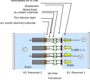
5.2.2 Self-assembled proton pumps on a porous substrate ... 70
5.3 Fabrication and Experimental Analysis ... 71
5.3.1 Preparation of the bio- functionalized porous substrate... 71
5.3.2 Fabriation of the polydimethylsiloxane (PDMS) microfluidic chip .... 71
5.3.3 Surface coverage of the Au-PAA substrate with biotinylated bR... 74
5.3.4 Photo-electro-chemical response based on pH- gradients... 77
5.4 Molecular Mechanism for the Light Driven pH Gradient Generation ... 84
5.5 Monitoring Solution Acidity Using Phenolphthalein Dye... 86
5.6 Concluding Remarks... 88
CHAPTER 6 Smart Hydrogel Microactuator ... 89
6.1 Introduction ... 89
6.2 Hydrogel Microactuators ... 89
6.2.1 Physics and chemistry of the pH-sensitive hydrogels ... 90
6.2.2 Synthesis techniques of hydrogels ... 92
6.3 PH-Sensitive Hydrogel Microvalve ... 93
6.3.1 Fabrication of pH-sensitive hydrogel ... 93
6.3.2 Fabrication of the microfluidic chip ... 94
6.4 Response of the pH-Sensitive Hydrogel Microvalve... 96
6.5 Results and Discussion ... 97
ix
CHAPTER 7
Light Driven Hydrogel Microactuator ... 99
7.1 Introduction ... 99
7.2 Photo-Responsive Hydrogel Microactuator ... 101
7.3 Fabrication of bR Activated Hydrogel Microactautor ... 102
7.3.1 Fabrication of the HEMA-AA Hydrogel ... 103
7.3.2 Fabrication of the bR functionalized porous substrate... 104
7.4 Microactuator Response... 106
7.4.1 Photoelectrochemical characteristics of the microactuator ... 107
7.4.2 Swelling chracteristics of the microactuator ... 109
7.4.3 Rsponse reversibility of the hydrogel microactuator ... 114
7.4.4 Microfluidic valve activated by the bR based pH-gradient generator 116 7.5 Concluding Remarks... 118
CHAPTER 8 Conclusions ... 120
8.1 Thesis Summary... 120
8.2 Thesis Contribution... 123
8.2.1 Production of bacteriorhodopsin ... 123
8.2.2 The Development of bR self-assembled monolayer ... 124
8.2.3Fabrication of the optically driven pH gradient generator…………...125
8.2.4Fabrication of the optically driven microactuator………...126
8.3 Limitations and Recommendations for Future Work ……….127
8.3.1 Purple membranes distribution on the substrate ………127
8.3.2 Optimizing the generated pH gradient ... 127
8.3.3 Swelling dynamics of the microactuator hydrogel ……..…………..128
8.4 Final Thought ………..128
Bibliography ... 129
x
LIST OF TABELS
Table 3.1 Materials and media used to grow Halobacterium Salinarium cells ... 31
LIST OF FIGURES
Figure 2.1 Schematic diagram of a Halobacterium salinarium cell. A two-dimensional crystalline purple membrane provides proton transport and membrane-bound ATP synthase enables the photosynthesis of ATP from ADP.. ... 16 Figure 2.2 General Structural description of purple membrane patches containing bR. A bR protein in the membrane. Its carboxy terminus is located inside the cell and the amino terminus is located outside the cell.... ... 17 Figure 2.3 Detailed structural description of the bR. The seven transmembrane helices are labelled from A to G. The retinal Schiff-base and the amino acids are most relevant for proton transport.. ... 19 Figure 2.4 Photo- isomerization of the bR retinal from (a) all-trans to (b) 13-cis in bR. The retinal has covalent bond with Lys-216 via a protonated Schiff base. When absorbing a photon, the retinal isomerizes around the C13=C14 bond. (Based on Gai et al., 1998). ... 20 Figure 2.5 Basic photochemical cycles of bacteriorhodopsin, when exposed to visible light (h), for both the aqueous (a) and dry forms (b) (Groma et al., 2001). The subscripts refer to the peak wavelength for the identified intermediate.. ... 21 Figure 2.6 Spectral absorbance of the bR produced at the University of Western Ontario.
xi
Figure 3.1 Illustration drawing of the Bacteriorhodopsin in its biological Halobacterium salinarium cell... 30 Figure 3.2 Photograph of the 1.0 L fermentation process of the Halobacterium salinarium
cells ... 32 Figure 3.3 Photograph of the 10.0 L fermentation process of the Halobacterium salinarium cells ... 33 Figure 3.4 Photograph of the density gradient formed the purple membrane band. ...
... 35 Figure 3.5 Spectral optical density of the bR produced at the University of Western Ontario. Note that visible light ranges from approximately 400nm to 700nm. ... 36 Figure 3.6 Fabrication of oriented bR films by the EPS technique. PM patches transport onto the anode due to its more negatively charged cytoplasmic side. ... 37 Figure 3.7 Preparation of a bR film by the LB technique. The cytoplasmic side of PM is more hydrophilic (facing into the water) than the extracellular side (facing the air). ... 39 Figure 3.8 Schematic drawing of multiple PDAC/PM layers as fabricated by the LBL technique (He et al., 1998)... 40 Figure 3.9 Illustration of the self-organized and self- assembled photoelectric dry layer on Au substrate. ... 41 Figure 3.10 AFM analysis of the bare gold coated substrate. ... 43 Figure 3.11 The simplified structure of the self-assembled
bR-biotin-streptavidin-biotin-thiol-Au film ... 44
xii
Figure 4.2 Fundamental photochemical cycles of bacteriorhodopsin, for (a) the aqueous phase, where the proton pumping starts with the release of a proton during the L→M transition and finish with a proton uptake during the M→N transition (Edman et al., 1999) and (b) the dry bR, where only K, L and M intermediates are observable and no proton pumping across the protein membrane. ... 50 Figure 4.3 SEM photograph of the self-assembled ultra-thin bR layer, with a magnification factor of 200. Enlarged area shown in upper right corner (taken in the Nanofabrication facility, the University of Western Ontario) ... 52 Figure 4.4 AFM analysis of the self-assembled ultra-thin bR layer. Two cross-sectional profiles are shown (takn in the surface science facility in the University of Western Ontario). ... 53 Figure 4.5 Experimental apparatus used to test the photoelectric properties of the photocell and equivalent electronic circuit for the light responsive bR thin layer. .. 55 Figure 4.6 Small- amplitude random photoelectric response of the bare Au substrate. .... 56 Figure 4.7 Measured voltage difference across the bR layer on a 0.185 cm2 substrate as a function of light exposure time. Six pulses with increased time durations of light exposure are shown. ... 57 Figure 4.8 Enlarged view of the photocell response to the second light pulse.
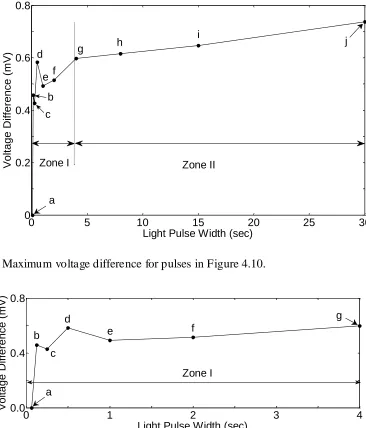
Illumination was intermittently reduced to 50% of intensity at 1800 s. ... 58 Figure 4.9 Enlarged view of the photocell response showing voltage buildup toward steady-state over the duration of the sixth light pulse in Figure 4.7. ... 59 Figure 4.10. Measured voltage difference across the 0.0925 cm2 pixel for a sequence of
varying input pulse widths (0.0625s, 0.125s, 0.25s, 0.5s, 1s, 2s, 4s, 8s, 15s, and 30s). ... 60 Figure 4.11. Relationship between the width of the light pulse and maximum voltage
xiii
xiv
of the bR-PAA pH gradient generator cause the solution in R1 to transform from a
light greenish blue (pH 5.10) to a darkened, more intense blue (pH 5.31). ... 87
Figure 6.1 Volume transitions of acidic and basic hydrogels under pH gradient activation ………91
Figure 6.2 Schematic drawing showing the hydrogel network structure...………92
Fig. 6.3 Steps in micromolding a PDMS microfluidic chip ... 94
Figure 6.4 pH-sensitive (HEMA-AA) microvalve at pH 3 and pH 10, respectively (Nanofabrication Facility UWO). ... 96
Figure 6.5 Swelling of 155µm (HEMA-AA) pH sensitive microvalve. ... 97
Figure 7.1 Schmatic drawing of the bR activated hydrogel structure. ... 101
Figure 7.2 Schmatic drawing of the ions flow through the activated hydrogel. ... 102
Figure 7.3 SEM photograph of the Au-coated PAA substrate. ... 105
Figure 7.4AFM photograph of bio- functionalized Au-coated substrate showing surface topology. ... 105
Figure 7.5 Measured voltage difference across the bR layer on a 0.096 cm2 substrate under continuous light exposure. ... 108
Figure 7.6 Recorded photocurrent based on the measured voltage difference across the bR layer on a 0.096 cm2 substrate under continous light exposure. ... 109
Figure 7.7 Schmatic drawing of the test chip. ... 110
Figure 7.8 Photograph of the test setup ... 111
Figure 7.9 Schematic drawing of the (a) deswelled hydrogel, and (b) swelled hydrogel valve. ... 112
xv
Figure 7.11 The change in diameter of the HEMA-AA hydrogel actuator. ... 113
Figure 7.12 The change in length of the HEMA-AA hydrogel actuator. ... 113
Figure 7.13 The change in volume of the HEMA-AA hydrogel actuator. ... 113
Figure 7.14 Photograph of the actuator in the microfluidic channel. ... 114
Figure 7.15 Change in microactuator cross-sectional area. ... 115
Figure 7.16 The reversibility of the microactuator response in the micro fluidic channel. ... 116
Figure 7.17 Photographs of the actuator in the microfluidic channel in configuration (a) deswelled phase and (b) swelled phase. ... 117
xvi
NOMENCLATURE
A Area of substrate
AbR Area of one bacteriorhodopsin of the hexagonal molecule
AA Acrylic acid
ADP Adenosine Diphosphate AFM Atomic Force Microscopy
SEM Scanning Electron Microscopy ATP Adenosine Triphosphate
Arg Arginine acid Asp Aspartic acid Au Gold
BR, bR Bacteriorhodopsin BS Basal Salt
(Ct) Total Capacitance
CID Charge Injection Device CM Culture Media
DNase Deoxyribonuclease (Eph) Photovoltage Source
EFS Electrophoretic Sedimentation
FIB-SEM Focused Ion Beam Scanning Electron Microscopy Glu Glutamic Acid
HEMA 2- hydroxyethyl methacrylate HCl Hydrochloric acid
xvii ITO Indium Tin Oxide
K216, Lys 216 Lysine 216
KCl Potassium chloride mH Mass of the hydrogen ion MS Media Salts
npho Number of photons received by one bR hexagonal molecule
nmax Maximum number of photons processed by One bR hexagonal
molecule
NaCl Sodium chloride NaOH Sodium hydroxide
N'P Maximum rate of proton pumping by one bacteriorhodopsin
molecule
LB Langmuir-Blodgett
LBL Layer-by-Layer adsorption LOC Lab-on-a-Chip
PAA Porous Anodic Alumina PBS Phosphate Buffer Saline
PDAC Poly(diallyldimethylammonium chloride) PDMS Polydimethylsiloxane
pKa Phase transition point PM Purple Membrane RM Rich Media
xviii UV Ultra Violet Beam
VC Volume of Fluid in the Reservoir
WT Wild Type bR
CHAP TER 1
INTRODUCTION
1 WTF!
1.1
The Problem
Microactuators are critical components of microelectromechanical systems because they convert the input signal into mechanical motion needed to perform useful work. These microactuators or the transducers as they often called are made from functional structures that can receive and process the input signal and then act with the suitable response. Their actuation has been considered to be the building block of mechanical work generators such micro-objects manipulators and flow control valves of microfluidic chips. One of the important technological challenges is how effectively drive microactuators such as microfluidic valves. The microfluidic valves are necessary active components of microfluidic systems that control the micro- fluid supply for the chemical and biochemical processes that are performed on the microfluidic chips. However their operating techniques require accessing the system physically to activate these valves such as the electrical and chemical techniques. In the electrically driven microvalves the electric leakage is highly expected, whereas triggering the chemically operated microfluidic valves can only be done by changing the chemical composition of the surrounding fluid. Electrical based techniques might make an interaction or cross-talk with biochemical processes on the chip as most of biochemical processes are electrical sensitive processes.
photo-thermal effect. Unfortunately the photophoto-thermal effect necessitates keeping the microvalve in very narrow temperature ranges which is not suitable for many practical applications. Looking towards the biological systems for inspiration, the optical system of the human vision for example is based on the photo-electro-chemical interaction between the light beam and the visual rhodopsin. In this manner it can be considered an optical to electro-chemical transducer. Whereas the biological muscles produces mechanical work when activated with an electro-chemical input. These two inspiring biological concepts interested the author to bring them together in one complete mechanism that generates mechanical work when powered with optical signal. This thesis supports the viability of employing one of the biologically synthesised visual rhodopsins known by bacteriorhodopsin protein and artificially fabricated smart material called hydrogel as a building block of novel optically driven microsystems. This system can work only by developing an effective technique for building monolayers of the self-assembled bacteriorhodopsin proton pumps, fabricating a suitable hydrogel and then creating the overlap working zone that can operate these different constituting components in the same time with degrading the performance of each other. Only then is it possible to demonstrate its effectiveness by driving smart hydrogels to control fluid flow on microfluidic chips as proof of concept.
1.2
Optofluidics
advances in broad technological challenges. Heng et al. in 2006 generated novel non-expensive optofluidic microscope, which is able to capture on-chip high resolution imaging (Heng et al., 2006). The integration of the microfluidics with the optics has also facilitate the fabrication of reconfigurable three dimensional microlenses (Rosenauer et al., 2008), and the fabrication of in-situ magnetic microactuators in microfluidic channels (Chung et al., 2010). Furthermore, the ability of trapping bio- functionalized micro-objects has been demonstrated (Domachuk et al., 2007).
The biological organisms undergo in large number of interactions with the environment throughout their lifecycle. Their high sensitivity allows them to detect and discriminate between very small changes in their surroundings. The small size and energy efficiency of the biological materials can make them a promising choice for developing unique transducers for sensing and actuation. The integration of bio- materials with microsystems has leaded to significant advances in wide ra nge of challenging applications such as creating flexible photocells (Wang et al., 2005) and self healing materials (Ratner 2007). During the biological generation of bio- materials, the natural evolution optimized their constituting bio- molecules and assembled them in three dimensional structures, allowing them to process and store large amounts of information in very small volumes.
becoming an advanced building block material for fabricating bioelectronic systems, as it exhibits high applicability potentials in light sensing, artificial vision and parallel associative memories, and optical actuation.
1.3
Optical Actuation in Microfluidic Systems
Microfluidic systems are integrated technologies used to manipulate very small amounts of fluid for a variety of medical and industrial applications. The design and fabrication of complex microfluidic systems requires various building blocks that enable fluid transport, directional flow, pumping, sample preparation, separation, mixing, detection, and in situ chemical reaction. One of the greatest design challenges has been controlling the directional flow of fluid through the constituent microchannels. Flow control is achieved by microactuating devices, such as valves, that perform mechanical work in response to an external command or control signal. The device can be separated into two parts: the actuator shell and the method of actuation. The shell is the basic mechanical structure of the actuator and, often, contains deformable or moving parts. The main function of any shell design is to provide a mechanism for the desired actuation method to produce useful work. The actuation method is the means by which a control signal is converted to a force that is applied to the actuator shell and generates physical movement. The output of the overall system is the desired response given as a displacement, force, or pressure value. An example of a micro-valve is a flexible diaphragm driven by piezoelectric, electrostatic, electromagnetic, or thermo-pneumatic actuator (Kovacs 1998). These micro- valves typically offer small displacements, in range of tens of microns. However, they are large in size, expensive to fabricate, and not easily integrated into existing microfluidic channels (Liu et al., 2002).
heat dissipation, and friction forces that greatly diminish the performance and efficiency of conventional electronic or electro- mechanical systems. The negative effects of current leakage and power loss are greatly amplified as design engineers strive for product miniaturization through the exploitation of nanotechnology.
Hydrogels are composed of hydrophilic or hydrophobic cross- linked polymeric networks with a fluid filling the interstitial spaces of the network. Due to their hydrophilic, hydrophobic, and elastic nature these hydrogels are primarily used for drug delivery systems and biomedical instrumentation. In general, hydrogels represent a promising class of materials for microfluidic applications that demand low stressing components and the formation of tight seals that prevent fluid leakage.
Light sensitive hydrogels are often driven by one of three approaches. The first approach is to exploit molecules that can undergo volume changes when exposed to specific wavelengths of light. Ishihara et al (Ishihara et al., 1984) investigated the swelling properties of 2-Hydroxyethyl methacrylate with azobenzene molecules as the side groups. The azobenzene molecule is a UV sens itive molecule that can make a 180° rotation around a carbon double bond. The rotation of the azobenzene group around the carbon double bond induces structural rearrangement. The maximum volume change that was observed with this structural rearrangement was 14% in 6 hours at 25°C. An alternative approach exploits light ionized molecules in a neutral hydrogel network. In this context, Ishikawa and Kitamura (Ishikawa et al., 1994) used polyacrylamide microgels that have triphenylmethane leuco cyanide as the light ionizable molecule. UV light ionizes this molecule inducing repulsive forces that drive the hydrogel to swell. It was reported that the photo-dissociation of this light sensitive chromophore occurs in less than 60 seconds. The equilibrium time for an 11μm particle is about 1 hour, while for a 180 μm particle it takes more than 55 hours.
hydrogel contained N- isopropylacrylamide as the main constitute and trisodium salt of copper chlorophyllin as a light sensitive chromophore. Discontinuous volume transition was observed around 31.5°C. The diameter of the sample shrank from abo ut 240μm to 100μm in response to a temperature increase from 25°C to 40°C. Mamada et al (Mamada et al., 1990) investigated the response of a N-isopropylacrylamide gel with the photosensitive molecule bis(4-(dimethylamino)phenyl) (4-~inylphenyl) methylelucocyanide as a side group. Juodkazis et al (Juodkazis et al., 2000) studied the effect of laser radiation on the temperature responsive hydrogel N-isopropylacrylamide. The experiments confirmed that radiation forces can alter the phase transition process by shifting back the volume transition temperature in the range of 10°C. Sershen et al (Sershen et al., 2005) incorporated optical absorption particles to drive the temperature responsive hydrogel poly[N-isopropylacrylamide-co-acrylamide]. The optical absorbing particles that were used are gold-colloid nanocomposites hydrogel that collapse when illuminated under green light and gold nanoshell hydrogel that collapses in response to near IR light. Both hydrogel composites have fast responses and can reach final state in 5s when fabricated in micron scale.
1.4
Motivation of the Present Work
Over the last three decades, optical driven systems using biologically synthesised materials have evolved into very efficient transducers. Biological transducers can be extremely sensitive; for example, the retina of human eyes can respond to individual photons (Baylor et al., 1979). These optical transducers can also respond to wide range of light intensities through the range of the light spectrum. Transmitting signals of the control information and power signals through bio-processors facilitate the natural and optimum data compression and processing, and minimizing the external wiring and hardwares of the electrical, chemical and mechanical systems.
then hybrid photoelectric using bioelectronics photosensitive proteins and then recently photomechanical transducers based on the photo-thermal phenomenon. Most of the reported photo mechanical systems exploited the photo-thermal effect. In essence the reported photomechanical transducers are activated by the heat associated with the light illumination where the focus was to generate enough heat from the light radiation to drive temperature sensitive polymers, or to induce thermal expansions. In micro-scale the most successfully optical driven transducers are systems that integrated with the temperature sensitive hydrogels based on the poly(N- isopropylacrylamide) monomers. These hydrogel actuators exhibited good functionality in microfluidic valves (Sershen et al., 2005; Richter et al., 2008; Lou et al., 2003).
However, in order to extend the thoughts for designing optically driven micro transducers without experiencing the photo-thermal effects, the biological organisms were considered as examples of complete integrated systems. One of the interesting examples is the conversion of the optical beams received by the eye receptors into mechanical motion in its muscles. The eye receives the incident light beam by the retinal rhodopsins. These rhodopsins release protons to make an electrochemical gradient. The generated electrochemical gradient is considered as the input signal to activate the eye muscles to make mechanical motions (Forrester et al., 1996; Batterbury et al., 2009 Malmivuo et al., 1995). This photomechanical energy conversion technique is taken in the present research as an inspiring point for designing true photomechanical system.
oriented methods, which can lead to contrasting findings. Furthermore in engineering, the designers study the theory and applicability in artificial systems. The core motivation of studying the bR protein is to investigate its performance at the monolayer level to understand the reality of the bR behaviour because it is found in the living bacterial cell in the form of monolayers. The study of the bR performance at the monolayer level not only enhances its applicability in engineering practices, but also facilitates fabrication of the thinnest photosensitive layer in the domain of nanometers.
The constituting a component that processes the received signal from the bR monolayer in the introduced optically driven microactuator is hydrogel actuating shell, which converts the electro-chemical signal into mechanical work to be used in microscale actuation and control. The objective of this part of the research is to develop a suitable pH gradient hydrogel that can function in a common electrochemical domain with bR monolayer, so that a directed light source can be used to activate and power the micro-actuator. This photo-responsive material should not genera te a significant temperature increase that can affect the surrounding fluid. Finally, this micro-actuator should form a seal that prevents undesirable leakage when blocking a channel and conform to all possible channel geometries.
1.5
Objectives and Scope of this Work
1. What is the signal pathway of the photoelectric effect in the dry bR monolayer? 2. What is the signal pathway of the photoelectrochemical effect in the wet bR
monolayer?
3. How the generated signal be converted into mechanical work?
4. What are the parameters affect the photosensitive monolayer response? 5. How the photosensitive monolayer response can be optimized?
6. How can the photosensitive monolayer response be exploited in a practical sensing and actuating application?
Understanding the bR and theory behind the signal conversions when exposing the bR monolayer to light beams in the dry and wet environments is underlying objective of this research. By doing so, it provides a means of developing clear understanding to describe the signal generated by the dry and wet bR monolayer photoreceptor, which in turn allows standard engineering design and analysis methodologies to be employed. For engineers and technologists to effectively integrate bR monolayer based photoreceptor with microfluidic chip, an appropriate theoretical model must be available. Because this is the first time bR monolayer based photoreceptor is introduced, no readily applicable model has been described in prior literature and therefore, the development of a meaningful model is a primary objective of this dissertation.
The fundamental component that affects the generated pH gradient is the bR photosensitive monolayer. The main key factors that contribute in the final response are subdivided into fabrication factors and operation parameters. The fabrication method and its affecting factors to create photosensitive monolayer are studied to produce high surface coverage, and therefore to be able to generate measurable absolute electrochemical response without using amplification circuits. On the other hand the operation parameters such as light beam parameters (intensity, wavelength), area of the photosensitive surface, volume, and strength of the ionic solution are optimized. The fabrication process, and the associated factors, and operation parameters are optimized to create an optofluidic transducer able to convert the incident light into electro-chemical potentials to be used as an input signal to the hydrogel actuator shell.
The final question is related to the applicability of the generated electrochemical potentials in sensing and actuation. In the living cell, the generated pH gradients provide the necessary energy for synthesizing adenosine triphosphate (ATP) from adenosine diphosphate (ADP) which is required for the bacterial cell activities and cell movements (Wang et al., 2005; Hampp, 2000; Voet et al., 2006). The fabrication bR photocell in the form of monolayer based photo-electro-chemical transducer makes it much closer approximation to a biological system. This molecular architecture makes bR a well-suited material for light sensing and for actuation of pH sensitive hydrogels. The final investigation of the created photo sensitive nano layer is exploring the limits and capability of activating microfluidic valves.
1.6
Overview of the Thesis
The remainder of this thesis is organized into eight chapters. Chapter 2 provides a detailed overview of bacteriorhodopsin proton pump. Its structure and photosensitive characteristics are described in detail, providing deep view on the material’s characteristics, and behaviours. This background knowledge establishes the foundation needed for the design and analysis described in subsequent chapters. The property most relevant to this work is bR’s photoelectric and photo-electrochemical responses, which are generated by the phototransduction processes.
In order to fabricate bR based photo-responsive transducer, the bR proton pumps must be directionally organized and irreversibly immobilized onto Au coated substrate. The methods of organizing and immobilizing bR layers are reviewed in Chapter 3. However, the biotin labelling method was used to recognize and label the extracellular side of the bR, and self assembly technique based on biotin-thiols and sterptavidin was used to allow linking the bR purple membranes directionally to the Au substrate. The introduced methodology was used for the dried, and the wet bR monolayer. This chapter also provides details of the fabrication method.
Chapter 4 investigates the photovoltaic characteristics of the molecularly organized bR based monolayer. The phototransduction process of the dried bR monolayer is investigated. The photo-cycle of the dried bR is significantly different from wet or aqueous bR because it is based on charge separation, whereas the wet bR photo-cycle is based on the transport of protons. The photovoltaic stability, power intensity and exposure duration are studied. The transient and steady state responses are investigated. Mechanical deformations of the bR structure are explained based on the photovoltaic characteristics. Factors that affect the fabrication process and operation performance are optimized for creating high performance photo-responsive monolayer without using signal amplification circuits.
medium. The affect of the input power and the distance between the pumping station and the measuring point are investigated to make clear understanding of the pH kinetics in the microchannels. A mathematical model is formulated to describe the phenomenon. This chapter further validates the proposed theoretical model and provides a basis for optimizing photodetector performance for a give n application.
The fabrication procedures and response characteristics of the pH- gradient activated hydrogel micro actuator are described in Chapter 6. As proof of applicability, the chapter describes how the pH-gradient activated hydrogel can be operated in larger microsystem with very specific electro-chemical environment so that it can be integrated with the bR based photo-electro-chemical transducer. It was fabricated in a comparable geometry, and operated in electro-chemical environment similar to the targeted environment with the based bR photo-electro-chemical transducer. The tests are performed in Polydimethylsiloxane (PDMS) microfluidic chip because of its optical properties, so that it can be integrated in the optically driven microfluidic systems. The swelling response characteristics of the pH-sensitive hydrogel are investigated and the limitations are discussed.
Chapter 8 summarizes the contributions of this thesis. It also recognizes the limits of the project and provides suitable recommendations for the future studies, and investigations.
CHAP TER 2
REVIEW OF BACTERIORHODOPSIN
2 WTF!
2.1
Photosensitive Proteins
A variety of biological and electrochemical processes are powered and controlled with solar radiation. The light triggered organisms generate photoreceptors that detect and process light beams into usable form of energy. Among the photosensitive structures, nourishing chlorophylls and visual rhodopsins are the most know photo triggered structures. In plants the protein complex chlorophylls which is found in their green leaves photosynthesizes glucose and starch to be part of their life cycle. On the other hand most of the mammalians have photosensitive proteins in their eyes known by visual rhodopsins. The light driven rhodopsins are used for sensing.
exposed to the photonic energy, basically, they convert the photonic energy into electrochemical potential used in each particular organism uniquely.
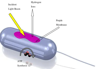
Figure 2.1 Schematic diagram of a Halobacterium salinarium cell. A two-dimensional crystalline purple membrane provides proton transport and membrane-bound ATP syntheses enable the photosynthesis of ATP from ADP.
2.2
General Structure of Purple Membrane and Bacteriorhodopsin
Bacteriorhodopsin proton pump is the only protein in the purple membranes of Halobacterium salinarium living cell. Under oxygen limited conditions the cell membrane grows purple membrane patches in the form of hexagonal two-dimensional crystalline lattice of bR, Figure 2.2. The bR molecules are arranged in directionally oriented arrays with lipid filling the inter- molecular spaces forming monolayer-thick PM patches. The molar ratio of the lipid to the bR molecules is 10:1 (Blaurock and Stoeckenius, 1971).
The PM patches are characterized their irregular lateral dimensions, where the average diameter can be up to 5µm, whereas they have uniform thickness of 5nm. The PM proved high functional and structural stability over several years of light exposure. In the wet or aqueous medium it keeps its stability at relatively high temperature of 80°C
Incident
Light Bea m
Purple
Membrane
Bea m Hydrogen
Ions
ATP
Synthesis
Membrane
and at harsh chemical environments for pH values from 0-12 in the presence of high ionic strengths up to 3 M NaCl (Hampp, 2000). The PM preserves its photoelectric ac tivity in dry environment and can keep functioning up to 140 °C (Shen et al., 1993).
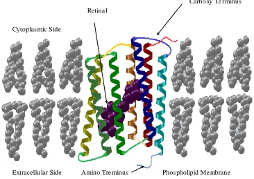
Figure 2.2 General Structural description of purple membrane patches containing bR. A bR protein in the membrane. Its carboxy terminus is located inside the cell and the amino terminus is located outside the cell.
The two dimensional structure of lipid-bR in the PM is the base of the thermal and chemical stability in the dry and wet environments. Each bR molecule converts the light radiation into electrochemical energy by pumping protons from the cytoplasmic side to the extracellular side of the membrane without allowing passive diffusion of protons back into the cell.
The bR is a retinal protein complex that consists of two main components: a protein molecule body and the retinal molecule. The retinal is simply known by Vitamin A aldehyde. The bR structure consists of 248 amino residues in a polypeptide chain arranged in seven α-helices (Henderson et al., 1990). The α-helices are labelled in the
Carbo xy Te rminus Retina l
Cytoplasmic Side
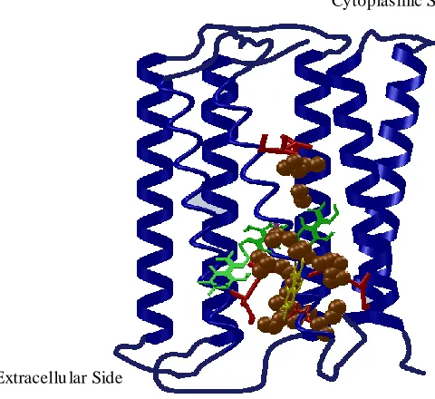
Figure 2.3 Detailed structural description of the bR. The seven transmembrane helices are labelled from A to G. The retinal Schiff-base and the amino acids are most relevant for proton transport.
2.3
Photoelectrochemical Cycles of Bacteriorhodopsin
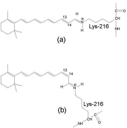
The retinal of bacteriorhodopsin proton pump is a natural chromophore that exists only in one of two configurations: all-trans and 13-cis (Maeda et al., 1977). In the dark, the bR molecules contain mixture of these two retinal configurations; it is called “dark-adapted”. Once bR molecules exposed to the light radiation the molecules that are in the 13-cis configuration ground their state to be at the all-trans retinal configuration. This is considered as the starting point of the photocycle, which is also known by “light-adapted” state. Basically the proton pumping process does not occur in the dark-adapted bR. When the bR molecules receive the activating light beams, the retinal undergo in an isomerisation process around the C13=C14 double-bond to transform the retinal configuration from all-trans to 13-cis configuration, as shown in Figure 2.4 (Gai et al., 1998). The retinal isomerisation is followed by proton transport from the cytoplasmic to the extracellular side of the membra ne, which is interpreted as converting the incident light energy into chemical energy (Lanyi, 1993). The proton pumping process is accompanied by a thermal structural relaxing photocycle with several intermediate states.
Extracellu lar Side
Figure 2.4 Photo- isomerization of the bR retinal from (a) all-trans to (b) 13-cis in bR. The retinal has covalent bond with Lys-216 via a protonated Schiff base. When absorbing a photon, the retinal isomerizes around the C13=C14 bond. (Based on Gai et al., 1998)
Prior studies have shown that the kinetics of the bR photocycle depend on the level of humidity in the sample (Váró et al., 1983; Korenstein et al., 1977). The photocycle and proton transfer kinetics of dried bR film differ from aqueous, or wet, bR because of dehydration (Korenstein et al., 1977; Cao et al., 1991; Groma et al., 2001; Wang 2006; Wang et al., 2007). In the bulk aqueous form, Figure 2.5a, the bR molecules act as a light-driven proton pump.
(a) Multistate intermediates of the aqueous bR photocycle.
(b) Limited intermediate of the dried bR photocycle.
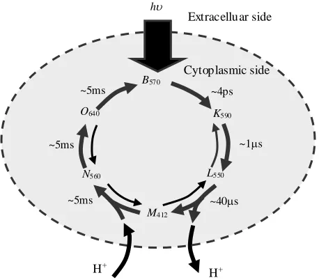
Figure 2.5. Basic photochemical cycles of bacteriorhodopsin, when exposed to visible light (h), for both the aqueous (a) and dry forms (b) (Groma et al., 2001). The subscripts refer to the peak wavelength for the identified intermediate.
When the bR molecules are exposed to light illumination, the y all return to the ground state B. This step is considered as the start point of the photocycle. The photocycle consists of photo-driven thermal intermediates with distinct photo-absorption
Cytoplasmic side Extracelluar side
h
H+
B570
K590
L550
M412
N560
O640
~4ps
~1s
~40s ~5ms
~5ms ~5ms
H+
h
Cytoplasmic side
B570
K590
L550
M412
~3s ~ns
~s
~ms
Extracelluar side
maxima. Fundamentally, the photocycle’s intermediates are wavelength driven transitions, where each intermediate can proceed by thermal relaxation to the next state or switch back to the ground state B, based on the received photo excitation. All of the intermediates are before the proton release and after the proton uptake. The transition of the bR that occur between releasing proton and up-taking another proton is considered as an irreversible transition, where the nitrogen atom in the Schiff base becomes no more accessible to the extracellular side of the proton way half-channel, then open the cytoplasmic side of the half-channel and close the extracellular half. The sequence of the proton release- uptake is a pH-based process, where at pH 7.0 the proton release precedes the proton uptake, whereas at pH 7.0 the proton uptake precedes the proton release (Ludmann et al., 1992). These simultaneous structural re-arrangements are considered to be the origin of the vectorial proton transport through the bR protein. In the wild-type bR proton pump a complete photo-cycle needs approximately 10 ms (Dancshazy et al., 1986).
However, when the humidity level in the bR film falls below 90%, the later N560 and O640 intermediates are no longer observed and fewer protons are transferred across the PM (Ganea et al., 1997). As shown in Figure 2.5b, only the K590, L550 and M412 intermediates of the dry bR are involved in the photochemical cycle. The dry bR protein returns to its ground state (B570) through several paths, each with a different lifetime (Varo et al., 1991). Furthermore, the only available ions for dry bR film are those enclosed within the structure of the bR purple membrane proton pumps (retinal ions).
0 0.5 1 1.5 2
350 400 450 500 550 600 650 700 750 800
Light Wavelength (nm)
Ab
s
o
rb
a
n
c
e
Spectral Peak at 568nm
Figure 2.6 Spectral absorbance of the bR produced at the University of Western Ontario. Note that visible light ranges from approximately 400nm to 700nm.
2.4
Photoelectric Properties of Bacteriorhodopsin
The photoelectric characteristics and response of bR based systems is the resultant of the light-bR interaction and its photocycle kinetics. In other words the charge dynamics within the bR protein can be directly related to the intermediate kinetics of the photocycle. When bR receives light beams, it starts the charge translocation process, and the sub sequential structure deformations needed to generate charge gradients (Lanyi, 1993). This charge activity can be detected with monitoring the potential difference. The existence of such voltage difference and the temporary changes in the polarity and amplitude can be directly related to the proton transfer across the PMs.
recombination within the bR molecule. This component is essential in the dry bR based photoelectric films where it is less sensitive to the moisture content. The charge displacement and recombination does not lead to net voltage difference as it occurs inside the bR pump channel.
The organized PMs generates electric signal once exposed to light radiation. The generated light signal contains three major components with different lifetimes. The fastest component shows its viability within 100 ps in the opposite direction of the proton pumping pathway. The other two photoelectric components take place in microsecond and millisecond range and are in the same direction of the proton transport (Liu, 1990). The internal charge dynamics, proton dynamics, and non-proton ion dynamics may all contribute in the photoelectric components. Theoretically, the photocycle lifetimes s hould coincide with the photoelectric components. However, the lifetimes of the PM photocycle components are not in full agreement with the detected responses (Wang, 2006). This might occurred due to the use of classical methods to organize and adsorb the PMs on the electrodes surface. The other possible reason, which is believed to have less contribution in the photoelectric activities, is the influence of the surrounding conditions such as salt concentration.
shows great potential candidate for high-speed photoelectric applications. The second expected reason is that: the protonated Schiff moves away from the negative counterion in the stage of forming the K state intermediate. This second process dominates the photoelectric response. Similar photoelectric responses are found in the visual rhodopsin of vertebrates which is known by the early receptor potential (ERP).
Figure 2.7 An electrostatic map of the primary photochemical event of bR.
2.5
Applications of Bacteriorhodopsin
2.5.1 Photochromic applications
The unique photochromic characteristics of bR have enabled creating all-optical logic gates, optical memories, parallel associative processors, and holographic interferometry (Sharmar and Roy, 2004; Renner and Hampp, 1993; Cullin et al., 1995; Birge et al., 1999). These techniques are based on the photochromic characteristics of bR, where each bR proton pump individually absorb the incident photons and launch a reversible photocycle. Bacteriorhodopsin can go in reversible transitions between the photocycle intermediates by exploiting the photo-responsivity of bR at different wavelengths. Furthermore bR can be genetically engineered to generate different spectral shifts and longer lifetimes at certain intermediates for improved photo-responsivity (Bräuchle et al., 1991). Organizing bR within the photoresponsive film remains at this point a technological challenge for generating higher performance bR based systems.
2.5.2 Photoelectric applications
The photo-resposivity of bR enabled producing bio-photocells for sensing and imaging technologies. However, bR proton pumps have to be directionally organized as pumping sense is a fundamental condition for detectable responses. The photoelectric characteristics of bR immobilized on conductive substrates have been investigated with the different substrates. Indium tin oxide (ITO) coated glass and plastics, gold, Platinum are the most used electronic substrates (Saga et al., 1999). However, for the micro and nano applications different substrates are proposed as potential candidates for immobilizing bR such as the gate terminal of a GaAs-based MOSFET and nano-black lipid membranes (Xu et al., 2004; Horn and Steinem, 2005).
using the detection grid of a charge injection device (CID) as the substrate for a bR-polymer film. This structure integrates processing ciruit to monitor the bR photoresponse. Libertino et al., (2003) deposited non-oreinted bR film on Si and SiO2 substrates.
The bR-based photoelectric and photochromic systems have shown functional mimic of the biological vision as the bR good degree of functional similarity with the biological eye rhodopsin. The bR showed inherent of processing simple vision information. Several research groups focused on two essential functions including feature extraction and pattern recognition. The function of feature extraction involves detection and position of spatial and temporary variations in the image intensity which is very important in the edge enhancement and motion detection applications. In the biological vision systems, the basic edge detection systems are formed from the ganglion cell reception field. This formation is referred to a zero-crossing filter. An artificial receptive field based on bR photodetector has been developed by Takei et al. (1991) and Yang et al. (1998). BR is also used in building parallel processing units based on the neurobiological principles (Haronian and Lewis, 1991). The researchers introduced bR based rapid reprogrammable neural network architecture with the capability of including large synapse matrix.
3 WTF! TF!
CHAP TER 3
PRODUCTION AND IMMOBILIZATION OF
BACTERIORHODOPSIN
3.1
Introduction
Bacteriorhodopsin (bR) is a biologically produced protein in a living organism called Halobacterium salinarum (formerly Halobacterium halobium) from the Archaea domain of life. The Halobacterium salinarum is a unicellular organism that grows and produces optimally in high salt concentration. This light-sensitive protein which is biologically formed in the marsh archaebacteria H. salinarum has been extensively studied as an organic photosensitive material for a variety of engineering applications including photocells and optical memory (Hampp 2000; Wang et al. 2005). The biological growth protocols in the laboratory and the molecular level organizing of the bR molecules have been established and presented in this chapter.
3.2
Production of Bacteriorhodopsin (bR) Protein
(ADP) which is required for the living cell activities (Wang et al., 2005; Hampp, 2000). Biologically, the ATP is known to be the energy transfer mechanism agent that diffuses in the cell to energize its cellular molecular processes, and cell movements (Voet et al., 2006). Interestingly enough, the ATP can be considered as the driver mechanism that transfers energy currency of life to the bacterial cell. This instantaneous process is repeatable as long as there is a continuous generation of pH gradients across the cell walls.
Unlike non-automated events, the process of generating pH gradients is understood to be consists of a fully automated sequence of photo-electro-chemical events at molecular level that is triggered by photon reception and completed with the pumping of hydrogen atoms to the extracellular medium. These processes are physically hosted by the purple membrane (PM) patches. In essence, each PM patch works as a hardware unit that contains an array of biological nano-processors known by bacteriorhodopsin (bR) protein which has an approximate volume of 56.7nm3 (Henderson et al., 1990; Henderson, 2007) and lipid filling to seal the bR intermolecular spaces. Structural analysis of the PM patches showed that, the bR proteins are arranged in parallel hexagonal crystalline lattice forming two-dimensional arrays. Each array is molecularly built by a multiple of the ratio of ten lipid molecules per one bR molecule (Sumper et al., 1976). This qualitative and quantitative assembly acquired the purple membrane patches at that low-scale their unique mechanical flexibility and robustness and their chemical and thermal stability (Wang et al., 2005; Hampp, 2000), Figure 3.1.
H+
H+
Figure 3.1. Illustration drawing of the bacteriorhodopsin in its biological Halobacterium salinarium cell. (This is similar to Figure 2.1)
3.2.1 Materials for fermentaion of Halobacterium salinarum growth media
The Halobacterium salinarum wild type (WT) strain was a generous gift from Dr. Robert Birge (Department of Chemistry, University of Connecticut, Storrs, Connecticut, USA). Uracil (item number U1128) provided by Sigma-Aldrich; Peptone Bacteriological (item number LP0037) provided by Oxoid Ltd; Tryptone (Stoke number Fulka T7293) provided by Sigma-Aldrich; Sodium chloride NaCl (Item number S640) provided by Fisher Scientific, MgSO4 (anhydrous) (Item number M63) provided by Fisher Scientific, KCl (Stoke number 7447-40-7) provided by Fisher Scientific, tri-Sodium citrate·2H2O (Stoke number 0754-12) provided by Mallinckrodt, 5N NaOH (item number SS254) provided by Fisher Scientific; yeast extract; Agar (Stoke number A360) provided by Fisher Scientific; DNase (Deoxyribonuclease, 541 units/mg (item number AMPD1 – Amplification grade) provided by Sigma-Aldrich; Percoll (item number 17-0891-01) provided by Amersham Bioscience. Dylasis tubes of 20µm wall thickness, flat width 25m, and volume of 1.98 ml (item number 21-152-18) provided by Fisher Scientific.
Hydrogen ion pumping by
bR in Purp le me mb rane Light beam
3.2.2 Fermentaion and growth media
The Halobacterium salinarum wild type (WT) have been grown in four phases with specific medium suitable for the growth process for each phase. The mediums used are: media salts, culture media, and rich media. Table 3.1 shows the standard composition for the scales used in this research work. The medias can be scaled up or down with the same ratios.
Table 3.1 Materials and media used to grow Halobacterium salinarum cells.
Media name Composition
Media Salts (MS) (10.0 L) 2.5 kg NaCl, 97.7 g MgSO4 (anhydrous), 30 g
tri-Sodium citrate·2H2O, 20 g KCl, 10L of
MilliQ water.
Culture Media (CM) (1.0 L) 1.0 L media salts, 3 g yeast extract, 5 g tryptone.
pH adjusted to 7.2 using 5N NaOH.
Autoclave for 30 min.
Rich media (RM) 1.0 L media salts, 10 g peptone
pH adjusted to 7.2 using 5N NaOH
Autoclave for 30 min.
Uracil stock solution 0.5 m uracil added to 200 mL de- ionized water Basal salt (BS) Rich media without peptone
Culture Media Plates (1.25 L) Should be done each two to three months to transfer the cell line so that keep it life for longer
1L Culture Media, 18.8 g Agar, 250ml ddH2O
pH set to 7.2 using 5N NaOH
Autoclave
Dry plates 12-17 hrs at 37 ºC before using
Phase I (one colony in 5mL): one pick of the Halobacterium salinarum colony cells picked using toothpick and placed in 5 mL of c ulture media in a test tube. 100 µL of 2.5 mg/mL uracil stock solution is immediately added. The mixture is exposed to continuous white light illumination and the temperature is adjusted to 39 ºC for 7 days. The swirly and cloudy appearance can be noticed in 4 days when the growth is going well.
Phase II (100 mL fermentation): 1 mL of phase I cells added to 100 mL of rich media. 2 mL of 2.5 mg/mL uracil stock solution is immediately added. The mixture is exposed to continuous white light illumination and the temperature is adjusted to 39 ºC. This phase is conducted in 1.0 L bioreactor with stirring speed of 50 rpm for 7 days.
Figure 3.2 Photograph of the 1.0 L fermentation process of the Halobacterium salinarium cells.
Phase III (1.0 L fermentation): 1.0 L of phase I cells added to 9.0 L of rich media. 200 mL of 2.5 mg/mL uracil stock solution is immediately added. The mixture is exposed to continuous white light illumination and the temperature is adjusted to 39 ºC. This phase is conducted in 1.0 L bioreactor with stirring speed of 50 rpm for 7-10 days, Figure 3.2.
Phase IV (10.0 L fermentation): 10.0 L of phase I cells added to 900 mL of rich media. 18 mL of 2.5 mg/mL uracil stock solution is immediately added. The mixture is exposed to continuous white light illumination and the temperature is adjusted to 39ºC. This phase is conducted in 10 L bioreactor with stirring speed of 50 rpm for 7-10 days, Figure 3.3.
Figure 3.3 Photograph of the 10.0 L fermentation process of the Halobacterium salinarium cells.
3.2.3 Extraction and purification of bacteriorhodopsin (bR)
The produced Halobacterium salinarum cells were harvested using 1.0 L centrifuge tube volume at 5000 rpm for 30 minutes cycles at 4ºC. The collected cells from the harvesting process were re-sespended in 300 mL of basal salt solution. The suspension was treated by adding 5 µL of DNase. The DNase reduces the viscosity of the lysate which reduces the possible deterioration of DNA by hydrolysis. Then, the resulted suspension was stirred on stirring plate with magenetic bar for 3 to 4 hours at room temperature.
The resultant solution was transferred into dialysis tube with minimum air bubles, and then immediately well sealed using the tube clips. The sealed tubing was placed in flask containing 0.1 M NaCl solution for 15 hours at 4ºC. The flask was completely covered with aluminum foil to prevent the light-cells interaction. The difference in the salt concentration from 4M to 0.1M generates osmotic pressure difference between the intra-cellular and the extracellular sides of the H. S. cell walls. This amount of osmotic pressure was found to be enough to break the cell walls without damaging the bacteriorhodopsin (bR) proton pumps. Once the microorganism was lysate the purple colour can be noticed clearly.
Figure 3.4 Photograph of the density gradient formed the purple membrane band.
The final suspension was centrifuged and suspended in 25% percoll solution and then centrifuged for 2 hours at 50,000 g (22,000 rpm Bekman) and 4 ºC. The percoll is linear gradient self- forming when centrifuged at greater than 10,000 g. The purple band was extracted and then suspended in 25% percoll for one more time to get ride of the cell wall fragments and the unwanted molecules when centrifuged for 2 hours at 4ºC, Figure 3.4. The purple band was suspended in de-ionized water, and then centrifuged at 50,000 g (22,000 rpm Sorval centrifuge with ro tor T-1270) for 30 minutes at 4ºC. The last step of centrifuging and re-suspending in the de- ionized water was repeated number of times until the solution conductivity was around 10µS /cm, which means clean protein suspension. Once it is purified, the optical response of the bR suspension was tested in the spectrophotometer µQuant from BIO-TEK Instruments, Figure 3.5. The optical density peaks shows a ratio of 1.8422:1. This ratio indicates that the used bR suspension is within the acceptable range of purification. Oesterhelt and Stoeckenius considered the bR suspension of peaks ratio 2:1 is acceptable, so that, the used bR suspension is within
the acceptable range. In general, it is recommended to have the bR purified within the peaks ratio that ranges (2.5-1.5):1.0. Low purification leads to the genrtaion of weak light to electrochemical energy conversion, and over-purification (peaks ration less than 1.5:1.0 may lead to degrading bR electrochemical properties.
0 0.4 0.8 1.2 1.6 2
260 360 460 560 660
Wavelength (nm)
O
p
ti
c
a
l
D
e
n
s
it
y
(O
D
)
Figure 3.5 Spectral optical density of the bR produced at the University of Western Ontario. Note that visible light ranges from approximately 400nm to 700nm.
3.3
Review of Immobilization Methods
Incorporating bacteriorhodopsin (bR) biomaterials into practical devices is a critical technological challenge because bR is a directionally functioning proton pump. Whenever there is non-even directional organization, signal cross-talk occurs and signal deterioration is expected. On the other hand thickness of the photo-sensitive layer plays significant role in determining the final dimensions and performance of the photo-responsive devices. The capability of achieving ultra-thin photo-photo-responsive layer enables fabricating ultra-thin photo-responsive devices.
Peak at 280nm