University of South Carolina
Scholar Commons
Theses and Dissertations
2015
Molecular Mechanisms Underlying Environmental
Enrichment-Induced Neuroprotection in
Vulnerability to Nicotine Addiction
Adrian Michael GomezUniversity of South Carolina - Columbia
Follow this and additional works at:https://scholarcommons.sc.edu/etd Part of theOther Pharmacy and Pharmaceutical Sciences Commons
This Open Access Dissertation is brought to you by Scholar Commons. It has been accepted for inclusion in Theses and Dissertations by an authorized administrator of Scholar Commons. For more information, please [email protected].
Recommended Citation
Gomez, A. M.(2015).Molecular Mechanisms Underlying Environmental Enrichment-Induced Neuroprotection in Vulnerability to Nicotine
M
OLECULAR MECHANISMS UNDERLYING ENVIRONMENTAL ENRICHMENT -INDUCED NEUROPROTECTION IN VULNERABILITY TO NICOTINE ADDICTIONby
Adrian Michael Gomez
Bachelor of Science College of Charleston, 2008
Submitted in Partial Fulfillment of the Requirements
For the Degree of Doctor of Philosophy in
Pharmaceutical Sciences
College of Pharmacy
University of South Carolina
2015
Accepted by:
Jun Zhu, Major Professor
Kim Creek, Committee Member
Steven Harrod, Committee Member
Douglas Pittman, Committee Member
Kennerly Patrick, Committee Member
ii
iii
A
CKNOWLEDGEMENTSFirst and foremost I would like to thank my mentor, Dr. Jun Zhu. Your guidance,
patience, and care have helped shape my initial career as a scientist. My experience in
your laboratory is an unforgettable one and will always be cherished more than words can
even express.
I would also like to thank my research family (in no particular order): Narasimha
Midde, Dr. Wei-Lun Sun, Pamela Quizon, Ivory Chen, Richard McCain, Dr. Steven
Harrod, Dr. Ryan Lacy, Dr. Rosemary Booze, Dr. Charles Mactutus, Dr. Kim Creek, Dr.
Diego Altomare, Dr. Michael Shtutman, Hao Ji (Emily), Dr. Douglas Pittman, Dr.
Kennerly Patrick, Dr. Jill Turner, Dr. Lawrence Reagan, and Dr. Jim Fadel.
In addition, I would like to thank my non-research family (in no particular order):
Dr. Lauren Richmond, Gabriel Gomez, Richard Gomez, Lynn Gomez, Donte Stewart,
Victor and Liz Sahijram, Daris and Claire Brown, David Elliott, Dr. Emily Stanley, Dr.
Brittany Law, Denise Grosenbaugh, and Sarah Bertrand.
Everyone listed above will never realize the enormous impact they have had on
me in some shape or fashion. A thank you will never be enough to express my gratitude
iv
A
BSTRACTTobacco use is the number one preventable cause of death in the world. Nicotine
is the principal, addictive component in tobacco responsible for its reinforcing properties
that drive the behavioral manifestations of nicotine addiction. Environment is becoming
increasingly implicated in nicotine susceptibility. However, the molecular mechanisms
that underlie susceptibility to nicotine addiction remain unknown. An ideal animal model
that addresses environmental factors uses rats raised in an enriched condition (EC), a
standard condition (SC), and an impoverished condition (IC); which differ in novelty,
social cohorts, handling, and physical activity. EC rats exhibit a neuroprotective-like
phenotype in the behavioral resistance to drugs of abuse; however, the impact of
enrichment on nicotine-mediated behaviors and the subsequent molecular mechanisms
underlying these behavioral adaptations remain unidentified.
EC rats were found to have increased behavioral sensitization, an indirect measure
of drug-mediated motivation, in response to repeated experimenter-delivered nicotine in
comparison to IC and SC rats. Additionally, EC rats have decreased nicotine-maintained
responding compared to IC rats in a self-administration paradigm, the most reliable
experimental model to evaluate the reinforcing properties of drugs of abuse. One
commonality we observed in both behavioral paradigms was that phosphorylated
extracellular signal-regulated kinase1/2 (pERK1/2), an intracellular signaling protein
v
were significantly increased in response to nicotine in IC rats, whereas nicotine-mediated
increases in pERK1/2 activity were attenuated in EC rats. MicroRNAs (miRs) are
post-transcriptional regulators of gene expression that have recently been implicated in
drug-mediated neuroadaptations. MiR-221 was found to be highly enriched strictly in the PFC
of EC rats in response to repeated nicotine administration. Lentiviral overexpression of
miR-221 in the PFC of IC rats enhanced nicotine-mediated locomotor sensitivity while
attenuating nicotine-mediated increases in pERK1/2 activity. However,
enrichment-induced decreases in intake were associated with an attenuation in
nicotine-mediated orexin receptor-1 (OX1R) upregulation within the PFC. Additionally, although
not as robustly as in the repeated model of repeated nicotine administration, miR-221 was
increased significantly within the PFC of EC rats that underwent nicotine
self-administration. Collectively, these studies implicate miR-221-dependent regulation of
ERK1/2 within the PFC in response to nicotine in mediating the increased behavioral
sensitivity observed in EC rats, as well as an OXR1-dependent regulation of ERK1/2
within the PFC in decreasing nicotine-intake in EC rats. Moreover, these studies suggest
that miR-221 may be a universal mediator of the enrichment-induced protective-like
phenotype in response to nicotine exposure. Future studies examining the upstream and
downstream mechanisms in which miR-221 is mediating its effects will better clarify the
exact mechanism of miR-221 and lead to potential therapeutic targets for nicotine
vi
T
ABLE OFC
ONTENTSACKNOWLEDGEMENTS ... iii
ABSTRACT ... iv
LIST OF FIGURES ... viii
LIST OF SYMBOLS ...x
LIST OF ABBREVIATIONS ... xi
CHAPTER 1.GENERAL INTRODUCTION ...1
1.1SIGNIFICANCE ...1
1.2NICOTINE ADDICTION ...2
1.3DRUG-INDUCED LOCOMOTOR SENSITIZATION ...3
1.4DRUG-INDUCED CONDITIONED PLACE PREFERENCE ...4
1.5DRUG SELF-ADMINISTRATION ...5
1.6MESOCORTICOLIMBIC DOPAMINERGIC SYSTEM ...6
1.7NEUROCHEMICAL SUBSTRATES OF NICOTINE ADDICTION ...7
1.8MICRORNAS ...10
1.9MICRORNAS AND ADDICTION ...11
1.10ENVIRONMENTAL ENRICHMENT ...13
1.11ENRICHMENT AND DRUG-MEDIATED BEHAVIORS ...14
1.12PREFRONTAL CORTEX: KEY AREA FOR ENRICHMENT-INDUCED BASAL ADAPTATIONS ...17
vii
1.14SIGNIFICANCE REVISITED ...23
CHAPTER 2.EFFECTS OF ENRICHMENT ON NICOTINE-MEDIATED SENSITIZATION...26
2.1INTRODUCTION ...26
2.2METHODS ...29
2.3RESULTS ...36
2.4DISCUSSION ...48
CHAPTER 3.ROLE OF MICRORNAS IN ENRICHMENT-INDUCED ALTERATIONS IN NICOTINE -MEDIATED SENSITIZATION ...72
3.1INTRODUCTION ...72
3.2METHODS ...75
3.3RESULTS ...85
3.4DISCUSSION ...91
CHAPTER 4.EFFECTS OF ENRICHMENT ON NICOTINE SELF-ADMINISTRATION ...108
4.1INTRODUCTION ...108
4.2METHODS ...110
4.3RESULTS ...117
4.4DISCUSSION ...120
CHAPTER 5.GENERAL DISCUSSION...133
5.1SUMMARY ...133
5.2CONCLUSIONS ...135
5.3FUTURE DIRECTIONS ...137
viii
L
IST OFF
IGURESFigure 1.1 Diagram of nicotine signaling within the mesocorticolimbic circuitry ...24
Figure 1.2 Integrated model of nicotine-mediated intracellular signaling cascades ...25
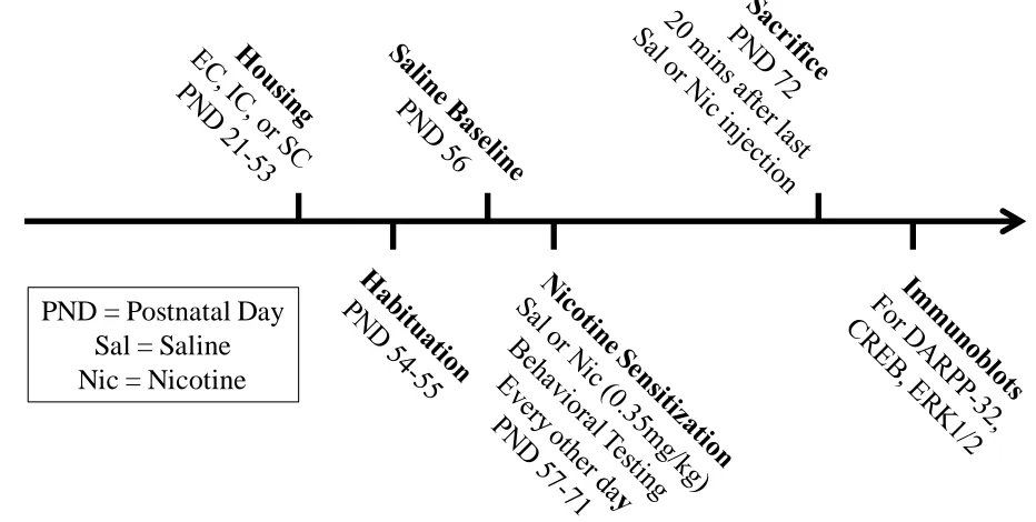
Figure 2.1 Timeline for nicotine sensitization experiments...61
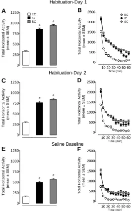
Figure 2.2 Baseline activity in EC, IC, and SC rats ...62
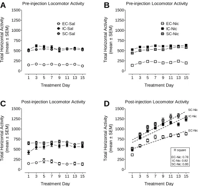
Figure 2.3 Effect of repeated nicotine across sessions on locomotor sensitization in EC, IC, and SC rats ...63
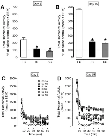
Figure 2.4 Effect of repeated nicotine within session on locomotor sensitization in EC, IC, and SC rats ...64
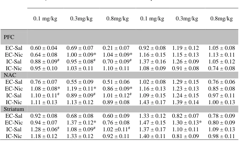
Figure 2.5 Basal and repeated nicotine-mediated pDARPP-32 activity within the mesocorticolimbic circuitry in EC, IC, and SC rats...66
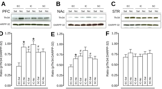
Figure 2.6 Basal and repeated nicotine-mediated pCREB activity within the mesocorticolimbic circuitry in EC, IC, and SC rats...67
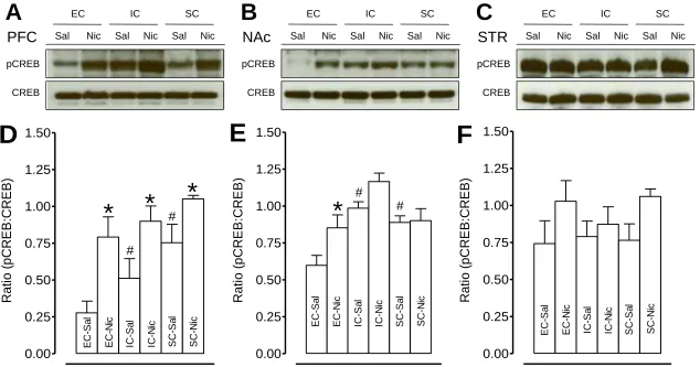
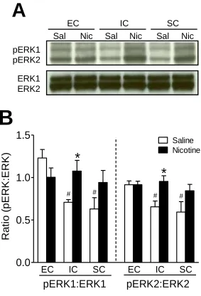
Figure 2.7 Basal and repeated nicotine-mediated pERK1/2 activity within the PFC of EC, IC, and SC rats ...68
Figure 2.8 Correlations of pDARPP-32 and pERK1/2 expression with basal locomotor levels of EC, IC, and SC rats ...69
Figure 2.9 Effect of SL327 on acute nicotine-mediated locomotor levels in EC, IC, and SC rats ...70
Figure 2.10 Effect of SL327 on acute nicotine-mediated pERK1/2 levels in EC, IC, and SC rats ...71
Figure 3.1 Timeline for identification of prefrontal miR expression in EC, IC, and SC rats in response to repeated nicotine ...100
Figure 3.2 Profile of prefrontal miR expression in EC, IC, and SC rats in response to repeated nicotine ...101
ix
activity...103
Figure 3.4 Timeline for nicotine sensitization experiments involving in vivo miR-221
overexpression ...104
Figure 3.5 Validation of in vivo miR-221 overexpression in the PFC...105
Figure 3.6 Effect of in vivo miR-221 overexpression in the mPFC of EC, IC, and SC rats .
on nicotine-mediated locomotor sensitization ...106
Figure 3.7 Effect of in vivo miR-221 overexpression on nicotine-mediated pERK1/2
activity in the mPFC of EC, IC, and SC rats ...107
Figure 4.1 Timeline for nicotine self-administration experiments ...129
Figure 4.2 Nicotine self-administration behavior in EC and IC rats ...130
Figure 4.3 Effect of nicotine self-administration on pERK1/2 activity within the PFC of EC and IC rats ...131
Figure 4.4 Effect of nicotine self-administration on OXR1 expression within the PFC of EC and IC rats ...132
Figure 5.1 Pathway analysis for differential prefrontal mRNA expression in EC and IC rats in response to repeated nicotine administration ...138
Figure 5.2 Pathway analysis for differential prefrontal mRNA expression in EC and IC rats in response to nicotine self-administration ...139
x
L
IST OFS
YMBOLS± Plus/minus
µ Micro
Δ Delta
° Degree
n Number of samples / subjects
ΔΔ Delta delta
V Volts
C Celsius
h Hour (time)
s Second (time)
min Minutes (time)
mg Milligram
g Gram
kg Kilogram
µl Microliter
xi
L
IST OFA
BBREVIATIONSAcH ... Acetylcholine
ANOVA ... Analysis of Variance
CAMKII ... Calcium/Calmodulin-dependent Protein Kinase II
cAMP ... Cyclic Adenosine Monosphosphate
cDNA ... Complementary DNA
CPP ... Conditioned Place Preference
CREB ...cAMP response-element binding protein
D1 ... Dopamine receptor D1
D2. ... Dopamine receptor D2
DA ... Dopamine
DARPP-32 ... Dopamine- and cAMP-regulated Phosphoprotein
DAT ... Dopamine Transporter
DOPAC ... 3,4-Dihydroxyphenylacetic Acid
EC ... Enriched Condition
ELK-1 ... Ets-like protein-1
ERK1/2 ... Extracellular signal-regulated kinase 1/2
FR ... Fixed Ratio
GFP ...Green Fluorescent Protein
i.p. ... Intraperitoneal
IC...Impoverished Condition
LV ... Lentivirus
xii
miR ... MicroRNA
mPFC ... Medial Prefrontal Cortex
NAc ...Nucleus Accumbens
Nic ... Nicotine
NMDA ... N-methyl-D-aspartate
OXR1 ... Orexin Receptor-1
PBS ... Phosphate Buffer Saline
PC12 ... Pheochromocytoma cell line
pCREB ... Phosphorylated CREB
pDARPP-32 Thr34... Phosphorylated DARPP-32 at Threonine 34
pDARPP-32 Thr75... Phosphorylated DARPP-32 at Threonine 75
pERK1/2 ...Phosphorylated ERK1/2
PFC ... Prefrontal Cortex
PKA... Protein Kinase A
PND... Postnatal Day
PP-1 ... Protein phosphatase-1
PR ... Progressive Ratio
qPCR ... Quantitative Reverse-Transcriptase PCR
s.c. ... Subcutaneous
S.E.M. ... Standard Error of the Mean
Sal ...Saline
STR ... Striatum
1
CHAPTER
1
G
ENERALI
NTRODUCTION1.1SIGNIFICANCE
Tobacco use is the number one preventable cause of death (Benowitz, 2010). Yet,
tobacco use is linked to 1 in 5 deaths (~440,000/year) in the United States every year and
tobacco-related disease is estimated to cost the United States $160 billion annually
(Benowitz, 2008, Mathers and Loncar, 2006). These staggering numbers contribute to the
grim statistic that globally, 5.4 million people die each year from tobacco use, and this
statistic is estimated to rise to 8 million deaths per year in the next 30 years. (Mathers and
Loncar, 2006, Benowitz, 2008, Ezzati and Lopez, 2003). Nicotine is the primary
psychoactive, addictive component responsible for the reinforcing properties in tobacco
products (Stolerman and Jarvis, 1995). Approximately 1/3 of the world’s adult population
uses a form of tobacco, thus making nicotine the most widely used, addictive drug of
abuse (Peto et al., 1996). Notably, of the 60-70% of people who try tobacco products,
only 20-25% progress to become daily dependent users (Benowitz, 2010). As a result,
there is a critical need to identify the mechanism(s) underlying the initiation and
development of nicotine addiction.
Gene-environment interactions contribute to nicotine abuse vulnerability (i.e.
whether a person becomes addicted or not). In particular, previous research has
2
(Leshner, 2000, Rhee et al., 2003, Compton et al., 2005). Additionally, shared genetic
factors involved in addiction vulnerability can be influenced by environmental conditions
(Hopfer et al., 2003, McGue et al., 1996). Therefore, one key mechanism to unlock what
underlies nicotine addiction is to understand how environmental factors shape individual
vulnerability to nicotine addiction.
1.2NICOTINE ADDICTION
Nicotine addiction is characterized as a relapse disorder consisting of a vicious
cycle of: intoxication (initial drug intake), bingeing (loss of control in drug intake),
withdrawal and emergence of a negative emotional state, craving of the drug, and then
relapse of the drug (Koob and Volkow, 2010, Koob and Le Moal, 1997). There also
appears to be two shifts in the progression of addiction. First, there is a shift from
impulsive to compulsive use of the drug. And second, there is a shift from positive
reinforcement (i.e. engage in the drug due to the euphoric-like effects) to negative
reinforcement (i.e. engage in the drug to relieve the unpleasant withdrawal-like effects)
(Koob et al., 2004, Koob, 2004). Thus, nicotine addiction within an individual is a
constant struggle to maintain an equilibrium in receiving the euphoric-like properties of
the drug, yet avoid the withdrawal-like symptoms of the drug. However, the
neurobiological mechanisms underlying each component of the nicotine addiction cycle
remains infantile at best, and future research is desperately needed to aid in therapeutic
strategies for prevention in the initiation, maintenance, and relapse of nicotine addiction.
The molecular adaptations in response to drug exposure serve as a gateway to the
3
development of rodent behavioral models of addiction has served as a preclinical method
for evaluating addiction in humans.
1.3DRUG-INDUCED LOCOMOTOR SENSITIZATION
One common behavioral adaptation in response to repeated administration of
addictive drugs is a sensitized behavioral or locomotor response. Following repeated
injections of a drug, rodents will display hyperlocomtor activity upon each subsequent
injection. Although locomotor sensitization does not measure reward or reinforcement
directly per se, this behavioral paradigm can enhance aspects of both conditioned place
preference (CPP) and drug self-administration. Because locomotor sensitization reflects
the sensitized dopaminergic activity within the motivational circuitry, this paradigm
represents an indirect measure of addiction in rodents with an emphasis on measuring the
incentive salience or wanting of the drug, and the subsequent drug-related stimuli
(Robinson and Berridge, 1993, Robinson and Berridge, 2008). Additionally, many of the
neural correlates of the locomotor sensitization paradigm overlap with the neural
correlates of CPP and SA. Therefore, locomotor sensitization is a more “simpler” tool for
dissecting how repeated exposures of a drug influence neuroadaptations. Notably,
locomotor sensitization persists for months and potentially years, even in the absence of
the drug during this timeframe (Robinson and Becker, 1986, Paulson et al., 1991).
Moreover, there is evidence that sensitization does indeed occur in humans (Strakowski
et al., 1996, Strakowski et al., 2001, Boileau et al., 2006). But due to the indirect nature
of this behavioral measurement there are limitations surrounding behavioral sensitization.
4
supporting the notion that the sensitization may be a critical process in initiating addition,
whereby drug addiction seems to involve a more long-term drug exposure. Secondly,
experimenter-delivered drug appears to increase the wanting of non-drug reinforcers (i.e.
food and sexual stimuli) (Nocjar and Panksepp, 2002, Nocjar and Panksepp, 2007).
However, this model is very useful in examining molecular changes in response to drugs
of abuse as all animals receive the same amount of the drug. To conclude, locomotor
sensitization may not include all aspects that encompass addiction, but this behavioral
paradigm has amassed immense validation in dissecting the neuroadaptations
accompanying exposure to drugs of abuse.
1.4DRUG-INDUCED CONDITIONED PLACE PREFERENCE (CPP)
CPP reflects the rewarding properties of drugs of abuse. Although CPP
experiments were not performed directly in the studies outline here, CPP will be referred
to throughout this document, and is needed to understand some of the mentioned
behavioral effects to drugs of abuse. As with locomotor sensitization, CPP does not
reflect the full spectrum of addiction as both paradigms employ experimenter-delivered
drugs. Animals are injected with the drug in one-context specific chamber, and also
injected with the control/vehicle in a separate context-specific chamber. After multiple
injection-context pairings, animals learn to associate the euphoric-like properties of the
drug within a specific context. When post-tested in an injection-free environment, where
animals can opt to stay in the drug-paired chamber (animals display CPP) or the
vehicle-paired chamber (animals display conditioned place aversion), animals will spend more
5
effects of the drug. Hence, CPP is thought of as an indirect measure of addiction by
characterizing the reward-like properties of addictive drugs.
1.5DRUG SELF-ADMINISTRATION
Self-administration measures the reinforcing properties of drugs of abuse. Unlike,
locomotor sensitization or CPP which involves experimenter-delivered drugs, the
self-administration paradigm allows the animals to control one’s own drug intake. For this
reason, the drug self-administration procedure is the best preclinical model of addiction
and is considered the most reliable and predictive experimental model for evaluating the
reinforcing effects of drugs in animals (Panlilio et al., 2008). Indwelling intra-jugular
catheters are surgically placed in the animals. Animals are then placed in an operant box
and learn to perform an instrumental action (lever press or nose poke) to receive a drug
infusion via the intravenous catheter. This allows researchers to assess the reinforcing
effects of drugs of abuse through a variety of different constructs by altering schedules of
reinforcement and doses. Fixed-ratio (FR) schedules require animals to make a set
number of responses to receive the drug (i.e. FR-5, animal has to make 5 lever presses to
receive drug) and at its simplest, measures drug-intake behavior (Koob, 1992).
Additionally, a progressive-ratio (PR) schedule reflects how hard an animal is willing to
work for the drug (motivation) by exponentially increasing the number of actions
required to obtain the drug after each successful drug infusion (Richardson and Roberts,
1996). Moreover, experimenters can emulate the consumption of large quantities of the
drug seen in humans via the escalation model (Ahmed and Koob, 1998). Here, animals
6
prolonged group will gradually begin to increase drug responding, therefore representing
the binge intake reflected in human drug addicts. For these reasons, self-administration
appears to represent the most advantageous pre-clinical model for studying drug
addiction.
1.6MESOCORTICOLIMBIC DOPAMINERGIC SYSTEM
The mesocorticolimbic dopaminergic (DAergic) system, which is responsible for
the transmission of the catecholinergic neurotransmitter dopamine (DA), consists of
initial projections from the ventral tegmental area (VTA) that innervate the nucleus
accumbens (NAc) as well as the prefrontal cortex (PFC) (Tzschentke, 2001). The
mesocorticolimbic DAergic system participates in a variety of processes including
motivational salience, motor functions for initiating goal-directed behaviors,
attention/cognition, and encoding the cues and values that regulate the rewarding and
reinforcing-like properties of external stimuli (Horvitz, 2000, Tzschentke, 2001,
Salamone and Correa, 2012). However, recent research has also found that this rewarding
system is intertwined with the aversive-like nature of events (Salamone and Correa, 2012,
Volman et al., 2013, Brooks and Berns, 2013). Thus, for all intents and purposes, this
DAergic circuit maximizes the ability to assign “value” or update the current “value” to
both appetitive and aversive stimuli for future actions.
The basal activity of DAergic neuronal firing within this circuitry consist of a
tonic, steady state, however, when salient or aversive stimuli is encountered (i.e. food,
sex, etc.) DAergic neuronal firing is altered to strengthen the behaviorally-relevant
7
topic, drugs of abuse prey on this circuitry to mediate the rewarding and reinforcing
properties of drugs of abuse that underlie addiction.
1.7NEUROCHEMICAL SUBSTRATES OF NICOTINE ADDICTION
For the purpose of the present topic, the review of the neurochemical substrates of
nicotine addiction will focus on the pharmacology of nicotine’s actions and the initial
neurochemical events leading up to the neuroadaptations underlying addiction. Nicotine
initially acts on nicotinic acetylcholine receptors (nAChRs) found abundantly throughout
the brain. NAChRs are pentameric receptor complexes consisting of a heteromeric or
homomeric assortment of 12 subunits (α2-α10 and β2-β4) that differ in pharmacokinetic
properties (McGehee and Role, 1995, Role and Berg, 1996). These receptors are
distributed throughout the entire brain, and thus determining the exact mechanism that
causes nicotine addiction is very difficult to examine. Nicotine binds ligand-gated ion
channels allowing for the entry of sodium or calcium, and which mediate subsequent
presynaptic- and postsynaptic-dependent neurotransmitter release (Dajas-Bailador and
Wonnacott, 2004). Corresponding with all psychostimulants, nicotine’s rewarding and
reinforcing properties are due to nicotine’s actions within the mesocorticolimbic DAergic
circuitry (Berridge and Robinson, 1998, Laviolette and van der Kooy, 2004). The basal
activity of DAergic neuronal firing within this circuitry consist of a tonic, steady state;
however when salient-stimuli is encountered (i.e. food, sex, etc.) DAergic neuronal firing
shifts to a more rapid, phasic/burst state to shape and strengthen the reward-associated
stimuli to further promote goal-directed actions (Grace, 2000). Nicotine “hijacks” this
8
GABAergic cell bodies that inhibit VTA DAergic neuronal firing. These nAChRs are
highly concentrated in VTA cell bodies, and allow GABAergic neurons to quickly
desensitize allowing for the disinhibition and increased firing of VTA DAergic neurons
(Mansvelder and McGehee, 2002, Mansvelder et al., 2002). These DA increases are
compounded by the fact that nicotine also binds to low affinity, slow desensitizing
α7-containing nAChRs on glutamatergic-α7-containing neurons that are highly concentrated
within the PFC (McGehee et al., 1995). Besides receiving dense dopaminergic
projections from the VTA and NAc, the PFC has reciprocal glutamatergic connections to
these same brain regions (Grace et al., 2007). This allows nicotine to induce glutamate,
the main excitatory neurotransmitter in the brain, release onto NAc and VTA
dopaminergic cell bodies and allowing for increased DAergic excitation (Girod et al.,
2000). Thus, the PFC modulates the activity of the NAc and VTA in response to nicotine
(see figure 1.1). These nicotine-dependent DAergic and glutamatergic alterations appear
critical in shaping the neuroadaptations responsible for the initial addiction process.
Further, these neuroadaptations are thought to be dependent upon the activity of complex
intracellular signaling systems between the cell surface and the nucleus (Dajas-Bailador
and Wonnacott, 2004) (see figure 1.2). Thus, nAChRs may not directly affect many of
the implicated intracellular signaling systems such as DA- and cAMP-regulated
phosphoprotein-32 (DARPP-32), extracellular signal-regulated kinase1/2 (ERK1/2, two
isoforms with overlapping structure and function), and cAMP-response element-binding
protein (CREB), but rather indirectly through nAChR-mediated dopamine and glutamate
9
Activation of the DA/D1 receptor/cAMP/protein kinase A (PKA) pathway
increases phosphorylation of DARPP-32 at the site Threonine 34 (pDARPP-32 Thr34),
but decreases phosphorylation of DARPP-32 at Threonine 75 (pDARPP-32 Thr75) (Nishi
et al., 2000). In contrast, phosphorylation of DARPP-32 at Thr75 by cyclin-dependent
kinase 5 has a major inhibitory effect on pDARPP-32 Thr34, thereby reducing D1 DA
signaling through the DARPP-32/ protein phosphatase-1 (PP-1) cascade (Nishi et al.,
2000, Bibb et al., 1999). In addition, activation of this PKA pathway enhances
phosphorylation of the transcription factor CREB at serine 133, (Nairn et al., 2004,
Svenningsson et al., 2005, Dash et al., 1991). In an opposing fashion, activation of Ca2+-
dependent calcineurin by D2 receptors and glutamate-activated N-methyl-D-aspartate
(NMDA) receptors results in dephosphorylation of pDARPP-32 Thr34 (Lindskog et al.,
1999). ERK1/2, a member of the mitogen-activated protein kinase (MAPK) signaling
pathway also signals to CREB at Ser133 via glutamate-dependent NMDA receptor
activation of calcium-mediated calcium/calmodulin-dependent protein kinase II
(CAMKII) activation, in addition to DARPP-32 being able to control ERK activation via
PP-1 (Valjent et al., 2000, Valjent et al., 2005). Consequently, these intracellular
signaling proteins are essential for neuronal plasticity in response to repeated nicotine
exposure. Therefore, long-term use of nicotine mediates signaling-induced
neuroadaptations responsible for the manifestation of the addicted phenotype. However,
research into nicotine’s regulatory processes on intracellular signaling-induced gene
10
1.8MICRORNAS
MicroRNAs (miRs) are small (~22 nucleotides in length) non-coding RNAs
encoded by the genome that function as gene regulators (Bartel, 2004). Biogenesis of
mature miRs are first derived from nuclear transcription by RNA polymerase II/III to
form hairpin structures known as primary miRs (pri-miR); which are then further cleaved
by the Drosha RNAse III endonuclease to give rise to a smaller hairpin structure known
as precursor miR (pre-miR) (Lee et al., 2002, Lee et al., 2003). The pre-miR is then
exported via exportin-5 into the cytoplasm, where another RNase III endonuclease
(Dicer) further cleaves the pre-miR into a miR duplex consisting of a “guide” strand
(miR) and a “passenger” strand (miR*) (Lee et al., 2003, Yi et al., 2003). The mature
miR or miR* strand is then translocated to the RNA-induced silencing complex (RISC),
where the miR-RISC complex is guided to the target mRNA transcript to facilitate either
mRNA degradation or mRNA silencing depending on the seed region (nucleotides 2-7 on
the 5’ region) of the miR (Sempere et al., 2004, Bartel, 2004, He and Hannon, 2004).
Although both methods serve a similar outcome by preventing translation, perfect
complimentarity in base-pairing interactions between the miR and the target mRNA
results in mRNA degradation, where as imperfect complimentarity in base-pairing
interactions between the miR and the target mRNA results in mRNA silencing. After
translational disruption, miRs can actually take place in multiple rounds of cleavage,
thereby allowing for the effects of miRs to be amplified. Adding to the complexity of
miRs, is the fact that multiple miRs can target the same mRNA transcript, and a single
miR may target multiple mRNA transcripts (Esteller, 2011). Additionally, the non-seed
11
mRNA (Orom et al., 2008, Elcheva et al., 2009). Since their discovery in 1993 (Lee et al.,
1993), the complex nature of miRs seems to be ever-growing. However, due to the ability
of miRs to regulate gene expression, miRs are rapidly being implicated in the
neuroadaptations involved in neurological diseases, where miRs are now being touted as
potential therapeutic targets (Kosik, 2006).
1.9MICRORNAS AND DRUG ADDICTION
Due to the nature of drug addiction, it should come as no surprise that miRs are
now implicated in the pathological process of addiction. The root of drug addiction is the
ability of drugs of abuse to impinge on the brain’s gene expression networks to produce
subsequent neuroplastic alterations. In fact, very recently, there have been several
seminal studies implicating miRs as regulators of not only drug-induced plasticity within
the reward circuitry, but of drug-induced behaviors as well. A study from the Kenny
laboratory (Hollander et al., 2010, Im et al., 2010), recently found that miR-212 was
drastically upregulated in the striatal regions of rats undergoing a cocaine-escalation
model. Interestingly, overexpressing exogenous miR-212 in the striatum decreased
intake, whereas blocking endogenous miR-212 expression increased
cocaine-intake; suggesting a regulatory switch of miR-212 in controlling the motivational
properties of cocaine. This behavioral manifestation was later found to be due to
homeostatic interactions of miR-212 with MeCP2 to regulate cocaine-induced of
brain-derived neurotrophic factor (BDNF) expression within the striatum. Chandrasehkar and
Dreyer (Chandrasekar and Dreyer, 2009, Chandrasekar and Dreyer, 2011) also
12
miR-124 and let-7d within the NAc of rats. Subsequent overexpression of miR-124 and
let-7d into the NAc attenuated cocaine-induced CPP, whereas overexpression of
miR-181a within the NAc enhanced cocaine-induced CPP. Although not as specific as the
previously mentioned study in determining the mRNA targets of the identified miRs, the
cocaine-induced CPP alterations depended on the ability of the three miRs to
differentially target multiple mRNA plasticity genes that have been implicated in cocaine
addiction including, but not limited to, DAT, FosB, D2 receptor, BDNF, MeCP2, and
pCREB levels. However, the actions of miRs are not exclusive to cocaine exposure. In a
study by Tapocik and colleagues (Tapocik et al., 2014), miR-206 was found to be
upregulated in the mPFC of rats after long-term exposure to alcohol vapor. When
miR-206 was overexpressed into the mPFC of drug-naïve rats undergoing alcohol
self-administration, rats that received exogenous miR-206 were found to significantly
consume more alcohol and induce escalation within these rats. Further analyses
determined miR-206 inhibits BDNF expression within the mPFC, thereby preventing
BDNF-induced neuroadaptations within the mPFC that may underlie the increased
alcohol consumption. Relevant to the current studies outlined here, nicotine exposure has
been recognized to differentially regulate a multitude of miRs depending on in vitro or in
vivo models, dose, and length of exposure (Huang and Li, 2009, Lippi et al., 2011, Taki
et al., 2014); however, the involvement of these nicotine activated miRs in mediating
nicotine-induced neuroplasticity and behavior remain unexplored. MiRs, therefore
represent critical mediators in regulating the behavioral consequences of drugs of abuse
that correspond to the homeostatic mechanisms involved in synaptic signaling and
13
1.10ENVIRONMENTAL ENRICHMENT
An enriched environment is “a combination of complex inanimate and social
stimulation” (Rosenzweig et al., 1978) typically employed to dissect the role of
environmental factors in inbred animals within a given experimental paradigm. By and
large, an experimental setting consists of an enriched condition (EC) consisting of novel
objects, social cohorts in a large-scale cage, and an enhancement of physical activity.
This EC condition is subjected to comparison to either both or one of the controlled
environments; an impoverished condition (IC) consisting of no novelty and no social
cohorts in a small-scale cage, and a standard condition (SC) consisting of social cohorts
with no novelty in an NIH-standard cage. These housing conditions are typically utilized
during the animals’ adolescence period before experimental testing to prevent an
experimental outcome, or utilized after an experimental paradigm to attenuate or reverse
the results of an experimental outcome. The complexity of the enriched environment
paradigm can have its limitations as this model comprises of multiple components: a
large space, physical exercise, novel objects, and social cohorts. Although it appears that
each factor may contribute to the enrichment-induced protective effect in response to
drugs of abuse (Kanarek et al., 1995, Meeusen and De Meirleir, 1995, Lynch et al., 2010,
Solinas et al., 2008, Gipson et al., 2011); isolating and determining which factor
contributes to the neuroprotective-like phenotype is necessary for further understanding
the role of environmental factors in nicotine abuse vulnerability. In fact, due to the
variety of factors involved in an enriched condition, some experimenters have adopted
14
impoverished rats are housed with novelty, and a social condition consisting of social
cohorts with no novelty in a large-scale cage (Gipson et al., 2011). However, these may
not prove feasible in every laboratory as this drastically increases the number of animals
and groups needed for the experiment. Nevertheless, the environmental enrichment
paradigm has been critical in establishing how environment influences behavioral and
neurochemical adaptations across multifaceted research fields.
1.11ENRICHMENT AND DRUG-MEDIATED BEHAVIORS
Because both genetic and environmental factors mediate whether or not an
individual progresses from a habitual drug user into an addict, it is not surprising that
differential rearing conditions in genetically identical rodents affect subsequent
addiction-related behaviors. There has been a vast amount of research into multiple
preclinical paradigms demonstrating that rodents raised in an enriched environment
display a neuroprotective-like phenotype to drug-induced behaviors in comparison to rats
raised in impoverished or standard conditions, with the majority of studies focusing on
psychostimulants.
In comparison to IC and SC rats, rodents raised in an enriched environment
appear to have increased sensitivity to the locomotor-activating effects of acute
amphetamine and cocaine administration (Bowling et al., 1993, Smith et al., 1997),
however, enrichment decreases the locomotor response to acute nicotine (Green et al.,
2003a) administration. When examining the locomotor response to repeated
administration of the drug, EC rodents exhibit reduced locomotor sensitization to
amphetamine (Bardo et al., 1995), cocaine (Smith et al., 1997), nicotine (Green et al.,
15
1997), and ethanol (Rueda et al., 2012). Interestingly, there were no differences in either
heroin- or methamphetamine-mediated locomotor sensitization (El Rawas et al., 2009,
Thiriet et al., 2011), however this may be due to rodent differences as these studies were
focused on mice, whereas the former studies focused on rats. In a CPP paradigm,
enrichment in rats increases the rewarding-like properties of amphetamine (Bowling and
Bardo, 1994), cocaine (Green et al., 2010), and morphine (Bardo et al., 1997). When
evaluating the effect of enrichment on the reinforcing properties of drugs of abuse, EC
rats, in contrast to IC and SC rats, have decreased responding on FR schedules of
reinforcement for amphetamine (Bardo et al., 2001), cocaine (Green et al., 2010), ethanol
(Deehan et al., 2011) and methylphenidate (Alvers et al., 2012), in addition to decreased
responding on PR schedules of reinforcement for cocaine (Green et al., 2010) and
amphetamine (Green et al., 2002). Moreover, enrichment also decreases cocaine-taking
behavior in an escalation paradigm in comparison to IC and SC rats (Gipson et al., 2011).
However, it should be mentioned that the majority of the findings regarding EC-induced
reductions in drug-taking behavior appear at low unit doses for the drug, where as the
responding rates in EC and IC rats were found to be not significantly different at high
unit doses. These behavioral adaptations are intriguing as enrichment appears to actually
increase the euphoric effect of drugs of abuse, yet somehow blunts the drug-taking
behavior. Overall, enrichment appears to have a preventative effect in the behavioral
responses to drugs of abuse. Although the focus for the before mentioned work focuses
on enrichment exposure before and during the drug exposure itself, it should be noted
that enrichment has a potential curative effect as well. After drug-mediated behaviors
cocaine-16
mediated locomotor sensitization (Solinas et al., 2008), cocaine-induced CPP (Solinas et
al., 2008); and cue- and stress-induced reinstatement of cocaine CPP (Chauvet et al.,
2009) and self-administration (Thiel et al., 2010, Thiel et al., 2011).
Collectively, exposure to environmental enrichment increases acute locomotor
sensitivity to drugs of abuse, decreases repeated drug-induced locomotor sensitization,
increases the rewarding properties of drugs of abuse, and decreases the reinforcing
properties of drugs of abuse. Accordingly, environmental enrichment produces powerful
behavioral adaptations in response to drugs of abuse, which are likely controlled by
enrichment-induced neuroadaptations. The environmental enrichment paradigm allows
researchers to critically examine how extreme conditions, as represented by the IC
condition, contribute to drug abuse vulnerability preclinically, while also allowing
researchers to control every aspect of the environmental conditions. Although,
environmental conditions are very diverse across humans, studies have shown clinical
relevance with regard to environmental influence in individual vulnerability to drug
abuse. In particular, previous research has determined that vulnerability to drug abuse is
highly dependent on environmental factors (Leshner, 2000, Rhee et al., 2003, Compton et
al., 2005). Additionally, shared genetic factors involved in addiction vulnerability can be
influenced by environmental conditions (Hopfer et al., 2003, McGue et al., 1996). Thus,
the environmental enrichment paradigm is able to extend the clinical findings of the
effect of environmental conditions on the behavioral aspects of individual susceptibility
17
1.12PREFRONTAL CORTEX:KEY AREA FOR ENRICHMENT-INDUCED BASAL ADAPTATIONS
The neurochemical correlates of an enriched environment stretch across numerous
brain regions, but one critical “hotspot” that is targeted by enrichment is the PFC. The
PFC is of particular importance for this review in that the PFC is a heterogenous structure
involved in regulating reward processing, habit learning, impulsivity, and drug seeking
behaviors (Perry et al., 2011). The PFC receives dense ascending dopaminergic
innervations from the NAc and VTA, while sending reciprocal descending glutamatergic
projections back to these areas (Grace et al., 2007). Thus, by altering the basal state of the
PFC, environmental enrichment may allow animals to better adapt to the drug-induced
response.
Structurally, enrichment increases dendritic spine density, dendritic length, and
dendritic branching within the PFC of primates. Importantly, there were no significant
differences in the number of neurons in PFC regions, suggesting that increases in
dendritic arborization play a relatively stronger role (Kozorovitskiy et al., 2005). These
prefrontal structural modifications have also been supported in rats as
enrichment-induced alterations in spine density, dendritic branching, and dendritic length were found
in the PFC of male and female rats (Mychasiuk et al., 2014). Additionally, indirectly
measuring the number of synapses through synaptophysin levels, enrichment increases
the number of synapses in the PFC (Kozorovitskiy et al., 2005, Nithianantharajah et al.,
2008). Together, these studies provide convincing evidence that enrichment modifies
prefrontal structure. What remains relatively underexplored is the way in which
18
The dopaminergic system within the PFC is highly vulnerable to
enrichment-mediated alterations. IC gerbils have reductions in DA innervations into the medial and
orbital portions of the PFC in comparison to EC gerbils (Winterfeld et al., 1998, Neddens
et al., 2001). Enrichment has been shown to have no effect on DA levels in the rodent
PFC (Naka et al., 2002, Zhu et al., 2004). While examination of the DA metabolite,
3,4-Dihydroxyphenylacetic acid (DOPAC) has been shown to be decreased in rats in
response to enrichment (Zhu et al., 2004); there appears to be no differences in DOPAC
or homovanilic acid (HVA) content found in mice (Naka et al., 2002). The seemingly
disparate results of decreased DOPAC content in the PFC of EC rats with no changes to
the DA levels, may be a result of modifications to the dopamine transporter (DAT). In
fact, enrichment decreased the maximal velocity of [3H] DA uptake with no differences
in Bmax or Kd values of DA binding sites (Zhu et al., 2004), suggesting a potential DAT
trafficking-dependent mechanism induced by enrichment. In support of this view, while
no DAT immunoreactivity was observed in the PFC of EC or IC rats, cell surface
biotinylation assays revealed a reduction in cell surface DAT expression in EC rats with
no differences seen in total DAT levels (Zhu et al., 2005b). Additionally, EC rats have
been shown to be more sensitive to prefrontal DAT internalization induced by protein
kinase C-dependent mechanisms (Wooters et al., 2011). While there were no differences
found in baseline DA clearance from the synapse between EC and IC rats, there does
appear to be some evidence that the DA clearance rate is slower in EC rats in comparison
to IC rats (Zhu et al., 2007a). Thus, these data heavily supports the idea that enrichment is
capable of modifying DAT function as a result of a DAT trafficking-dependent
19
dopaminergic system at the cell surface. EC rats have decreased D1 receptor density in
the PFC (Del Arco et al., 2007a). This was accompanied by experiments showing that
perfusion of the D1 agonist, SKF38393, into the PFC increases local acetylcholine (AcH)
release in IC rats only, suggesting that EC rats decrease the expression and function of
prefrontal D1 receptors. Interestingly, we have found that EC rats have increased D2
receptor mRNA compared to IC rats within the PFC (unpublished data), while others
have shown that compared to SC rats, IC rats have decreased D2 receptor expression
(Fitzgerald et al., 2013). These data support the idea that an enriched environment likely
increases the dopaminergic inhibitory tone in the PFC, while potentially decreasing the
dopaminergic stimulatory tone within the PFC.
In opposition, enrichment appears to increase the glutamatergic tone within the
PFC. Microdialysis experiments have determined that there are no differences in the
basal levels of PFC glutamate levels between EC and IC rats (Melendez et al., 2004;
Rahman and Bardo, 2006); however, the function and expression of cell surface
glutamate receptors appear susceptible to enrichment-mediated adaptations. Enriched
mice have been shown to have increased AMPA receptor subunit GluR1, and NMDA
receptor subunits NR2B and NR2A expression, with no change to NR1 expression in the
mouse forebrain compared to standard mice (Tang et al., 2001). Yet, others have found
no differences in AMPA GluR1 levels (Nithianantharajah et al., 2008) and NR2A or
NR2B expression (Shum et al., 2007) between enriched and non-enriched mice.
However, the latter study did show an enrichment-induced enhancement of the
NR2B:NR2A current ratio, which further corresponded to increased AMPA-mediated
20
Moreover, enrichment alters metabotropic-glutamate receptor (mGluR) subunit
composition in a region-specific manner within the PFC (Melendez et al., 2004). Due to
the fact that a group 1 mGluR agonist and a mGluR2/3 antagonist failed to elevate
extracellular glutamate in IC rats, it is likely that EC rats have enhanced prefrontal
mGluR transmission compared to IC rats (Melendez et al., 2004). In conclusion,
enrichment increases glutamatergic tone within the PFC in addition to decreasing the
dopaminergic tone within the PFC.
Although the emphasis of enrichment-induced alterations in the literature appears
to be concentrated on the dopaminergic and glutamatergic systems, enrichment is not
limited to these systems. Enrichment has been found to have no effect on norepinephrine
levels or its metabolites in the rodent PFC (Naka et al., 2002, Brenes et al., 2008).
Additionally, enrichment’s effects on the serotonergic system is conflicting with one
group reporting no changes in prefrontal serotonin levels or its metabolites (Naka et al.,
2002), and another reporting that enrichment increased serotonin levels in the PFC of
rodents (Brenes et al., 2008). Also, no changes in basal levels of acetylcholine in the PFC
have been found (Del Arco et al., 2007b, Segovia et al., 2008). Aside from the classical
neurotransmitter systems, corticosterone levels and glucocorticoid receptors remain
unchanged in the PFC between EC and IC rats (Garrido et al., 2013). Moreover, studies
of the PFC have revealed that enrichment decreases basal BDNF levels in mice (Rueda et
al., 2012); however, in rats BDNF levels have been found to be unaltered between EC
and IC rats (Chen et al., 2005).
Enrichment also mediates downstream targets beyond the cell surface. EC rats
21
pDARPP-32 Thr75 or total DARPP-32 levels in the PFC (Gomez et al., 2012).
Additionally, EC rats have lower pCREB levels, with no change in total CREB, within
the PFC compared to IC and SC rats (Gomez et al., 2012). The enrichment-mediated
alterations in basal DARPP-32 and CREB activity may reflect the dysregulated
dopaminergic and glutamatergic systems within the PFC of EC rats. There also appears to
be strong evidence that enrichment increases prefrontal basal levels of immediate early
genes including c-fos, ΔFosB, zif-268, and Arc (Pinaud et al., 2001, Shum et al., 2007,
Lehmann and Herkenham, 2011, Venebra-Munoz et al., 2014). In summary, while there
has been a concerted effort to understand the enrichment-induced neuroadaptations that
occur within the PFC, the fact that enrichment-dependent alterations appear at the cellular
and sub-cellular levels leave researchers with a great deal still left to understand.
1.13PREFRONTAL CORTEX:POTENTIAL KEY AREA FOR ENRICHMENT-INDUCED PROTECTION
TO DRUG-INDUCED NEUROADAPTATIONS
The PFC receives dense ascending dopaminergic innervations from the NAc and
VTA, while sending reciprocal descending glutamatergic projections back to these areas
(Grace et al., 2007). Through this circuitry the PFC has the ability to modulate the
behavioral output induced by drugs of abuse. As mentioned previously, enriched rats
have a neuroprotective-like phenotype to the behavioral effects of drugs of abuse, and
also have dramatic basal neuroadaptations in the PFC. As a result, the PFC represents a
potential culprit in mediating the behavioral-induced adaptations observed in rats raised
22
underpinnings of environment-mediated neuroadaptations within the PFC in response to
addictive drugs.
As seen with enrichment’s effect basally, the DAT appears to be altered in EC
rats in response to psychostimulants. After rats were prenatally treated with cocaine, and
then placed postnatally in EC or IC conditions, post-enrichment was shown to attenuate
the nicotine-mediated response of the DAT within the PFC (Neugebauer et al., 2004).
Furthermore, in response to acute nicotine EC rats had dramatic increases in the
dopamine clearance within the PFC in comparison to IC rats, suggesting that EC rats
have altered DAT function in response to nicotine (Zhu et al., 2007a). Moreover, acute
methylphenidate administration was shown to decrease DA uptake in the PFC of EC, but
not IC rats (Wooters et al., 2011). In this fashion, environmental enrichment appears to
modulate the dynamics of the DAT in response to psychostimulants as well as basally.
Moreover, additional research has determined that EC mice further decrease the low
basal BDNF levels in the PFC in response to repeated ethanol exposure compared to SC
mice (Rueda et al., 2012). With respect to examining intracellular activity, there have
been very few studies. In response to chronic nicotine exposure, enrichment increases the
sensitivity of pDARPP-32 Thr34 and pCREB activity in the PFC of rats (Gomez et al.,
2012); as well as diminishes the enrichment-induced baseline increase in ΔFosB within
the PFC in comparison to standard mice (Venebra-Munoz et al., 2014). This is the current
extent of the research regarding drug-induced neuroadaptations in the PFC of rats raised
in an environmental enrichment. Accordingly, more extensive studies are needed to better
characterize the neurobiological consequences of enrichment on drug-mediated
23
1.14SIGNIFICANCE REVISITED
These findings provide a framework whereby environmental enrichment may
serve as a neuroprotective factor in nicotine-mediated behaviors by mediating
environmental-induced neuroadaptations, which are likely to be mediated by
miR-dependent intracellular signaling processes in response to nicotine exposure within the
PFC. The overarching goal of these studies was to identify whether environmental
enrichment alters mediated behaviors via distinct alterations in
nicotine-mediated intracellular signaling (please see figure 1.2 for overview). As a follow-up,
these studies sought to determine the potential regulatory mechanisms controlling
enrichment-dependent changes in nicotine-mediated intracellular signaling. Thus, by
dissecting the prefrontal molecular mechanisms of an enriched environment in
nicotine-mediated behaviors, we may better understand individual vulnerability to nicotine
24
Figure 1.1. An integrated model depicting the role of nicotinic acetylcholine receptors in the mesocorticolimbic brain circuitry. Nicotine binds to nicotinic acetylcholine receptors allowing for the release of numerous
neurotransmitters which regulates the rewarding and reinforcing properties of nicotine. Repeated exposure to nicotine produces neuroadaptations within this brain circuitry leading to nicotine addiction.
DA
GABA
Glutamate
VTA
NAc
PFC
= α7
= α4β2
25
Figure 1.2. An integrated signaling cascade involved in nicotine-mediated intracellular signaling cascades responsible for the neuroadaptations that underlie nicotine-induced behavioral alterations. The central hypothesis is that environmental
enrichment alters nicotine-mediated signaling which is responsible for the enrichment-induced behavioral adaptations in response to repeated nicotine administration.
Ca2+
PKC/CAMK
pERK1/2
cAMP
PKA
pCREB / pELK-1
Enrichment-induced
nicotine-mediated behavior
ERK1/2
Dopamine / D2
PP2B
pDARPP-32 Thr34 / Thr75
Nucleus
26
CHAPTER
2
E
FFECTS OF ENRICHMENT ON NICOTINE-
MEDIATED SENSITIZATION2.1INTRODUCTION
Environmental factors contribute to individual vulnerability for drug abuse
(Leshner, 2000, Rhee et al., 2003). The environmental enrichment paradigm has been
widely used to address how environmental factors influence susceptibility to drug abuse.
In this animal model, rats are raised in one of the three different conditions during
adolescence: an enriched condition (EC), an impoverished condition (IC) or a standard
condition (SC); which differs in novelty, handling, social cohorts, and physical activity.
Exposure within the environmental enrichment paradigm results in robust
neurobiological adaptations, particularly within the prefrontal cortex (PFC) of the
mesocorticolimbic dopaminergic system (Zhu et al., 2004, Bowling et al., 1993, Hall et
al., 1998, Del Arco et al., 2007a). This neural circuit, in part, organizes motivated
behavior and environmental enrichment-dependent alterations to this system are
suggested to be protective against drug-induced maladaptive behaviors (Gomez et al.,
2012, Zhu et al., 2013, Stairs and Bardo, 2009, Bardo et al., 2001).
The behavioral effects of nicotine are primarily mediated by its stimulating
actions on nicotinic acetylcholine receptor-mediated dopamine (DA) release within the
mesocorticolimbic system (Laviolette and van der Kooy, 2004). Long term exposure to
27
behavioral sensitization (Clarke and Kumar, 1983b, Kalivas, 1995, Clarke and Kumar,
1983a). Although behavioral sensitization is not a direct measure of drug reward or
reinforcement, this procedure is sensitive to the behavioral and molecular changes
produced by the psychostimulant effects of abused drugs (Robinson and Berridge, 1993,
Berridge and Robinson, 1998, Wise and Bozarth, 1987). EC rats exhibit reduced
nicotine-mediated locomotor activity compared to IC and SC rats (Green et al., 2003b), which
may be mediated by enriched environment-induced alterations of dopaminergic
pathways. Indeed, drug-naïve EC rats exhibit diminished DA transporter function (Zhu et
al., 2004), less synaptic DA levels in medial prefrontal cortex (Bowling et al., 1993, Hall
et al., 1998), and show decreased D1 receptor function and expression in the PFC
compared with IC and SC groups (Del Arco et al., 2007a). In contrast, repeated nicotine
administration profoundly increases DA clearance and 3,4-Dihydroxyphenylacetic acid
(DOPAC) levels in the PFC of EC rats but not in IC rats (Zhu et al., 2007b). Therefore,
EC rats may have a lower dopaminergic tone compared to IC rats under basal conditions,
which may contribute to differential behavioral responses to psychostimulants. Acute and
repeated nicotine administration activates the DA/D1 receptors/cAMP/protein kinase
pathway, including DA- and cAMP-regulated phosphoprotein-32 (DARPP-32),
extracellular signal-regulated kinase1/2 (ERK1/2, two isoforms with overlapping
structure and function), and cAMP-response element-binding protein (CREB)
(Nakayama et al., 2001, Hamada et al., 2004, Valjent et al., 2004a). Moreover, alterations
in the phosphorylation states of these proteins have been shown to alter the expression of
nicotine-induced behavioral sensitization (Valjent et al., 2006b, Addy et al., 2007). As a
28
are the result of nicotine’s indirect action on the plasticity of these intracellular signaling
proteins via nAChR-dependent increases in dopamine and glutamate release
(Dajas-Bailador and Wonnacott, 2004).
Activation of the DA/D1 receptor/cAMP/protein kinase A (PKA) pathway
increases phosphorylation of DARPP-32 at the site Threonine 34 (pDARPP-32 Thr34),
but decreases phosphorylation of DARPP-32 at Threonine 75 (pDARPP-32 Thr75) (Nishi
et al., 2000). In contrast, phosphorylation of DARPP-32 at Thr75 by cyclin-dependent
kinase 5 has a major inhibitory effect on pDARPP-32 Thr34, thereby reducing D1 DA
signaling through the DARPP-32/ protein phosphatase-1 (PP-1) cascade (Nishi et al.,
2000, Bibb et al., 1999). In addition, activation of this PKA pathway enhances
phosphorylation of the transcription factor CREB at serine 133, (Nairn et al., 2004,
Svenningsson et al., 2005, Dash et al., 1991). In an opposing fashion, activation of Ca2+-
dependent calcineurin by D2 receptors and glutamate-activated N-methyl-D-aspartate
(NMDA) receptors results in dephosphorylation of pDARPP-32 Thr34 (Lindskog et al.,
1999). ERK1/2, a member of the mitogen-activated protein kinase (MAPK) signaling
pathway also signals to CREB at Ser133 via glutamate-dependent NMDA receptor
activation of calcium-mediated calcium/calmodulin-dependent protein kinase II
(CAMKII) activation, in addition to DARPP-32 being able to control ERK activation via
PP-1 (Valjent et al., 2000, Valjent et al., 2005). Consequently, these intracellular
signaling proteins are essential for neuronal plasticity in response to repeated nicotine
exposure. By acting as multifunctional platforms that mediate the activity between
dopaminergic and glutamatergic receptor activation at the cell surface with downstream
29
2004, Beaulieu and Gainetdinov, 2011), these proteins play a critical role in drug-induced
neuroplasticity.
Understanding the molecular mechanisms of enrichment-induced changes in these
intracellular signaling cascades may provide important insights into how environmental
enrichment reduces susceptibility to psychostimulant drugs. Collectively, these studies
aimed to determine the molecular underpinning(s) of enrichment-induced alterations in
the intracellular signaling events implicated in nicotine-mediated locomotor sensitization.
Ultimately, I proposed that environmental enrichment-induced alterations in basal and
nicotine-induced locomotor activity are associated with adaptations in the basal and
nicotine-mediated phosphorylation states of DARPP-32, ERK1/2, and CREB within the
mesocorticolimbic circuitry.
2.2METHODS
Animals
Male Sprague-Dawley rats were obtained from Harlan Laboratories, Inc.
(Indianapolis, IN, USA). Rats arrived at the age of 21 days and were housed with food
and water ad libitum in a colony room in the Division of Laboratory Animal Resources at
the University of South Carolina. The colony room was maintained at 21 ± 2 ºC, 50 ±
10% relative humidity on a 12-h light/dark cycle with lights on at 07:00 AM. All of the
experimental procedures using animals were performed according to the National
Institute of Health guidelines for AAALAC accredited facilities. The experimental
30
Committee (IACUC) at the University of South Carolina in compliance with animal
welfare assurance.
Environmental conditions
Upon arrival at postnatal day 21, rats were randomly assigned to EC, IC, or SC
groups. EC rats were group-housed (10-15 per cage) in a metal cage (120 cm length × 60
cm width × 45 cm height). Twelve hard non-chewable plastic objects were randomly
placed in the cage. On a daily basis half of the objects were replaced with new objects,
and the remaining objects were rearranged. IC rats were individually housed in wire
mesh hanging cages (25 cm length × 18 cm width × 17 cm height) with solid metal sides
and wire mesh floor. SC rats were pair-housed in a clear polycarbonate cage (43 cm × 20
cm width × 20 cm height) with a wire cage top. EC rats were handled each day as to
change the novelty of the environment on a daily basis. IC and SC rats were neither
handled nor exposed to any object except food and water; however, all rats were handled
extensively throughout behavioral testing so that novelty and the number of cohorts were
the only factors that differed among the groups throughout behavioral paradigms. The
SC condition represents the standard housing conditions set in the NIH Guide for the
1996 version of the NIH Guide for the Care and Use of Laboratory Animals.Rats were
raised in these conditions from 21 to 53 days of age and were maintained in these
conditions throughout all experiments.
Nicotine administration and locomotor activity
Nicotine hydrogen tartrate salt (Sigma-Aldrich, St. Louis, MO, USA) was
dissolved in sterile saline (0.9% sodium chloride). The nicotine solution (freebase) was
31
irritation. Locomotor activity was assessed using Digipro System Software (v.140,
AccuScan Instruments) to detect movement in 16 square (40 × 40 cm) chambers
(Hamilton-Kinder Inc., Poway, CA). Movement was detected by infrared photocell
interruptions; each chamber has 32 emitter/detector pairs capable of measuring horizontal
and vertical (rearing) activity. Each beam was spaced 2.5 cm apart and 7.0 cm above the
chamber floor. The chambers were converted into round (~ 40 cm diameter)
compartments by adding clear Plexiglas inserts; photocell emitter/detector pairs were
tuned by the manufacturer to handle the extra perspex width. Horizontal activity was
measured as all beam breaks in the horizontal plane, and rearing activity was measured as
all beam breaks in the vertical plane. All activity monitors were located in an isolated
room that is separate from the animal colony.
Beginning at 54 days of age, all animals were habituated to the locomotor activity
chambers for two 60 min sessions, once/day with no injection. Twenty-four hours after
the second habituation session, all rats were habituated to the locomotor chambers for 30
min prior to injection, and then injected subcutaneously with saline and placed into the
activity chambers for 60 min to measure baseline activity. The behavioral sensitization
procedure began 24 h after the saline baseline measurement. All rats received a 30 min
habituation period in the testing chamber prior to nicotine (0.35 mg/kg) or saline injection
based on previous studies from our laboratory and others showing a dose of 0.35 mg/kg,
s.c produces robust behavioral sensitization across sessions to repeated nicotine (Addy et
al., 2007, Midde et al., 2011, Gomez et al., 2012). This was done so that the onset of
nicotine's effects did not overlap with the period that rats showed the most exploratory
32
and Van Horn, 2009). After the 30 min habituation session, rats were administered
nicotine (0.35 mg/kg; s.c.) or saline. Subsequently, horizontal and rearing activities were
assessed during the subsequent 60 min session. Rats received saline or nicotine
injections once/day for a total of 15 days; however, locomotor activities with regard to 30
min pre-injection sessions and 60-min post-injection sessions were recorded every other
day, i.e., on days 1, 3, 5, 7, 9, 11, 13, and 15. During the “off” days of locomotor testing,
rats were still transported to the same room where rats were injected for locomotor testing
and then returned to home cages after nicotine or saline injection. On day 16 after
completion of the behavioral sensitization phase, all rats were injected with saline or
nicotine and brains were removed by rapid decapitation 20 min after the last injection.
Brain regions were dissected in a chilled matrix for Western blot analyses.
Western blot analysis
Rats were sacrificed by rapid decapitation and brain regions were dissected in a
chilled matrix and sonicated immediately on ice in a homogenization buffer containing
20 mM HEPES, 0.5 mM EDTA, 0.1 mM EGTA, 0.4 M NaCI, 5 mM MgCI2, 20%
glycerol, 1 mM PMSF, phosphatase inhibitor cocktails I (P2850, Sigma-Aldrich, St.
Louis, MO, USA) and protease inhibitors (P8340, Sigma-Aldrich, St. Louis, MO, USA).
Homogenates were then centrifuged at 12,000 g for 15 min at 4°C. The supernatants
were collected and stored at -80°C. Protein concentrations were determined in triplicate
using Bio-Rad DC protein detection reagent. In brief, proteins (30 µg per PFC and NAc,
15 µg per STR tissue samples) were loaded and separated by 10% SDS-polyacrylamide
gel electrophoresis (SDS-PAGE). Membranes that were transferred with proteins were
Tween-33
20) and then incubated overnight at 4 ºC in blocking buffer with primary antibodies:
DARPP-32 (1:2000, 374-DARPP, PhosphoSolutions, Aurora, CO), pDARPP-32 Thr34
(1:1000, 34, PhosphoSolutions, Aurora, CO), pDARPP-32 Thr75 (1:1000,
p1025-75, PhosphoSolutions, Aurora, CO), CREB (1:1000, 9104, Cell Signaling, Danvers,
MA), pCREB (1:1000, 9196L, Cell Signaling, Danvers, MA),total ERK1/2 (1:5000,
V114A, Promega, Madison, WI, USA), and pERK1/2 (1:1000, SC-16982R, Santa Cruz
Biotechnology, Inc., Santa Cruz, CA, USA). Blots were washed 10 min × 3 times with
wash buffer (PBS containing 0.5% Tween 20) at room temperature,and then incubated in
blocking buffer containing the following secondary affinity-purified, horseradish
peroxidase-conjugated antibodies: anti-rabbit IgG (111-035-144, Jackson
ImmunoResearch, West Grove, PA; 1:20000 for Total DARPP-32, 1:7500 for
pDARPP-Thr34 and pDARPP-Thr75); anti-rabbit IgG; (1:20,000) from Jackson ImmunoResearch
(West Grove, PA, USA) for total ERK1/2 and 1:5000 for pERK1/2; anti-mouse IgG
(7076, Cell Signaling, Danvers, MA; 1:2000 for both Total CREB and pCREB). Blots
were then washed another 10 min × 3 times with wash buffer (PBS containing 0.5%
Tween 20) at room temperature. Immunoblots were detected using enhanced
chemiluminescence (ECL-plus) and developed on Hyperfilm (Amersham Biosciences
UK Ltd., Little Chalfont Buckinghamshire, UK). After detection and quantification of
these proteins, each blot was stripped in a Re-blot plus mild antibody stripping solution
(CHEMICON, Temecula, CA, USA) and reprobed for detection of β-tubulin (1:5000;
H-235, Santa Cruz Biotechnology, Inc, Santa Cruz, CA, USA) to monitor protein loading
34
and immunoreactive bands within the linear range of detection were quantified by
densitometric scanning using Scion image software (Scion Corp., Frederick, MD, USA).
Effect of SL327-induced blockage of ERK on acute nicotine-mediated activity
To determine the role of the ERK pathway in nicotine-mediated activity in rats
raised in different conditions, we determined the effects of ERK signaling blockage by
SL327, an inhibitor of ERK activation, on acute nicotine-mediated activity. A separate
group of rats were raised in different housing conditions as described above. At 54 days
of age, all rats were habituated to the locomotor activity chambers for two consecutive
days of 60 min sessions with no injections. Twenty-four hours after the second
habituation session, all rats were placed in the locomotor chambers for 30 min and
horizontal activity was recorded, and then injected subcutaneously with saline and placed
into the activity chambers for 60 min to measure baseline activity. Twenty-four hours
after the baseline activity measurement, all rats received a 30 min habituation period in
the testing chamber, and then administered DMSO or SL327 (50 mg/kg, s.c.)
intraperitoneally in a volume of 1.5 mg/kg. A dose of 50 mg/kg SL327 was chosen
because SL327 at this dose has been reported to reduce the number of active
ERK-positive neurons by 80% but had no effect on the spontaneous locomotor activity (Valjent
et al., 2006b, Shi and McGinty, 2006). One hour later, rats were administered saline or
nicotine (0.35 mg/kg, s.c.) and placed in the locomotor chambers for an additional 60 min
recording of horizontal activity. In the current study, rats were randomly assigned to four
treatment groups (DMSO/saline, DMSO/nicotine, SL327/saline and SL327/nicotine).