neuritis (CRION).4-7 Steroid dependency is a crucial point in distinguishing CRION from RION.
A decade ago, Kidd, et al., described CRION syndrome as a unilateral or bilateral recurrent isolated optic neuropathy characterized by painful visual loss in association with inflammatory pathology.8 Moreover, according to their study, treatment with corticosteroid eliminates pain and improves visual acuity but steroid withdrawal causes tendency to relapse. Long term immunosuppressive therapy is recommended in most cases for preventing steroid side effects.9-14
The Patients with CRION and RION should neither have clinical nor para clinical features of demyelinating disorders such as multiple sclerosis (MS) and neuromyelitis optica (NMO), sarcoidosis and systemic autoimmune disease. Testing for anti-aquaporin 4 antibody (anti-NMO) can be helpful in distinguishing NMO spectrum disorders from CRION.3,15-20
The accurate diagnosis of optic neuropathy as CRION is important because its treatment, recovery and prognosis are different from those of RION, MS and other demyelinating disorders. Despite MS, optic neuritis resulting from CRION may lead to blindness or sever visual loss.2,17,21,22
The present study reports the demographic features and clinical characteristics of some Iranian patients with RION and CRION who referred to our center from different parts of the country.
All patients presented with recurrent optic neuritis (at least two episodes) who referred to the MS Clinic at Sina Hospital, the major referral center for demyelinating disorders in Tehran, Iran, between 2003 and 2014, participated in the present study. The diagnosis of optic neuritis had been confirmed by an ophthalmologist as well as a neurologist. The patients were precisely assessed with lab tests and radiological evaluation to exclude those with demyelinating diseases of the central nervous system, and those with infiltrative, inflammatory, granulomatosis
and vasculitis disorders involving the
optic nerve.
Radiological studies were performed on the patients including orbital magnetic resonance imaging (MRI) and anterior optic pathway with fat suppression, brain, cervical and thoracic MRI and chest X-ray (for the detection of sarcoidosis).
Patients without any evidence of specific
lesion on brain and spinal MRI involvement were eligible to enter the study.
Blood sampling for complete blood count (CBC), electrolytes, thyroid function tests (TFT), erythrocyte sedimentation rate (ESR), C-reactive protein (CRP), antinuclear antibody (ANA), anti-double stranded DNA (anti dsDNA), vitamin B12, aquaporin 4-antibody (enzyme-linked immunosorbent assay method) and cerebrospinal fluid (CSF) sampling were obtained from all of the patients.
According to the data obtained from medical history, systemic and neurological examination, radiological, serological and spinal fluid investigation, only the patients with isolated idiopathic recurrent optic neuritis were included in this study.
Demographic findings of these cases, response to the treatment, and disease evolution during the survey were analyzed. The patients had some visits with every 6 months to 1 year interval. In addition, they were visited whenever required and after the attacks.
Recurrent attacks affecting the same eye were considered when there was at least 1-month interval between two attacks.
As indicated in the method, the patients diagnosed with a demyelinating, ophthalmologic,
nutritional deficiency, infiltrating and
inflammatory systemic disorders causing optic neuropathy, were excluded from the survey; thus only 16 women and 6 men had the criteria for CRION or RION.
Out of 22 patients, 14 clearly fulfilled the diagnostic criteria for CRION (corticosteroid dependency and attack recurring with steroid withdrawal) while the others were RION.
The women to men ratio was 2.6:1. The mean age of the patients was 31.7 ± 9.8 years.
In the women group, the mean age was 29.3 ± 9.7 while it was 37.7 ± 7.7 for men, suggesting no significant statistical difference between the two groups (P = 0.07). The most prevalent age of onset was in the third and fourth decades of life with 54% and 27% for each decade respectively, with the age range of 9-45 years.
Table 1. Demographic data of patients
P Total
Male Female
Total number Mean ± SD (median) Mean ± SD (median) Mean ± SD (median) 22
6 16
0.07 31.7 ± 9.8 (31.0)
37.7 ± 7.7 (39.5) 29.3 ± 9.7 (29.0)
Age at diagnosis (year)
0.07 29.1 ± 10.9 (29.9)
35.8 ± 7.2 (37.0) 26.4 ± 11.2 (26.8)
Age of onset (year)
0.54 2.95 ± 1.32 (3.0)
3.67 ± 2.65 3.06 ± 1.76
Number of attacks
0.57 6.24 ± 3.43 (6.0)
6.91 ± 3.10 (6.0) 5.99 ± 3.60 (5.7)
Duration of disease (year) SD: Standard deviation
P of < 0.05 are considered significant
The maximum number of episodes was 9 times in two patients; three patients experienced 4 attacks and it was less than 4 for the others. Four patients experienced bilateral simultaneous optic neuritis in one episode during the course of their disease. One patient had simultaneous bilateral optic neuritis at the onset and two further times during the follow-up. The demographic data is shown in table 1.
The demographic data is presented in detail for each group in table 2. Comparison of age of onset, duration of disease and follow-up between CRION and RION revealed no significant differences between the two groups, but the number of attacks was more in CRION before the beginning of treatment, compared to RION cases and the difference was significant (P = 0.02).
MRI, CSF, and serologic test findings: The CSF analyses for cell count, protein and glucose were
normal in all patients, and oligo clonal band (OCB) was negative in all patients except for one who showed just one unmatched band in CSF.
Para clinical tests for vasculitis/autoimmune disorders were negative in all patients.
Three patients had nonspecific T2 hyper intense lesions in white matter on brain MRI, the number of these T2 nonspecific brightness lesions remained unchanged during the brain MRI follow-up in the next 6 months and the following year. Spinal cord MRI was negative in all patients at the first evaluation.
NMO antibody was negative during the first evaluation in all patients.
Clinical course of the disease: In the RION group, all patients responded completely to intravenous corticosteroid during their attacks and after tapering corticosteroid, they remained free of exacerbation for a mean time of two-year follow-up.
Table 2. Demographic data of each group
Variable CRION RION Total
P
Mean ± SD Mean ± SD Mean ± SD
Age at diagnosis (year) 35.90 ± 3.40 34.00 ±6.60 31.70 ± 9.80
0.72
Male 44.70 ± 9.90 39.00 ± 9.90 37.70 ± 7.70
Female 32.40 ± 13.40 32.00 ± 4.90 29.30 ± 9.70
Age of onset (year) 29.30 ± 12.60 28.70 ± 7.20 29.10 ± 10.90
0.90
Male 37.00 ± 6.50 33.50 ± 10.60 35.80 ± 7.20
Female 26.20 ± 13.40 26.70 ± 5.70 26.40 ± 11.20
Duration of disease (year) 6.70 ± 4.07 5.43 ± 1.80 6.24 ± 3.43
0.55
Male 7.75 ± 3.59 5.25 ± 1.06 6.91 ± 3.10
Female 6.29 ± 4.36 5.50 ± 2.07 5.99 ± 3.60
Number of attacks 3.79 ± 2.32 2.25 ± 0.46 2.95 ± 1.32
0.02
Male 4.25 ± 3.20 2.50 ± 0.70 3.67 ± 2.65
Female 3.60 ± 2.06 2.17 ± 0.40 3.06 ± 1.76
Duration of follow up (year) 4.32 ± 2.91 3.35 ± 1.31 4.00 ± 2.54
0.54
Male 7.00 ± 3.55 4.75 ± 0.35 6.25 ± 2.99
Female 3.25 ± 2.01 2.80 ± 1.09 3.10 ± 1.73
Number of patients 14 8 22
Male 4 2 6
Female 10 6 16
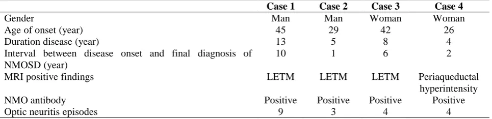
Table 3. Neuromyelitis optica spectrum disorder (NMOSD) cases
Case 1 Case 2 Case 3 Case 4
Gender Man Man Woman Woman
Age of onset (year) 45 29 42 26
Duration disease (year) 13 5 8 4
Interval between disease onset and final diagnosis of NMOSD (year)
10 1 6 2
MRI positive findings LETM LETM LETM Periaqueductal
hyperintensity
NMO antibody Positive Positive Positive Positive
Optic neuritis episodes 9 3 4 4
NMOSD: Neuromyelitis optica spectrum disorder; LETM: Longitudinally extensive transverse myelitis; MRI: Magnetic resonance imaging
In the CRION cases, two patients had one episode of optic neuritis who needed to be treated with plasma exchange in addition to the
intravenous corticosteroid [one of them
developed Neuromyelitis optica spectrum
disorder (NMOSD) later]. After initiating the long-term immunosuppressive treatment (13 patients received azathioprine and only one patient received low dose prednisolone), patients were evaluated for their response to drug and disease status in regular visits of every 6 months.
Four out of fourteen CRION cases developed NMO spectrum disease during follow-up course (Table 3); three of them presented paraplegia and spinal cord syndromes, with extensive cord lesions compatible to NMO spectrum disorders. Anti-aquaporin 4 was positive in these three patients. In the other patient, follow-up MRI revealed periaqueductal hyper intensity in the brain and positive NMO antibody in the serum.
The mean years between the onset of recurrent ON and final diagnosis was 4.75.
The prevalence of demyelinating disorders including MS is increasing. Although optic neuritis is one of the most common presenting symptoms in MS and NMO, there are still a small number of patients without any definite diagnosis even with the recurrence of the symptoms.23-25 RION and CRION are associated with those patients who manifest recurrent optic neuritis without a definite diagnosis of any other demyelinating diseases such as MS or NMO. They are more prevalent in women and may develop to other typical demyelinating diseases over time. Four of our cases developed NMOSD which is concordant with other studies.20,26,27 Except these converted cases, other CRION patients remained responsive to steroids and immunosuppressive therapy like the previous observations made on CRION.6,11 Only two of our
patients had recurrent episodes with less favorable responses to treatment.
Similar to the report presented here, other studies showed that the prevalence of CRION and RION is relatively higher among women.1,2,11,22,28 The strong point of this study is the comparison made between RION and CRION cases with respect to the age of onset, sex superiority, rate of attacks and disease conversion. Since the risk of blindness is high among CRION cases, recognition of this disease is of considerable importance.
Other probable differential diagnoses were also made in the present study. In contrast to studies conducted by Myers, et al.11 and Lin, et al.29 there were not any evidence of vasculitis disorders or granulomatous diseases in the cases of the present study; moreover, no abnormality was found in the screening serologic test performed for autoimmune disorders, suggesting the presence of autoimmune optic neuropathy in the absence of systemic autoimmune process.30
Brain MRIs were normal except for three patients who had non-specific white matter lesions. This finding is compatible with the findings of other studies about brain MRI in CRION.8,20,26,31 Besides, except for one patient, OCB was negative.
prognosis of recovery from optic neuritis is poorer among NMO and CRION cases compared to those with MS.2
The CRION cases in the present study (except for two cases who finally converted to NMOSD) were well controlled during the long term immunomodulatory treatment similar to the other studies.6,11 In addition to reports about azathioprine and methotrexate, cyclosporine, alkylating agents such as cyclophosphamide and chlorambucil, and intravenous immunoglobulin (IVIG) were among the choices of treatment.11,14,22 A case series of patients with RION reported failure of rituximab in controlling attacks in one patient, but a potent effect was reported for natalizumab on another patient.32 Thereafter, it can be concluded that due to the small data which is presented specially on more aggressive treatments, judgment about the choice of treatment is challenging.
The significant difference observed in the present study between CRION and RION in terms of means of ON attacks may be due to the higher number of attacks in CRION cases before the initiation of the therapy.
Limitation: Unfortunately, there was no OCT data of the patients, which could add documentary information about optic nerve and retinal damage in both groups. The cell-based assay anti-NMO technique does not exist in Iran; therefore, we had to utilize enzyme-linked immunosorbent assay (ELISA) technique. It is hoped that future studies apply this novel method instead.
Antibody against myelin oligodendrocyte glycoprotein is introduced as a diagnostic marker
and can be used as a prognostic factor in patients with a seronegative anti-NMO, facilitating diagnosis of a subgroup of NMOSD. However, this method was not employed it in this study; it would be one of the prospective issues in further studies.
Early immunomodulatory treatment is
recommended to prevent further optic neuritis attacks and consequent axonal damage. Isolated optic neuritis may be dependent on steroids and should be followed and managed properly.
These patients may fulfill the criteria for NMO or MS, but most of them remain isolated even after several years. Early proper treatment with steroids or cytotoxic agents is recommended to prevent further optic neuritis attacks and consequent axonal damage.
The authors declare no conflict of interest in this study.
This study was supported by Sina Multiple
Sclerosis Research Center and Research
Development Center of Sina Hospital.
1. Arndt C, Labauge P, Speeg-Schatz C,
Jeanjean L, Fleury M, Castelnovo G, et
al. Recurrent inflammatory optic
neuropathy. J Fr Ophtalmol 2008; 31(4): 363-7. [In French].
2. Petzold A, Plant GT. Diagnosis and
classification of autoimmune optic
neuropathy. Autoimmun Rev 2014; 13(4-5): 539-45.
3. Kim SM, Kim SJ, Lee HJ, Kuroda H,
Palace J, Fujihara K. Differential
diagnosis of neuromyelitis optica
spectrum disorders. Ther Adv Neurol Disord 2017; 10(7): 265-89.
4. Perez-Diaz H, Casado JL, Ucles-Sanchez
A, Saiz A. Chronic relapsing
inflammatory optic neuropathy (CRION)
without detection of IgG-NMO
antibodies. Neurologia 2007; 22(8): 553-5. [In Spanish].
5. Saini M, Khurana D. Chronic relapsing
inflammatory optic neuropathy. Ann
Indian Acad Neurol 2010; 13(1): 61-3.
6. Kaut O, Klockgether T. 51-year-old female
with steroid-responsive optic neuropathy: a
new case of chronic relapsing
inflammatory optic neuropathy (CRION). J Neurol 2008; 255(9): 1419-20.
7. Samra A, Ramtahal J. Recurrent subacute
visual loss presenting in a 52-year-old Caucasian woman with chronic relapsing inflammatory optic neuropathy: A case report. J Med Case Rep 2012; 6: 15.
8. Kidd D, Burton B, Plant GT, Graham
EM. Chronic relapsing inflammatory optic neuropathy (CRION). Brain 2003; 126(Pt 2): 276-84.
9. Kurne A, Karabudak R, Yalcin-Cakmakli
G, Gursoy-Ozdemir Y, Aydin P, Ilksen-Colpak A, et al. Recurrent optic neuritis: Clues from a long-term follow up study of recurrent and bilateral optic neuritis patients. Eye Brain 2010; 2: 15-20.
10. Kurz D, Egan RA, Rosenbaum JT.
Treatment of corticosteroid dependent
optic neuropathy with intravenous
immunoglobulin. Am J Ophthalmol 2005; 140(6): 1132-3.
11. Myers TD, Smith JR, Wertheim MS,
Egan RA, Shults WT, Rosenbaum JT. Use of corticosteroid sparing systemic immunosuppression for treatment of corticosteroid dependent optic neuritis not associated with demyelinating disease. Br J Ophthalmol 2004; 88(5): 673-80.
12. De Benito L, Munoz L, Martinez M,
Cortes C, De Andres C. Recurrent bilateral inflammatory idiopathic optic neuropathy treated with mycophenolate mofetil: Clinical and radiological course. J Neuroophthalmol 2005; 29(4): 149-59.
13. Plant GT, Sibtain NA, Thomas D.
14. Stiebel-Kalish H, Hammel N, van Everdingen J, Huna-Baron R, Lee AG. Intravenous immunoglobulin in recurrent-relapsing inflammatory optic neuropathy. Can J Ophthalmol 2010; 45(1): 71-5.
15. Matiello M, Lennon VA, Jacob A, Pittock
SJ, Lucchinetti CF, Wingerchuk DM, et al. NMO-IgG predicts the outcome of recurrent optic neuritis. Neurology 2008; 70(23): 2197-200.
16. Petzold A, Pittock S, Lennon V,
Maggiore C, Weinshenker BG, Plant GT. Neuromyelitis optica-IgG (aquaporin-4) autoantibodies in immune mediated optic neuritis. J Neurol Neurosurg Psychiatry 2010; 81(1): 109-11.
17. Jarius S, Frederikson J, Waters P, Paul F,
Akman-Demir G, Marignier R, et al. Frequency and prognostic impact of antibodies to aquaporin-4 in patients with optic neuritis. J Neurol Sci 2010; 298(1-2): 158-62.
18. Unsold R. Neuropathies of the optic
nerve in inflammatory systemic diseases and vasculitis. A frequently misdiagnosed early symptom. Ophthalmologe 1994; 91(2): 251-62.
19. Abstracts of the 28th Congress of the
European Committee for Treatment and Research in Multiple Sclerosis. October 10-13, 2012. Lyon, France. Mult Scler 2012; 18(4 Suppl): 9-542.
20. Falcao AB, Bichuetti DB, Oliveira EML,
Gabbai AA. NMO, CRION or MS: what we say when we have recurrent optic neuritis. Mult Scler 2012; 18(4 Suppl): 262.
21. Cheng H, Laron M, Schiffman JS, Tang
RA, Frishman LJ. The relationship between visual field and retinal nerve fiber layer measurements in patients with multiple sclerosis. Invest Ophthalmol Vis Sci 2007; 48(12): 5798-805.
22. Petzold A, Plant GT. Chronic relapsing
inflammatory optic neuropathy: A
systematic review of 122 cases reported. J Neurol 2014; 261(1): 17-26.
23. Etemadifar M, Izadi S, Nikseresht A,
Sharifian M, Sahraian MA, Nasr Z. Estimated prevalence and incidence of multiple sclerosis in Iran. Eur Neurol 2014; 72(5-6): 370-4.
24. Sahraian MA, Moinfar Z, Khorramnia S,
Ebrahim MM. Relapsing neuromyelitis optica: demographic and clinical features in Iranian patients. Eur J Neurol 2010; 17(6): 794-9.
25. Martinez-Lapiscina EH, Fraga-Pumar E,
Pastor X, Gomez M, Conesa A, Lozano-Rubi R, et al. Is the incidence of optic neuritis rising? Evidence from an epidemiological study in Barcelona (Spain), 2008-2012. J Neurol 2014; 261(4): 759-67.
26. Pandit L, Shetty R, Misri Z, Bhat S, Amin
H, Pai V, et al. Optic neuritis: Experience
from a south Indian demyelinating disease registry. Neurol India 2012; 60(5): 470-5.
27. Pirko I, Blauwet LA, Lesnick TG,
Weinshenker BG. The natural history of recurrent optic neuritis. Arch Neurol 2004; 61(9): 1401-5.
28. Benoilid A, Tilikete C, Collongues N,
Arndt C, Vighetto A, Vignal C, et al. Relapsing optic neuritis: A multicentre study of 62 patients. Mult Scler 2014; 20(7): 848-53.
29. Lin YC, Wang AG, Yen MY. Systemic
lupus erythematosus-associated optic
neuritis: clinical experience and literature review. Acta Ophthalmol 2009; 87(2): 204-10.
30. Frohman L, Dellatorre K, Turbin R,
Bielory L. Clinical characteristics,
diagnostic criteria and therapeutic
outcomes in autoimmune optic
neuropathy. Br J Ophthalmol 2009; 93(12): 1660-6.
31. Sharma A, Khurana D, Kesav P. MRI
findings in chronic relapsing
inflammatory optic neuropathy. BMJ Case Rep 2013; 2013: 1-2.
32. Waschbisch A, Atiya M, Schaub C,