Spring 2012, Volume 3, Number 3 Basic and Clinical
A Brief Look into Spike Sorting Methods
Mojtaba Kermani 1,2, Seyed Mohammad Noorbakhsh 2,*, Abbas Haghparast 1
1. Neuroscience Research Center, Shahid Beheshti University of Medical Sciences, P.O. Box 19615-1178, Tehran, Iran 2. ScienceBeam Institute, Tehran, Iran
* Corresponding Author:
Seyed Mohammad Noorbakhsh, M.D., Science Beam Institute, Tehran, Iran
Tel.: (+98912) 670-1773/ Fax: (+9821) 221-76125 E-mail: [email protected]
Spike sorting is a class of techniques used in the analysis of electrophysiological data. Studying the dynamics of neural activity via electrical recording relies on the ability to detect and sort neural spikes recorded from a number of neurons by the same electrode. This article reviews methods for detecting and classifying action potentials, a problem commonly referred to as spike sorting.
A B S T R A C T
Article info:
Received: 18 February 2012
First Revision: 19 April 2012 Accepted: 09 May 2012
Key Words: Spike,
Data Acquisition, Filtering,
Feature Extraction, Clustering.
1. Introduction
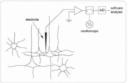
ne of the major questions contemporary neuroscience seeks to elucidate concerns the mechanisms used by dedicated parts of brains (our brain, vertebrate brains, in-sect brains) to perform specific tasks. Our brains are buzzing with electrical activity created by so-dium and potassium ions moving in and out of neurons through specialized pores. Classical methods for explor-ing the mechanisms of brain function involve recordexplor-ing the electrical activity of single nerve cells. Neurophysi-ologists often record the currents flowing across cell membranes using an insulated wire probe whose ‘listen’ to a few neurons close-by the electrode tip that fire action potentials or ‘spikes’ (Fig.1).Any such probe will record currents passing across all sorts of physiological mem-branes close to its tip, but we can approximate this as a small number of close, high-amplitude sources imposed on a background of synaptic, somatic and axonal cur-rents from more distant sources. Each neuron has spikes
O
than just detecting the spikes for each channel without caring from which neuron they come. It is already well established that complex brain processes are reflected by the activity of large neural populations and that the study of single-cells in isolation gives only a very limited view of the whole picture (Brown, Kass, & Mitra, 2004; Buzsáki, 2004)Therefore, progress in Neuroscience re-lies to a large extent on the ability to record simultane-ously from large populations of cells. The implemen-tation of optimal spike sorting algorithms is a critical step forwards in this direction, since it can allow the analysis of the activity of a few close-by neurons from each recording electrode. This opens a whole spectrum of new possibilities. For example, it is possible to study connectivity patterns of close-by neurons(Buzsáki, 2004; Harris, 2005; Harris, Henze, Csicsvari, Hirase, & Buzsáki, 2000), or to study the topographical orga-nization of a given area and discriminate the responses of nearby units(Lewicki, 1998; Quian Quiroga, 2009; Quiroga, 2007). It is also possible to have access to the activity of sparsely firing neurons, whose responses may be completely masked by other neurons with high firing rates, if not properly sorted. Separating sparsely firing neurons from a background of large multi-unit activity is not an easy job, but this type of neurons can show striking responses (Hahnloser, Douglas, & Hepp, 2002; Perez-Orive, et al., 2002; Wood, Black, Vargas-Irwin, Fellows, & Donoghue, 2004).
General Framework: Why is spike sorting important?
The short answer to this is that it is vital for extra-cel-lular recording from multiple cells. Obviously, sorting spikes from a single electrode can provide signals from more than one cell. But automatic recognition is also es-sential for any multiple electrodes recording as achieving and maintaining isolation on multiple probes simultane-ously can be highly impractical. The action potentials originating from different recorded neurons have distinct shapes, and based on these differences one can select (or discriminate) certain neurons among others.
2. Spike Sorting Steps
2.1. Data Acquisition
The first step in any spike sorting algorithm involves the acquisition of extracellular data in a form amenable to the detection of neuronal spikes. For many neurons,
cording electrode so that the spikes from the neuron of interest are maximally separated from the background activity (Quiroga, 2009) . Window discriminators can be implemented on-line, but have the main disadvan-tage that they require a manual setting of the windows by the user, which may need readjustment during the experiment. In this regard, manual procedures are of limited utility because shape parameters designed for human inspection are inefficient at representing com-plex waveforms. Moreover the labor-intensive process scales poorly to experiments performed with large num-bers of electrodes, and a subjective approach makes it difficult to design reproducible and reportable quality metrics. For these reasons, an algorithmic approach is desirable, and in fact, computational solutions with lim-ited human monitoring have been shown to generally outperform manual sorting (Harris, 2005).
2.2. Filtering
The word "filtering" refers to an attempt to extract the important part of some data while eliminating random contributions called "noise" or other unwanted features which obscure the ones that matter (Quian Quiroga, 2009). The first step when processing continuously re-corded data is to apply a band pass filters in order to avoid low frequency activity and visualize the spikes (Quiroga, 2007). This step is usually overlooked in the literature, but its implementation can dramatically change the spike shapes. In the example of Fig. 3, the continuous data was filtered with a band pass filter be-tween 300 and 3000Hz. Frequencies below 300Hz are filtered to delete the slow components of the raw data. The upper cutoff frequency of the filter is to diminish the noisy appearance of the spike shapes. As it is usually the case with filtering, a compromise has to be taken. On the one hand, one would like to have a narrow fil-ter band to betfil-ter visualize the spikes, but on the other hand, if the band is too narrow, the filter may hinder different features of the spike shapes (Quian Quiroga, 2009; Quiroga, 2007).
2.3. Spike Detection
Spring 2012, Volume 3, Number 3 Basic and Clinical
Although being simple and fast, due to some difficul-ties, using this method require special vigilance. Mov-ing threshold durMov-ing recordMov-ing considered as a next problem. The threshold level determines the trade-off between missed spikes (false negatives) and the number of background events that cross threshold (false posi-tives). In fact increasing the threshold level reduces the number of spikes that are misclassified (Type II error), but at the expense of many missed spikes. On the other hand, getting false positives due to noise crossing a low threshold (Type I error) (Quiroga, 2009; 2007). Another problem in spike detection is misclassification error due to overlaps. In addition to the background noise, the spike height can vary greatly if there are other neurons in the local region that generate action potentials of sig-nificant size. If the peak of the desired unit and the dip of a background unit line up, a spike will be missed. (Quian Quiroga, 2009; Quiroga, 2007). However, an automatic threshold is preferable, especially when pro-cessing large number of channels. Once spikes are de-tected, they have to be stored for clustering (Quiroga, 2007).
There are two issues concerning spike storage that need a brief description. The first one is how many data points to store. This of course depends on the sampling frequency and ideally one would like to store the whole spike shape; i.e. about 2 ms of data. With a sampling frequency of 30 KHz, this corresponds to 60 data points. Some methods for feature extraction, such as wavelets (using a multi-resolution decomposition implementa-tion), require that the number of data points is a power of 2 (Nenadic & Burdick, 2005). In this case, with 30KHz sampling, 64 data points would be optimal. The second issue has to do with the alignment of the spike shapes. Spikes can be aligned to their maximum. But due to insufficient sampling the maximum can be at different points of the spike shape. To avoid such misalignments, which could lead to over clustering, the spike shapes can be oversampled using interpolated waveforms, for example, using cubic splines. Then, the interpolated shapes can be aligned and later decimated to the original sampling rate (Quiroga, 2007).
For recordings with good signal to noise ratio (SNR) this is usually achieved by a simple thresholding (Kim & Kim, 2000). When the SNR is not good enough, differ-ent spike-detection algorithms can be applied, e.g. use of nonlinear energy operator (Kim & Kim, 2000) con-tinues wavelet transform (Nenadic & Burdick, 2005).
2.4. Feature Extraction
Transforming the input data into the set of features is called feature extraction which involves simplifying the amount of resources required to describe a large set of data accurately. As a third step, it measure features of shapes, such as spike height and width, peak-to-peak amplitude, energy (the square of the signal), mean or variance. In general, the more features we have, the bet-ter we will be able to distinguish different spike shapes. The result of this step is an M×K-matrix, where K is the number of detected spikes and M is the number of extracted feature. Principal component analysis (PCA), wavelet decomposition or some other techniques are commonly used to reduce the dimensionality of the M×K-matrix by extracting the most important features of the detected spikes. The result is a new matrix of re-duced dimension, L×K, where L < M is the number of extracted features per spike.(Tiganj & Mboup, 2011).
Ideally, one wants to extract those features that best separate the different clusters of spikes and get rid of all the dimensions dominated by noise.This step saves computational time and it is mandatory for some clus-tering algorithms that cannot handle too many inputs in a reasonable time (Quiroga, 2007). Although eliminat-ing inputs dominated by noise can certainly improve clustering outcomes, but the major challenge is still re-main, which two features are the best among extracted features?
A first choice would be to take basic characteristics of the spikes, such as their peak (or peak to peak) am-plitude, their width and energy (the square of the sig-nal). However, it has been shown that such features are not optimal for differentiating spike shapes in general (Quiroga, 2007; Sakowitz, Quian Quiroga, Schürmann,
usually containing more than 80% of the energy of the signal (Quiroga, 2007). However, principal component analysis (PCA) selects the directions of maximum vari-ance of the data, which are not necessarily the directions of best separation. In other words, it may well be that the information for separating the clusters is represented in one (or a combination) of principal components with low eigenvalues, which are usually disregarded (Quiro-ga, 2007).
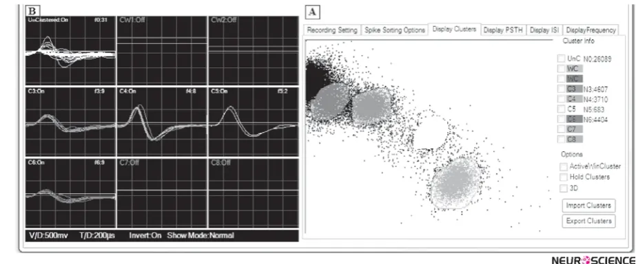
jects in the same cluster are more similar (in some sense or another) to each other than to those in other clusters. The fourth and final step of spike sorting is to group spikes with similar features into clusters, corresponding to the different neurons. A common method is a technique called cluster cutting. In this approach, the user defines a boundary for a particular set of features. If a data point falls within the boundary, it is classified as belonging to that cluster; if it falls outside the boundary, it is discard-ed. Figure 3(a) shows an example of boundaries placed around the primary clusters. In off-line analysis the clus-ter boundaries are declus-termined afclus-ter the data have been collected by looking at all (or a sample from) the data
Figure 2. The extracellular waveform shows several different action potentials generated by an unknown number of neurons. The data were recorded from Rat’s locus coeruleus witha Tungsten electrode by Electromodule and Neurocomet (sciencebeam institiute, Tehran, Iran). In this case, by amplitude discrimination method, neurons C and D are recorded as a same neuron by window 2.
Spring 2012, Volume 3, Number 3 Basic and Clinical
over the collection period. This allows the experiment to verify that the spike shapes were stable for the duration of the collection period (Lewicki, 1998). However, besides being a very time-consuming task, manual clustering in-troduces errors due to the limited dimensionality of the cluster cutting space and due to human biases(Harris, et al., 2000). In fact, in many cases clusters overlap and the manual setting of a boundary is very subjective.
4. Discussion
We have reviewed here the process of spike sorting. In addition to so called methods, there are some other methods for spike sorting including template match-ing, Bayesian methods and so on. Each method has some advantages and disadvantages itself. According-ly, template-based methods can fail for neurons that burst and can becomeincreasingly inaccurate if there is electrode drift or Bayesian methods are much more accurate for spike shapes that are similar. On the other hand, despite the simplicity ofamplitude discrimina-tion, this technique can be time consuming and biased. Not onlycan neurophysiologists waste hours search-ing for well isolated cells, but in the end thissearch is biased towards cells that produce the largest ac-tion potentials which may not berepresentative of the entire population. Hence according to pros and cons of each method, a more practical question might be: what is the simplest method that satisfiesexperimental demands? For many researchers this is still a single electrode with threshold detection despite mentioned difficulties.
References
Abeles, M., & Goldstein Jr, M. H. (1977). Multispike train analy-sis. Proceedings of the IEEE, 65(5), 762-773.
Brown, E. N., Kass, R. E., & Mitra, P. P. (2004). Multiple neural spike train data analysis: state-of-the-art and future challeng-es. Nature neuroscience, 7(5), 456-461.
Buzsáki, G. (2004). Large-scale recording of neuronal ensem-bles. Nature neuroscience, 7(5), 446-451.
Glaser, E., & Marks, W. (1968). On-line separation of inter-leaved neuronal pulse sequences. Data Acquisition Process Biol Med, 5, 137-156.
Hahnloser, R. H. R., Douglas, R. J., & Hepp, K. (2002). Atten-tional recruitment of inter-areal recurrent networks for selec-tive gain control. Neural computation, 14(7), 1669-1689.
Harris, K. D. (2005). Neural signatures of cell assembly organi-zation. Nature Reviews Neuroscience, 6(5), 399-407.
Harris, K. D., Henze, D. A., Csicsvari, J., Hirase, H., & Buzsáki, G. (2000). Accuracy of tetrode spike separation as deter-mined by simultaneous intracellular and extracellular meas-urements. Journal of Neurophysiology, 84(1), 401-414.
Kim, K. H., & Kim, S. J. (2000). Neural spike sorting under near-ly 0-dB signal-to-noise ratio using nonlinear energy operator and artificial neural-network classifier. Biomedical Engineer-ing, IEEE Transactions on, 47(10), 1406-1411.
Lewicki, M. S. (1998). A review of methods for spike sorting: the detection and classification of neural action potentials. Network: Computation in Neural Systems, 9(4), 53-78.
Nenadic, Z., & Burdick, J. W. (2005). Spike detection using the continuous wavelet transform. Biomedical Engineering, IEEE Transactions on, 52(1), 74-87.
Perez-Orive, J., Mazor, O., Turner, G. C., Cassenaer, S., Wil-son, R. I., & Laurent, G. (2002). Oscillations and sparsening of odor representations in the mushroom body. Science, 297(5580), 359-365.
Quian Quiroga, R. (2009). What is the real shape of extracellular spikes? Journal of neuroscience methods, 177(1), 194-198.
Quiroga, R. Q. (2007). Spike sorting. Scholarpedia, 2(12), 3583.
Sakowitz, O. W., Quian Quiroga, R., Schürmann, M., & Basar, E. (2005). Spatio-temporal frequency characteristics of in-tersensory components in audiovisually evoked potentials. Cognitive brain research, 23(2-3), 316-326.
Sambeth, A., Maes, J., Quian Quiroga, R., & Coenen, A. M. L. (2004). Effects of stimulus repetitions on the event-related potential of humans and rats. International journal of psy-chophysiology, 53(3), 197-205.
Tiganj, Z., & Mboup, M. (2011). A non-parametric method for automatic neural spike clustering based on the non-uniform distribution of the data. Journal of Neural Engineering, 8, 066014.