INTERNATIONAL RESEARCH JOURNAL OF PHARMACY
www.irjponline.com
ISSN 2230 – 8407
Research Article
EFFECT OF TRANSITION METAL DOPING ON THE STRUCTURAL, OPTICAL,
THERMAL PROPERTIES, AND ANTIMICROBIAL ACTIVITY OF ZINC OXIDE NANOPARTICLES
Alya'a Jabbar Ahmed *
Directorate Scholarship and Cultural Relations, Ministry of Higher Education and Scientific Research, Baghdad-Iraq
*Corresponding Author Email: [email protected]
Article Received on: 20/07/18 Approved for publication: 10/09/18
DOI: 10.7897/2230-8407.099181
ABSTRACT
Effect of transition metal dopant on structural, optical, thermal properties, and antimicrobial activities of synthesized zinc oxide nanoparticles (ZnO NPs) were investigated in this study. undoped, cobalt, and nickel doped ZnO nanoparticles are successfully synthesized using chemical co-precipitation method. The structure and morphology of these nanoparticles are characterized using X-ray diffraction (XRD), atomic force microscope (AFM), scanning electron microscope (SEM), energy dispersive spectroscopy (EDS), as well as an appropriate physical measurement; (Vibrational and electronic) spectroscopy. The Results obtained from XRD indicate that the particles are possessed in wurtzite structure , SEM and AFM studies reveal that the particle size is in the 45-65 nm range. The EDS confirmed the Zn, O, Co, and Ni as major elements of as-synthesized nanoparticles. Ultraviolet-visible (UV-Vis) spectra show that doping Co and Ni, lead to an increase in the absorption edge wavelength and a decrease in the band gap energy, Fourier- transform infrared (FT-IR) spectra give the presence of functional groups and chemical bonding as well as doped sub-lattice. To measure the biological activity and potential antimicrobial efficiency of these undoped, Co, and Ni-doped ZnO nanoparticles on the Candidates.
Keywords: Doped, ZnO nanoparticle, co-precipitation method, and antimicrobial activity.
INTRODUCTION
The nanotechnological applications in medicine have yielded a completely new field of technology that is set to bring significant
advances in the fight against a range of diseases1. Nanoparticles
have risen as novel microbial activity owing to the high surface area to volume ratio, which will become of utmost importance to the researcher due to the fact of the developing microbial resistances against metallic element ion, antibiotics, and the
advancement of safe strains2. Numerous nano- particles display
incredible antimicrobial productivity and used to control
medicate safe microbial populations3. Different inorganic metal
oxide nanoparticles such as ZnO, MgO, TiO2 and SiO2 exhibit
significant antimicrobial activities and used in therapeutics,
diagnostics, and Nanomedicine-based antimicrobial agents4,5.
Inorganic nanoparticles show greater effects on resistant strains of microbial pathogens, less toxicity, heat resistance and provide
mineral elements essential to human cells 6. Among these metal
oxide nanomaterials, ZnO should be given a special attention, due to its essential role in healthcare products, UV blocking
capability, biocompatibility and modest cost 7.
Furthermore, microbial contamination effects should not be underestimated in the healthcare and food industry so that the development of antimicrobial agents and surface coatings have been a motivating factor for many researchers in the recent
decades8. ZnO nanoparticles are known to be one of the
multifunctional inorganic nanoparticles with powerful antibacterial action. Antibacterial and antifungal activities of zinc oxide nanoparticles are observed even at very low concentrations and also the antifungal activity does not influence soil fertility
compared to the conventional antifungal agents9. A large number
of studies have been performed over the last decades on the
properties of ZnO that can be tuned by doping with various metals ions10, 11. The transition metals (Ni, Co, Al, Cu, Ag) doped ZnO
display best properties, as transition metal elements have close
ionic radius parameter to that of Zn2+, which means that these
elements can easily penetrate into ZnO crystal lattice or substitute
Zn2+ position in crystal12. Doped transition metals in zinc oxide
optimize the electronic, magnetic, optical, and electrical
characterization that is useful for various practical applications12.
Doped ZnO demonstrates a greater effect against pathogenic organisms when contrasted with ZnO, in this way using nanoparticles as an antimicrobial agent and considered one of the most useful techniques to minimize the cost and chemical waste
13. Ni is an essential depend on these magnetic materials. Besides,
Ni+2 (0.69 A ° ) has the same valence as Zn+2 and its radius is
close to that of Zn+2 (0.74 A ° ), so it is workable for Ni+2 to
replace Zn+2 in ZnO lattice. Some researchers reported that the
luminescence properties of zinc oxide are changed after doping of Ni 14.
In this study, an experimental investigation in order to synthesize and characterize the structure of zinc oxide nanoparticles doped with cobalt and nickel is performed using a chemical co-precipitation method. It highly predicted to become the useful tool to fight against microbial infections in the near future.
MATERIALS AND METHODS
All the starting materials, solvents as well as reagents, were purchased commercially and then used without any further purification.
Instrumentation
X-ray Diffraction (XRD)
The crystal structure and lattice parameters of the synthesized of undoped ZnO, Co-doped, and Ni-doped ZnO nanoparticles were used Philips (X-ray diffractometer XRD- 6000).
Atomic force microscopy (AFM)
The particle sizes and morphology of the nanoparticles were estimated by atomic force microscopy (AFM). The AFM images were performed with an (AFM AA3100) microscope.
Scanning electron microscopy (SEM)
The morphology of the prepared nanoparticles was estimated using an Oxford instrument attached to JSM-5300 scanning microscope equipment.
Energy dispersive X-ray Spectroscopy (EDS) Analysis
The elemental analysis of the particles was performed using the scanning electron microscope (SEM) that attached with an electron dispersion spectrometer (EDS).
Ultraviolet-visible (UV–Vis) and Fourier Transform Infrared, (FTIR) spectroscopy
Ultraviolet-visible Uv-visible spectra were measured using
Shimadzu Uv-vis. 160 A-Ultra-violet spectrophotometer in the range (200-1000) nm. Fourier transform infrared analyzed spectra
in the frequency range (400-4000) cm-1 were recorded as KBr
discs on FTIR.8300 Shimadzu spectrophotometer.
Thermal gravimetric analyses (TGA)
Thermal analyses (TGA) were achieved in N2 atmosphere with a
heating rate 10℃ min-1 using a Perkin- Elmer model 4000
instrument.
Synthesis of ZnO nanoparticles
Zinc Oxide nanoparticles were prepared using the chemical precipitation method. A suspension of (0.2 M) zinc acetate dihydrate dissolved in (50ml) deionized water and was stirred for a half hour. Then (1M) of sodium hydroxide was added dropwise to zinc acetate solution and mixed at 70℃ with constant stirring
to get the solution with the pH (12).A white gel precipitate was
formed, washed with deionized water and followed by ethanol. After that, a white powder was obtained after drying at 70 ℃ in the vacuum oven overnight.
Synthesis of ZnO: Co+2 and ZnO: Ni+2 nanoparticles
For the synthesis, transition metal doped ZnO nanoparticles, (0.2M) of transition metal salts dissolved in (50ml) deionized water was added dropwise to the zinc acetate solution after stirring for a half hour. Then (1M) of sodium hydroxide was added dropwise to the solution and mixed at 70℃ with constant stirring to obtain the solution to with the pH (12). The obtained precipitate was filtered, then washed with water and ethanol, and finally dried at 50℃. The prepared samples were then used for further experimental analysis.
Biological Studies
The in vitro antimicrobial activity of the synthesized compounds were tested against two types of fungi which are (Candida albicans and Candida tropicalis) using the disc diffusion method. Each one of the prepared compounds was first dispersed in methanol to form monodispersed and then each nanoparticles were was sonicated for 30min.
The antifungal test was performed according to the disc diffusion method. The culture was inoculated and then was well formed in the medium. Moreover, the plates were incubated at room temperature and after 48 hours of incubation, the growth inhibition was examined utilizing the zone formation.
RESULTS AND DISCUSSION
Characterization
X-ray diffraction (XRD) analysis
The XRD pattern of the prepared ZnO, Co- doped, and Ni-doped ZnO is shown in Figure 1 (a, b, and c). The XRD of the ZnO
shows a diffraction peak at 2𝜃 = 31.80°, 34.45°, 36.28°, 47.57°,
and 56.62° which can be indexed to (100), (002), (101) (102)
(110) and (200) crystal plane respectively. The position of these peaks indicates the formation of hexagonal wurtzite structure which generally is in agreement with standard JCPDS (NO-36-1451) (2157-7439). Similar characteristic peaks and assigned
lattice plane are observed in some other studies too15. The X-ray
diffraction pattern of Co-doped ZnOconsists of diffraction peaks
located in 2θ = 19.7785, 23.4242, 27.5667, 28.9447, 33.2112, 33.6991, 48.5689, 59.1846, and 69.5614 respectively as shown in (Figure 1 (b)). Furthermore, no peaks associated with Co are
detected in the XRD pattern indicating that Co2+ ions have
substitutionally replaced the Zn2+ 16. The XRD pattern of
Ni-doped ZnO( Figure 1 (c) ) show peaks in (100) and (101). No signals of the metallic Zn are detected by XRD. However, there
is no peak attributed to Ni, suggesting that the Ni+2 ion may be
doped into ZnO17. It can be concluded from the results that
incorporation of Co and Ni into ZnO lattice enhances crystalline quality.
The average crystalline size is calculated using Debye–Scherrer
equation18,
D = = >?@ A; < (1)
Where; D = is the average crystalline size of the nanoparticles, K = is a geometric factor (0.94), λ= is the wavelength of X-ray
radiation source, 𝛽 = is the angle (full-width at half maximum)
FWHM of the XRD peak at the diffraction angle. The calculated average crystalline size of the undoped, Co-doped,and Nidoped ZnO is about (2-13) nm. From the results in Table.1, the incorporation of Co and Ni into ZnO lattice improves crystalline
qualitythat calculated using Debye–Scherrer formula.
Atomic Force Microscopy (AFM)
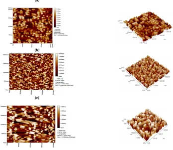
The Atomic Force Microscopy image is used to determine the distribution of the particle size and estimate the mean size of the particles. Figure (2a, 2b, and 2c), show the two-dimensional (2D) and three-dimensional (3D) AFM images of pure ZnO, Co doped ZnO, and Ni doped ZnO respectively. It can be observed that the
average size is about 50 nm,45nm and 65nm for ZnO, ZnO: Co+2
(a)
(b)
(c)
Fig. 1: The X-ray diffraction of (a) Undoped, (b) CO doped, and (c) Ni doped ZnO.
(a)
(b)
(c)
(b) (a)
(c)
Fig. 3: Scanning electron microscope image of (a) UndopedZnO, (b) CO doped ZnO, and (c) Ni doped ZnOnanoparticles
(b) (a)
(c)
(a)
(b)
(c)
Fig. 5: Fourier Transform Infrared spectrum of (a) UndopedZnO, (b) Co doped ZnO, and (c) Ni doped ZnO nanoparticles.
(b) (a)
(c)
(a)
(b)
(c)
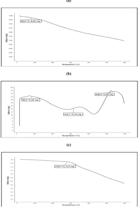
Fig. 7: The TGA curve of the prepared (a) undoped, (b) Co doped, and (c) Ni dopedZnO nanoparticles .
Table 1: The lattice constants as obtained from Rietveld refinement and particle size as deduced from X-ray line broadening.
Grain size (nm) d(h k l)
(A°) FWHM
(Radian) 2θ
(Deg) Lattice
Plane Nanoparticles
13 2.47356
0.21710 36.28
101 ZnO
3 2.65749
0.76000 33.6991
101 Co-Doped ZnO
2 2.65405
1.43000 33.7441
101 Ni-Doped ZnO
Table 2: The atomic percentage of elements in undoped ZnO, Co-doped and Ni-doped ZnO nanoparticles.
Atomic Percentage (%)
Element ZnO Co-Doped ZnO Ni- Doped ZnO
39.18 33.50
76.87 Zn
58.50 64.43
23.13 O
- 2.01
- Co
2.31 -
- Ni
Table 3: Evaluation of the antimicrobial activity of the undoped, Co, and Ni doped ZnO nanoparticles, and determination of the IZ (inhibition zone) in mm.
Diameter of inhibition zone growth (mm/mg sample)
Compound Candida albicans Candida tropicalis
8 9
ZnO
13 15
ZnO: Co+2
10 11
Scanning Electron Microscopy (SEM)
The morphology of the prepared nanoparticles was investigated by scanning electron microscope (SEM) as shown in the Figure 3 (a,b, and c) . In has been noticed from the SEM micrographs that the ZnO nanoparticles (Figure 3a) have a spherical nature which is in agreement with the shape of the surface plasmon resonance band of the UV–Vis spectra . While, Figure (3b) and Figure (3c) present SEM micrograph of cobalt and nickel doped ZnO and can clearly be seen that the sampls are composed of nanoparticles are uniformly distributed over the entire surface. The morphology of Co and Ni doped ZnO nanoparticles completely differ from that of undoped ZnO nanoparticles. This may be assigned to the effect of doping in ZnO nanoparticle
Energy dispersive X-ray spectroscopy (EDS) analysis
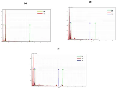
The EDS measurements were performed to emphasize the presence of cobalt and nickel ions in the prepared nanocrystalline ZnO particles, Figure 4 (a) shows the EDS spectrum proven that the nanoparticles suspension contains nothing but zinc and oxygen. The elemental composition of ZnO nanoparticles with two major peaks was observed to have an atomic percentage of 70.87 of Zinc and 29.13 of oxygen. Figure 4 (b) shows the EDS spectra of cobalt doped ZnO nanoparticles, and this spectrum confirmed the presence of zinc, oxygen , and cobalt. It was found that Co-doped zinc oxide nanoparticles have an atomic percentage of 13.73 of Zinc, 70.27 of oxygen, and 7 of cobalt. Figure 4 (c). Shows the atomic percentage of 36.61 of Zinc, 56.81 of oxygen and 6.58 of Nickel. This indicated the doping of Ni ion into the ZnO lattice.
Fourier transform infrared (FTIR) spectroscopy
Fourier Transform Infrared analyzed were performed in order to study the surface changes on the particles, as well as to determine the presence of functional groups and chemical bonding. These analyses can reveal the quality or consistency of samples. Figure 5 (a), 6 (b), and5 (c) show the FTIR spectrum of undoped and
doped ZnO with Co+2 and Ni+2. The spectrum of the samples,
show a band observed at (3350-3450) cm-1 assigned to the v
-OH which refers to the presence of H2O. Whereas, the presence
of a band in the range 1624-1635 cm-1 is due to bending vibration
of adsorbed H2O molecules19. Hence, the presence of an OH band
in the spectrum is owing to chemically and physically adsorbed
water molecule on the surface of nanoparticles 20. The band that
observed at 1338 cm-1 and 1546 cm-1 is assigned to the C=O
asymmetric and symmetric vibration mode respectively, probably
from the un-reacted acetates 21. The absorption band in the range
400-678cm-1 are mainly ascribed to ZnO stretching mode22.These
bands undergo slight shifting.
When Co doped in ZnO indicates the incorporation of Co dope in
the lattice structure of ZnO 22. The bands located near 790 and
844 cm-1 are assigned to the vibrations of ZnO-Ni local bonds and
defect states respectively 23.
UV-Vis spectroscopy
Electronic spectra of the prepared nano compounds were dispersed individually in methanol with 0.1% wt to form a monodisperse solution. After that, each nanoparticle was sonicated for 30min Figure 6(a) shown the electronic spectra of ZnO nanoparticles revealed one band at 378nm emerge from the electronic transition from valence band to the conduction band
(O2p → Zn3d) 24,25, as shown in Figure 6(a)). The Co-doped ZnO,
(see Figure 6(b)) shows the presence of three bands attributed to
d-d transition in the region 514,625, and 794 nm which were
assigned to 4A
2(F)→ 2A1(G) and 4A2→ 4T1(P) respectively.
These minima are in agreement with the Co+2 d-d (tetrahedral
symmetry) 26. For Ni-doped ZnO case the absorption band is
shifted to a lower wavelength at 214 nm. “ These electronic
transitions indicate that Ni2+ ions are incorporated Zn2+ ions in the
octahedral ZnO. Usually, divalent Ni ions in an octahedral symmetry show three main bands at about 8300, 14080, and
24995 cm-1. These bands are commonly attributed due to the
spin-allowed triplet-triplet transitions” 27:
3A
2g(F) → 3T2g(F), 3A2g(F) → 3T1g(F) and 3A2g(F) → 3T1g(P).
The obtained data detect that the single observed absorption band
is most probably due to 3A
2g (F) → 3T1g (P). Electronic
transitions occur between split d levels of the central atom giving rise to so-called d-d transition or ligand field spectra. Where this electronic transition attributed to d–d transition is localized at
(26525 cm-1) 28. The band gap energies are calculated using the
equation of the reference”29:
Eg = 1240 ⁄ λ max (2)
Where Eg: is the band gap energy, the value of (1240) is the Hz, and λ max is the wavelength in nanometer. The calculated values are 3.27 eV (378 nm), 3.17 eV (390nm) , and 3.13 eV (396 nm) for undoped ZnO , Co and Ni- doped ZnO respectively. These values are obviously red-shifted. This shift might be due to the sp-d exchange interaction between the band electron and the
localized d-electron of the Co+2 ion and Ni+2ion.
Thermal analysis
The thermogravimetric analysis (TGA) carried out for further confirmation of cobalt and nickel doped ZnO nanoparticles. Figure 7 (a) shows the TG curve of ZnO which was heated from
room temperature to 600 ℃ at 10 ℃/min under a nitrogen
atmosphere. TGA curve shows that the decomposition started at
in the temperature range of 250–450℃, where a water molecule
(found 0.03%) was lost. The temperature was performed in TGA
of ZnO nanoparticles up to 400 ℃. Itindicates there is no change
in the structure and indicates the formation of nanocrystalline ZnO, as confirmed by XRD analysis. For cobalt doped ZnO, the result of the TG curve, Figure 7 (b) shows that the weight loss for
Co-doped sample decomposition stated at 85.5℃ and was
completed at 536.5℃. Whereas, the weight loss for Ni-doped
samples at 276.4 ℃ is assigned to the release of absorbed water
on the surface.
Biological activity
Many studies have concluded that the zinc oxide nanoparticles an
effective antimicrobial against pathogenic microorganisms 30,31.
In addition, the antifungal activity of ZnO nanoparticles against Botrytis cinerea and Penicillium expansum has been found in the
same studies of the references30, 31. “They proposed that the cell
functions of Botrytis cinerea were influenced by zinc oxide nanoparticles due to increased produce of nucleic acids through
the stress response in fungal hyphae leads to the death of cells 32.
Whereas the treat cells of Penicillium expansum by ZnO nanoparticles, the damaged cell membrane results in the decreased amount of proteins, carbohydrates and lipids in fungal
cells lead to death of the cells”33.
Candida albicans and Candida tropicalis. The Ni doped Zno nanoparticles show a slight effect compared with undoped ZnO in all organisms. While, Co doped ZnO nanoparticles reveal the maximum antifungal activity against all organisms that have been used in this study, and recorded high inhibition activity against Candida tropicalis comparable with undoped ZnO and Ni-doped ZnO. It is clear, from the experimental results, that the transition metal doping in ZnO nanoparticles shows higher antifungal activities as compared to undoped ZnO. In general, good agreement between our results and Sankara et al results 33 results, regarding to enhance the antimicrobial activity. Thus, It can be said that in this study, transition metal doped ZnO nanoparticles have shown the best antifungal behavior compared to undoped ZnO nanoparticles.
CONCLUSION
Undoped, Co doped, and Ni doped ZnO nanoparticles were synthesized by chemical co-precipitation method and the prepared nanoparticles are characterized by XRD, AFM, SEM, EDX, UV-vis, FTIR spectrophotometer. The zone of inhibition was observed against pathogenic fungal strains and suggests that
ZnO: Co+2and ZnO: Ni+2 showed good antifungal activity
adjacent to Candida albicans and Candida tropicalis than the
undoped ZnO nanoparticles.
REFERENCES
1. Ferrari M. Cancer nanotechnology: opportunities and
challenges. Nat Rev Cancer, 2005; 5: 161-171. https://doi. org/10.1038/nrc1566
2. Meruvu,H.,Vangalapati,M.,Chippada,S.C., Bammidi, S.R.
Synthesis and characterization of zinc oxide nanoparticles and its antimicrobial activity against bacillus subtilis and escherichla coli" .RASAYAN J.Chem. 2011;4(1) : 217-222.
3. Rai M, Yadav A, Gade A.Silver NPs as a new generation of
antimicrobials. Biotechnol Adv. 2009; 27:76-83. https://doi. org/10.1016/j.biotechadv.2008.09.002
4. Reddy KM, Kevin F, Jason B, Denise GW, Cory H, Alex P.
Selective toxicity of zinc oxide nanoparticles to prokaryotic and eukaryotic systems. J Appl Phys Lett.2007 ; 90:1-3. https://doi.org/10.1063/1.2742324
5. Jahanshahi,M., and Babaei,Z. Protein nanoparticle: a unique
system as drug delivery vehicles. African Journal of Biotechnology. 2008; (25): 4926-4934.
6. Sharma, N., Jandaik, S., Kumar, S., Chitkara, M.,Sandu,
I.S.Synthesis, charac- terisation and antimicrobial activity of manganese and iron doped zinc oxide nanoparticles. Journal of Experimental Nanoscience. 2016;11 (1): 54-71. https://doi.org/10.1080/17458080.2015.1025302
7. Mary, J.A., Vijaya,J.J. Influence of ZnO Co-Doping On the
Structural, Optical, and Antibacterial Properties of (Zn1-2xcexfex) O Nanoparticles. International Journal of Innovative Research in Science, Engineering and Technology.2015; 4 (1):9-14.
8. Karthik,A.D.,Geetha,K.Applications of transition metal
nanoparticles in antimicrobial therapy. Open Access Journal. 2016; 3 (1-4): 28 – 34.
9. Shah,R.K., Boruah,F., and Parween,N., Synthesis and
Characterization of ZnO Nanoparticles using Leaf Extract of Camellia sinesis and Evaluation of their Antimicrobial Efficacy. Int.J.Curr.Microbiol.Ap.Sci .2014; 4(8), 444-450.
10.Zhai,C., Zhang,R., Chen,X., Zheng,Y., Wang,S., Liu,J.,
Dai,N., and Chen,L. Effects of Al Doping on the Properties of ZnO Thin Films Deposited by Atomic Layer Deposition. Nanoscale Research Letters. 2016 ; 11:407. https://doi.org/ 10.1186/s11671-016-1625-0
11.Iqbal,A., Mahmood,A., Khan ,T.M., Ahmed,E. Structural and
optical properties of Cr doped ZnO crystalline thin films deposited by reactive electron beam evaporation technique. Progress in Natural Science: Materials International. 2013 ; 23(1):64–69. https://doi.org/10.1016/j.pnsc.2013.01.010
12.Aluri, J., Sudha Lakshmi P. B ., Mandava, B. R.Influence of
Ni Doping on Structural and Optical Properties of ZnO Nanopowders Synthesized by Sol-gel Process. Sensors & Transducers.2016; 7: 66-71.
13.Reddy,S., Reddy, V., Reddy,K., and Kumari, P.Synthesis,
Structural, Optical Properties and Antibacterial activity of co-doped (Ag, Co) ZnO Nanoparticles. Research Journal of Material Sciences.2013; 1(1): 11-20.
14.Salem, J. K ., Hammad, T. M., Harrison,R.R. Synthesis,
structural and optical properties of Ni-doped ZnO micro-spheres. Journal of Materials Science: Materials in Electronics. 2013;24 (5):1670–1676. https://doi.org/10.1007/ s10854-012-0994-0
15.Brintha, S.R., Ajitha, M.Synthesis and characterization of
ZnO nanoparticles via aqueous solution, sol-gel and hydrothermal methods. IOSR Journal of Applied Chemistry. 2015; 8(11):66-72.
16.Vanaja,A., Rao,K.S.,Effect of Co Doping on Structural and
Optical Properties of Zinc Oxide Nanoparticles Synthesized by Sol-Gel Method. Advances in Nanoparticles. 2016; 5: 83-89. https://doi.org/10.4236/anp.2016.51010
17.Bhuiyana, M.R.A., RahmanaM. K.Synthesis and
Characterization of Ni Doped ZnO Nanoparticles. J. Engineering and Manufacturing, 2014; 1: 10-17. https://doi.org/10.5815/ijem.2014.01.02
18.Husain, S., Rahman, F., Ali,N., Alvi, P. A.Nickel Sub-lattice
Effects on the Optical Properties of ZnO Nanocrystals. Journal of Optoelectronics Engineering.2013; 1 (1): 28-32.
19.Sundar.S.A., John,N.J.Synthesis and studies on structural and
optical properties of zinc oxide and manganesedoped zinc oxide nanoparticles. Nanosystem: physics, chemistry, mathmetics.2016 ; 7 (6): 1024–1030.
20.Shirsat, M. D. Optical and Structural Properties of Zinc Oxide
Nanoparticles. International Journal of Advanced Research in Physical Science.2015; 2 (1): 14-18.
21.Rahman F. Structural and optical properties of undoped ZnO
and Co doped ZnO nanoparticles. International Journal of Advance Research In Science And Engineering.2015; 4(0.1):257-262.
22.Mote,V.D., Huse, V.R. Dole, B.N. Synthesis and
Characterization of Cr Doped ZnO Nanocrystals. World Journal of Condensed Matter Physics. 2012; 2: 208-211. https://doi.org/10.4236/wjcmp.2012.24035
23.Dayakumar, S.U., Renuga, V., Kavitha K.Synthesis and
characterization of Ni - doped ZnO by chemical precipitation method. International Journal of Recent Scientific Research.2012; 3 (2):118 – 122.
24.Paul,S., KumaBan, D. Synthesis, characterization and the
application of Zno nanoparticles in biotechnology.Int'l Journal of Advances in Chemical Engg., & Biological Sciences (IJACEBS). 2014;1 (1):1-5.
25.Zak, A.K., Razali, R., Abd Majid, WH., Darroudi, M.
Synthesis and characterization of a narrow size distribution of zinc oxide nanoparticles. International Journal of Nanomedicine 2011; 6:1399–1403.
26.Naeem, M., Hasanain, S. K., Mumtaz, A.Electrical transport
and optical studies of ferromagnetic Cobalt doped ZnO nanoparticles exhibiting a metal-insulator transition. J. Phys.: Condens, 2008 ;20:025210. https://doi.org/10.1088/0953-8984/20/02/025210
27.Gadalla, A.A., Abood, I., M.ELOKR, M.Synthesis, structural
Research in Applied,Natural and Social Sciences . 2016; 4 (10): 5-16.
28.Gadalla, A.A., Abood,I, Elokr, M.MStructural, optical and
magnetic properties of Ni-doped ZnO synthesized by co-precipitation method. J Nanotech Mater Sci . 2017; 4 (1):19-26.
29.Shahpal,A.,Choudhary,M.A.,Ahmad,Z.An investigation on
the synthesis and catalytic activities of pure and Cu-doped zinc oxide Nanoparticles. Cogent Chemistry.2017;3: 1301241. https://doi.org/10.1080/23312009.2017.1301241
30.Kumar, G., Badoni,P.P.Antimicrobial Activity of Rhus
parviflora Roxb.: Leaves Extract Mediated Synthesized ZnO Nanoparticles. International Journal of ChemTech Research.2017; 10 (7): 377-381.
31.Jasim,N.O., Antifungal Activity of Zinc Oxide Nanoparticles
on Aspergillus Fumigatus Fungus &Candida Albicans Yeast. Journal of Natural Sciences Research. 2015; 5(4):23-27.
32.He, L., Liu, Y., Mustapha, A. and Lin, M. Antifungal activity
of zinc oxide nanoparticles against Botrytis cinerea and Penicillium expansum. Microbiological Research 2011; 166: 207—215. https://doi.org/10.1016/j.micres.2010.03.003
33.Peral, F. J. A., Zaragoza, O., Pedreno, Y. and Arguelles, J.C.
Protective role of trehalose during severe oxidative stress caused by hydrogen peroxide and the adaptive oxidative stress response in Candida albicans. Microbiology 2002; 148: 8.
Cite this article as:
Alya'a Jabbar Ahmed et al. Effect of transition metal doping on
the Structural, Optical, Thermal properties, and Antimicrobial activity of zinc oxide nanoparticles. Int. Res. J. Pharm. 2018;9(9):16-24 http://dx.doi.org/10.7897/2230-8407.099181
Source of support: Nil, Conflict of interest: None Declared