STRUCTURE-ACTIVITY CHARACTERIZATION OF NITRIC OXIDE-RELEASING DENDRIMERS AS DUAL-ACTION ANTIBACTERIAL AGENTS
Brittany Virginia Worley
A dissertation submitted to the faculty at the University of North Carolina at Chapel Hill in partial fulfillment of the requirements for the degree of Doctor of Philosophy in the Department
of Chemistry (Analytical Chemistry)
Chapel Hill 2016
ii © 2016
iii ABSTRACT
Brittany Virginia Worley: Structure-Activity Characterization of Nitric Oxide-Releasing Dendrimers as Dual-Action Antibacterial Agents
(Under the direction of Mark Schoenfisch)
The increasing prevalence of antibiotic-resistant bacteria coupled with the inherent resistance of biofilm-based infections have necessitated the development of new antibacterial agents capable of eradicating biofilms without fostering resistance. Nitric oxide (NO), an endogenously produced free radical, holds great promise as an antibacterial agent due to its broad-spectrum antimicrobial action. Combining NO with contact-based biocides on a macromolecular scaffold should further enhance bactericidal action. Herein, the synthesis of NO-releasing antibacterial dendrimers and their anti-biofilm capabilities as a function of exterior modification are described.
Dual-action antibacterial agents were synthesized through the functionalization of poly(amidoamine) dendrimer scaffolds with contact-based biocides and subsequent modification with N-diazeniumdiolate NO donors. Quaternary ammonium- and alkyl chain-modified dendrimers were designed with a range of generations and alkyl chain lengths. Nitric oxide storage was turned so that each set of modified dendrimers exhibited similar NO totals, allowing for the evaluation of antibacterial action independent of NO-release payloads.
iv
of the shorter chains was improved with higher dendrimer generations and the addition of NO release. Long alkyl chain dendrimers did not benefit from NO release due to the significant membrane damage they induced precluding intracellular NO buildup. The anti-biofilm action of alkyl chain-modified dendrimers was dependent on the biocide’s ability to penetrate into the biofilm and compromise cell membranes, with longer alkyl chains improving biofilm eradication due to greater membrane intercalation. The addition of NO release enhanced the efficacy of dendrimer biocides incapable of good biofilm penetration, indicating the utility of dual-action dendrimers as broad-spectrum anti-biofilm agents.
v
To my family –
vi
ACKNOWLEDGEMENTS
This dissertation was only made possible with the help of many people. First, I would like to thank my advisor, Dr. Mark Schoenfisch, for giving me the opportunity to work with him and always pushing me to grow and better myself as a scientist. I’d also like to thank him for providing both personal and professional support, especially over these last few months as I figured out my next step after grad school. Additionally, I would like to thank my undergraduate advisor, Dr. Mark Meyerhoff, for taking me into his lab and sparking my interest in analytical chemistry, as well as his continued support through both my undergraduate and graduate studies.
vii
weekends and snow days. She was a fantastic undergraduate researcher, and I wish her the best of luck in the future!
I would next like to acknowledge some of the amazing facilities we have at UNC. First, all of the individuals from Chapel Hill Analytical and Nanofabrication Lab (CHANL), including Dr. Carrie Donley (XPS), Dr. Amar Kumbhar (TEM), and Dr. Wallace Ambrose (microscopy). I would also like to thank Dr. Michael Chua and Dr. Neal Kramacy at the (now defunct) Michael Hooker Microscopy Facility for their assistance with confocal microscopy.
viii
TABLE OF CONTENTS
LIST OF TABLES ... xiv
LIST OF SCHEMES... xvi
LIST OF FIGURES ... xvii
LIST OF ABBREVIATIONS AND SYMBOLS ... xxii
CHAPTER 1: Design of Novel Antibacterial Agents for Enhanced Biofilm Eradication ...1
1.1 Biofilm-based infections ...1
1.1.1 Biofilm formation ...2
1.1.2 Protective mechanisms of biofilms ...5
1.2 Current research into anti-biofilm therapeutics ...8
1.2.1 Prevention of initial biofilm attachment ...8
1.2.2 Exopolysaccharide matrix disruption ...10
1.2.3 Quorum sensing inhibition ...11
1.2.4 Development of novel anti-biofilm agents ...12
1.3 Nitric oxide ...14
1.3.1 Physiological roles of nitric oxide ...15
1.3.2 Antimicrobial properties of nitric oxide ...16
1.4 Nitric oxide-releasing materials ...19
1.4.1 Nitric oxide donors ...19
ix
1.4.3. Effect of nitric oxide-releasing scaffold
properties on biofilm eradication ...23
1.5 Designing dual-action therapeutics ...25
1.6 Dendrimers as scaffolds for dual-action antibacterial agents ...30
1.6.1 Antibacterial dendrimer scaffolds ...30
1.6.2 Nitric oxide-releasing dendrimers...33
1.7 Summary of Dissertation Research ...35
1.8 References ...38
CHAPTER 2: Dual-Action Nitric Oxide-Releasing Poly(amidoamine) Dendrimers ...56
2.1 Introduction ...56
2.2 Materials and Methods ...58
2.2.1 Synthesis of quaternary ammonium-modified PAMAM dendrimers ...59
2.2.2 Synthesis of alkyl chain-modified PAMAM dendrimers...60
2.2.3 N-Diazeniumdiolation of QA- and alkyl chain-modified PAMAM dendrimers ...61
2.2.4 Characterization of single- and dual-action PAMAM dendrimers ...62
2.2.5 Characterization of NO storage and release ...63
2.3 Results and Discussion ...65
2.3.1 Synthesis and characterization of quaternary ammonium-modified dendrimers ...65
2.3.2 Synthesis and characterization of alkyl chain-modified dendrimers ...71
x
2.5 References ...82
CHAPTER 3: Antibacterial Action of Dual-Action Dendrimers against Planktonic Bacteria ...85
3.1 Introduction ...85
3.2 Materials and Methods ...88
3.2.1 Planktonic bactericidal assays...89
3.2.2 Confocal microscopy to assess dendrimer-bacteria association ...90
3.2.3 Confocal microscopy for detection of intracellular NO and cell death...91
3.2.4 In vitro cytotoxicity...92
3.3 Results and Discussion ...92
3.3.1 Antibacterial action of QA-modified PAMAM dendrimers...92
3.3.2 Antibacterial action of alkyl chain-modified PAMAM dendrimers ...103
3.3.3 In vitro cytotoxicity...107
3.4 Conclusions ...112
3.5 References ...114
CHAPTER 4: Anti-Biofilm Efficacy of Dual-Action Dendrimers ...117
4.1 Introduction ...117
4.2 Materials and Methods ...119
4.2.1 Biofilm eradication assays ...120
xi
NO and cell death...123
4.2.4 In vitro cytotoxicity...124
4.3 Results and Discussion ...125
4.3.1 Anti-biofilm efficacy of alkyl chain-modified PAMAM dendrimers ...125
4.3.2 In vitro cytotoxicity...138
4.3.3 Synergy of dual-action dendrimers with vancomycin ...142
4.4 Conclusions ...146
4.5 References ...148
CHAPTER 5: Nitric Oxide-Releasing Single-Component Electrospun Polyurethane Fibers ...153
5.1 Introduction ...153
5.2 Materials and Methods ...155
5.2.1 Synthesis of QA- and alkyl chain-modified PAMAM dendrimers ...156
5.2.2 N-Diazeniumdiolation of QA- and alkyl chain-modified PAMAM dendrimers ...158
5.2.3 Fabrication of single-component electrospun polyurethane fibers...159
5.2.4 Characterization of NO storage and release ...159
5.2.5 Characterization of electrospun polyurethane fibers ...160
5.2.6 In vitro cytotoxicity...162
5.2.7 In vitro cell proliferation ...162
5.2.8 Bacterial adhesion assays of single-component fibers ...163
xii
5.3.1 Synthesis and characterization of NO-releasing
G4 dendrimers ...164
5.3.2 Fabrication and characterization of single- component electrospun polyurethane fibers ...165
5.3.3 Fabrication and characterization of electrospun Tecoflex fibers with various dendrimer dopants ...174
5.3.4 In vitro cytotoxicity and cell proliferation ...183
5.3.5 Antibacterial action of NO-releasing single- component electrospun fibers ...187
5.4 Conclusions ...190
5.5 References ...191
CHAPTER 6: Active Release of Dual-Action Dendrimers from Co-Axial Electrospun Polyurethane Fibers ...195
6.1 Introduction ...195
6.2 Materials and Methods ...197
6.2.1 Synthesis of QA- and alkyl chain-modified PAMAM dendrimers ...198
6.2.2 N-Diazeniumdiolation of QA- and alkyl chain-modified PAMAM dendrimers ...200
6.2.3 Fabrication of electrospun polyurethane fibers...200
6.2.4 Characterization of NO storage and release ...201
6.2.5 Characterization of electrospun polyurethane fibers ...202
6.2.6 Zone of inhibition assays ...203
6.2.7 Bacterial log reduction assays ...204
6.2.8 Fluorescence microscopy for detection of intracellular NO and cell death ...205
xiii
6.3 Results and Discussion ...207
6.3.1 Synthesis and characterization of NO-releasing G4 dendrimers ...207
6.3.2 Fabrication and characterization of electrospun polyurethane fibers...207
6.3.3 Zone of inhibition ...219
6.3.4 Bacterial log reduction ...222
6.3.5 In vitro cytotoxicity...228
6.4 Conclusions ...230
6.5 References ...231
CHAPTER 7: Summary and Future Directions ...237
7.1 Summary ...237
7.2 Future Directions ...241
7.2.1 Studies to increase the antibacterial action of dual-action scaffolds ...242
7.2.2 Methods to extend nitric oxide release from electrospun fibers ...243
7.2.3 Antibacterial action against polymicrobial biofilms ...245
7.3 Conclusions ...247
7.4 References ...248
xiv
LIST OF TABLES
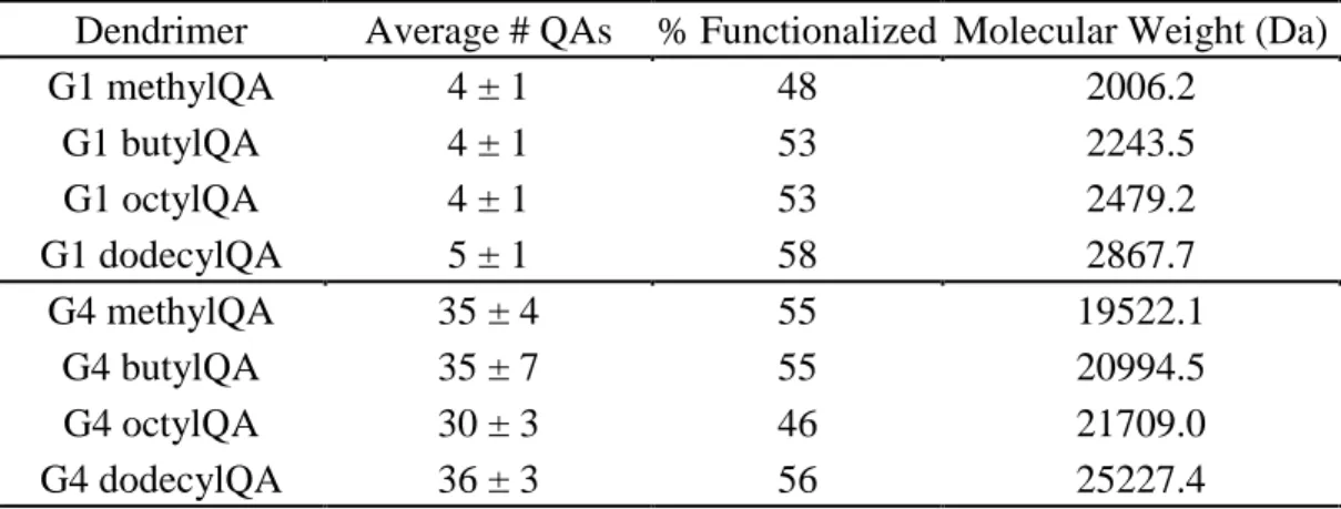
Table 2.1 Percent functionalization and corresponding molecular weight for
QA-modified G1 and G4 PAMAM dendrimers ...66 Table 2.2 Nitric oxide-release properties for G1 and G4 QA-modified
dendrimers in PBS (pH 7.4, 37 °C) as measured by a
chemiluminescence NO analyzer ...68 Table 2.3 Percent functionalization, corresponding molecular weight, and
critical vesicle concentrations for alkyl chain-modified PAMAM
dendrimers...75 Table 2.4 Nitric oxide-release properties of alkyl chain-modified dendrimers
in phosphate buffered saline (pH 7.4, 37 °C) as measured by a
chemiluminescence NO analyzer ...78 Table 3.1 Minimum bactericidal concentrations (MBC4h) and bactericidal
NO doses for single- and dual-action QA-modified dendrimers
against S. aureus and P. aeruginosa ...94 Table 3.2 Minimum bactericidal concentration (MBC24h) for single- and
dual-action alkyl chain-modified dendrimers against planktonic P.
aeruginosa, S. aureus, and MRSA...104 Table 4.1 Minimum biofilm eradication concentrations (MBEC24h) for
single- and dual-action alkyl chain-modified dendrimers against P.
aeruginosa, S. aureus, and MRSA biofilms ...128 Table 4.2 Inhibitory concentrations at 50% cell viability (IC50) for single-
and dual-action alkyl chain-modified dendrimers against L929
mouse fibroblast cells ...140 Table 4.3 Calculated therapeutic index (TI) for each dendrimer biocide
against P. aeruginosa, S. aureus, and MRSA biofilms ...141 Table 4.4 Combined biofilm eradication concentrations and calculated
fractional bactericidal concentration index (FBC24h) against S.
aureus and MRSA biofilms ...144 Table 5.1 Characterization and NO-release properties of G4 dendrimers in
PBS (pH 7.4, 37 °C) as measured by a chemiluminescence NO
xv
Table 5.2 Characterization of single-component electrospun polyurethane
fibers ...170
Table 5.3 Nitric oxide-release properties for G4 octyl/NO-doped electrospun polyurethane fibers in PBS (pH 7.4, 37 °C) ...172
Table 5.4 Characterization of electrospun Tecoflex fibers ...179
Table 5.5 Nitric oxide-release properties for NO-releasing electrospun Tecoflex fibers in PBS (pH 7.4, 37 °C) ...181
Table 5.6 Average log reduction of adhered bacterial viability ...189
Table 6.1 Characterization of electrospun polyurethane fibers ...215
Table 6.2 Total nitric oxide storage and dendrimer encapsulation by fiber mass...216
Table 6.3 Nitric oxide-release properties for NO-releasing electrospun fibers in PBS (pH 7.4, 37 °C) ...221
Table 6.4 Average zone of inhibition against planktonic bacteria ...223
Table 6.5 Average log reduction against planktonic bacteria ...226
Table A.1 Characterization and NO-release properties of G4 dendrimers ...253
xvi
LIST OF SCHEMES
Scheme 2.1 Reaction of PAMAM scaffold with either (A) QA or (B) alkyl chain epoxides to yield single-action dendrimers, followed by reaction with high pressures of NO to yield dual-action,
NO-releasing dendrimers ...64 Scheme 6.1 Reaction of G4 PAMAM scaffold with either (A) octyl alkyl chain
or (B) octylQA epoxides to yield G4 octyl and G4 octylQA
dendrimers, respectively, followed by reaction with high pressures
xvii
LIST OF FIGURES
Figure 1.1 Stages of pathogenic biofilm formation ...3
Figure 1.2 Schematic representation of biofilm protection mechanisms ...4
Figure 1.3 Schematic representation of the multi-mechanistic killing pathways of NO and its byproducts ...17
Figure 1.4 (A) Possible decomposition mechanisms for S-nitrosothiol (RSNO) NO donors, including heat, light, and copper-mediated decomposition. (B) Formation of N-diazeniumdiolate NO donors on secondary amines followed by proton-initiated decomposition to yield two moles of NO for every mole of NO donor ...21
Figure 1.5 Modes of combination therapies ...26
Figure 1.6 Mechanisms of action for quaternary ammonium (QA) compounds ...29
Figure 1.7 Structure of poly(amidoamine) (PAMAM) dendrimer scaffold ...32
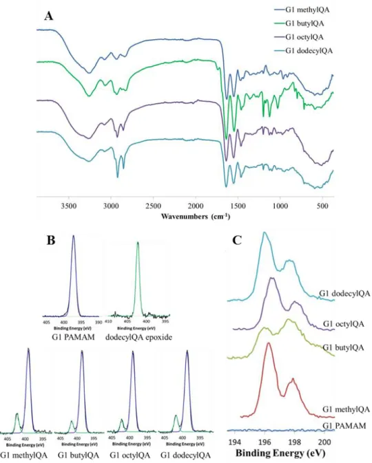
Figure 2.1 Representative (A) Fourier transform infrared spectra of G1 QA-modified dendrimers. Representative (B) N 1s and Cl 2p XPS spectra for G1 QA-modified dendrimers ...67
Figure 2.2 Representative UV-vis spectra for NO-releasing QA-modified dendrimers...69
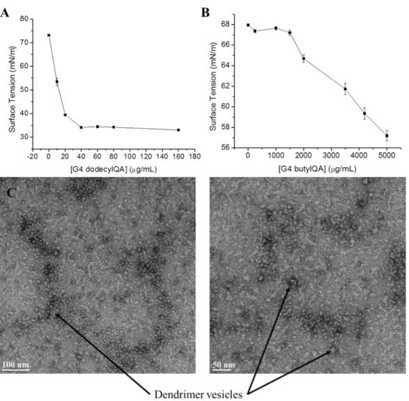
Figure 2.3 Formation of dendrimer vesicles at pH 7.4 ...72
Figure 2.4 Surface tension plots of (A) G4 dodecylQA and (B) G4 butylQA. Transmission electron microscopy images of (C) G4 dodecylQA dendrimer vesicles ...73
Figure 2.5 Fourier transform infrared spectra of G1 alkyl chain-modified dendrimers...76
Figure 2.6 Representative UV-vis spectra for NO-releasing alkyl chain-modified dendrimers ...79
xviii
Figure 3.2 Confocal microcopy images of S. aureus exposed to 50 µg/mL RITC-tagged G4 methylQA and dodecylQA dendrimers at (A) 0,
(B) 20, (C) 24, (D) 28, and (E) 32 min after dendrimer addition ...97 Figure 3.3 Confocal microcopy images of P. aeruginosa exposed 50 µg/mL
RITC-tagged (A) G4 methylQA and (B) G4 butylQA dendrimers
after 20 min exposure...100 Figure 3.4 Confocal microscopy images of P. aeruginosa and S. aureus
exposed to 20 µg/mL G4 dodecylQA/NO dendrimers at (A) 0, (B)
25, (C) 35, (D) 45, and (E) 60 min after dendrimer addition ...101 Figure 3.5 Viability (%) of L929 mouse fibroblast cells following 4 h
exposure to single- (blue) and dual-action (green) QA-modified dendrimers, as well as unmodified PAMAM dendrimer (red), at the MBC4h against (A) S. aureus and (B) P. aeruginosa compared
to untreated control cells ...109 Figure 3.6 Viability (%) of L929 mouse fibroblast cells following 24 h
exposure to single- (blue) and dual-action (green) alkyl
chain-modified dendrimers compared to untreated control cells ...110 Figure 4.1 Confocal microscopy images of (A) P. aeruginosa and (B) S.
aureus biofilms exposed to 100 µg/mL G1 hexyl/NO dendrimers
for 2 h ...129 Figure 4.2 Confocal microscopy images of P. aeruginosa biofilms exposed to
100 µg/mL G1 butyl/NO and G1 hexyl/NO dendrimers for (A) 1 h
and (B) 2 h ...131 Figure 4.3 Confocal microscopy images of S. aureus biofilms exposed to 50
µg/mL RITC-tagged (A) G1 butyl, (B) G2 butyl, (C) G4 butyl, (D)
G1 hexyl, (E) G2 hexyl, and (F) G4 hexyl dendrimers for 1 h ...132 Figure 4.4 Confocal microscopy images of S. aureus biofilms exposed to 250
µg/mL (A) G1 hexyl/NO and (B) G2 hexyl/NO dendrimers for 3 h ...136 Figure 4.5 Viability (%) of L929 mouse fibroblast cells following 24 h
exposure to individual (solid) and combined (hashed) dendrimer and vancomycin biofilm eradication concentrations against (A) S.
xix
Figure 5.1 Scanning electron micrographs of blank, control, and NO-releasing G4 octyl-doped electrospun Tecoplast, Tecoflex, and Hydrothane
fibers ...169 Figure 5.2 Representative total NO-release curves for (A) G4 octyl/NO-doped
electrospun Tecoplast, Tecoflex, and Hydrothane fibers and (B) NO-releasing dendrimer-doped electrospun Tecoflex fibers. Representative NO-release curves for (C) G4 hexyl/NO, (D) G4 octyl/NO, (E) G4 dodecyl/NO, and (F) G4 octylQA/NO-doped
electrospun Tecoflex fibers ...173 Figure 5.3 Electron micrographs of G4 RITC octyl-doped (A) Tecoplast, (B)
Tecoflex, and (C) Hydrothane fibers. (D) Fluorescence image of
Tecoplast-G4 RITC octyl fibers...175 Figure 5.4 Dendrimer delivery (%) for control (solid line) and NO-releasing
(dashed line) G4 octyl-doped electrospun Tecoplast (red), Tecoflex (blue), and Hydrothane (green) fibers after 2 h, 1 d, or 7 d soaking
in PBS (pH 7.4, 37 °C) ...176 Figure 5.5 Scanning electron micrographs of control and NO-releasing G4
hexyl-, G4 dodecyl-, and G4 octylQA-doped electrospun Tecoflex
fibers ...178 Figure 5.6 Dendrimer delivery (%) for control (solid line) and NO-releasing
(dashed line) G4 hexyl- (red), G4 octyl- (blue), G4 dodecyl- (green), and G4 octylQA- (purple) doped electrospun Tecoflex
fibers after 2 h, 1 d, or 7 d soaking in PBS (pH 7.4, 37 °C) ...182 Figure 5.7 Viability (%) of L929 mouse fibroblast cells following (A,C) 2 h
or (B,D) 24 h exposure to blank (solid), control (diagonal lines), and NO-releasing (horizontal lines) (A,B) G4 octyl-doped
electrospun fibers or (C,D) dendrimer-doped electrospun Tecoflex
fibers ...184 Figure 5.8 (A) Viability (%) of L929 mouse fibroblast cells adhered to control
(diagonal lines) and NO-releasing (horizontal lines) dendrimer-doped Tecoflex electrospun fibers following 24 h exposure. (B) Scanning electron micrographs of L929 mouse fibroblast cells adhered to blank, control, and NO-releasing G4 octyl-doped
xx
Figure 5.9 Viability of (A,B) P. aeruginosa or (C,D) S. aureus adhered to blank (solid), control (diagonal lines), and NO-releasing (horizontal lines) dendrimer-doped Tecoflex electrospun fibers
following 2 or 6 h exposure ...188 Figure 6.1 Scanning electron micrographs of blank, control, and NO-releasing
G4 octyl-doped electrospun SG 80A/HP 93A, TP 470/SG 80A,
and TP 470/HP 93A fibers ...210 Figure 6.2 Image of electrospun fiber substrates (1.267 cm2). From left to
right: SG 80A/HP 93A, TP 470/SG 80A, and TP 470/HP 93A ...211 Figure 6.3 Histograms depicting fiber diameter distribution in nm for blank,
control, and NO-releasing G4 octyl-doped electrospun SG 80A/HP
93A, TP 470/SG 80A, and TP 470/HP 93A fibers ...214 Figure 6.4 (A) Delivery of control (solid line) and NO-releasing (dashed line)
G4 octyl dendrimers from electrospun SG 80A/HP 93A fibers at 15 (blue) and 25 (purple) mg/mL initial dendrimer concentration. (B) Delivery of control (solid line) and NO-releasing (dashed line) G4 octyl dendrimers from electrospun SG 80A/HP 93A (blue), TP 470/SG 80A (red), TP 470/HP 93A (black) fibers. (C) Delivery of control (solid line) and NO-releasing (dashed line) G4 octyl (square) and G4 octylQA (triangle) dendrimers from electrospun
TP 470/HP 93A fibers ...220 Figure 6.5 Fluorescence microscopy images of P. aeruginosa exposed to TP
470/HP 93A-G4 octyl/NO fibers for 5 and 15 minutes ...227 Figure 6.6 Viability (%) of L929 mouse fibroblast cells following (A) 2 h or
(B) 24 h exposure to blank, control, and NO-releasing electrospun SG 80A/HP 93A (blue), TP 470/SG 80A (red), TP 470/HP 93A
(gray) fibers ...229 Figure A.1 Custom electrospinning apparatus in co-axial configuration ...255 Figure A.2 Scanning electron micrographs and fiber diameter histograms of
TP 470/HP 93A-G4 octylQA electrospun fibers ...256 Figure A.3 Scanning electron micrographs of RITC-tagged TP 470/HP 93A
xxi
Figure A.5 Representative zone of inhibition images against planktonic
bacteria ...259 Figure A.6 Viability of planktonic bacteria after 2 h exposure to TP 470/HP
93A electrospun fibers ...260
Figure A.7 Viability of planktonic bacteria after 24 h exposure to TP 470/HP
93A electrospun fibers ...261 Figure A.8 Viability (%) of L929 mouse fibroblast cells following 2 h (solid)
or 24 h (diagonal lines) exposure to blank, control, and
xxii
LIST OF ABBREVIATIONS AND SYMBOLS
~ approximately
°C degree(s) Celsius > greater than
≥ greater than or equal to < less than
≤ less than or equal to [NO]max maximum NO flux
% percent
± standard deviation of the mean ×g times the force of gravity
µg microgram(s)
µL microliter(s)
µm2 micrometers squared
µM micromolar
µmol micromole(s)
Abs absorbance
Ag silver
Ag+ silver ion(s)
AHAP3 N-(6-aminohexyl)amino-propyltrimethoxysilane AMP antimicrobial peptide
Ar argon
xxiii Ca2+ calcium ion(s)
cfu colony forming units
cGMP cyclic guanosine monophosphate Cl chlorine atom(s)
Cl+ oxidative chlorine CO2 carbon dioxide
cm centimeter(s)
cm-1 wavenumber(s)
cm2 centimeters squared
cNOS constitutive nitric oxide synthase CVC critical vesicle concentration
d day(s)
Da Daltons
DAF-2 4,5-diaminofluorescein
DAF-2 DA 4,5-diaminofluorescein diacetate
DETA/NO N-diazeniumdiolate-modified diethylenetriamine DMEM Dulbecco’s modified Eagle’s medium
DMF N,N-dimethylformamide DNA deoxyribonucleic acid
E. coli Eschericia coli E. faecalis Enterococcus faecalis
EDA ethylene diamine
xxiv et al. et alia (and others)
eNOS endothelial nitric oxide synthase EPS exopolysaccharide
eV electron volts
FBC24h 24 h fractional bactericidal concentration index FBS fetal bovine serum
Fe3+ iron ion(s)
FISH fluorescent in situ hybridization
FTIR Fourier transform infrared spectroscopy
Ga gallium
G1 generation 1
G2 generation 2
G3 generation 3
G4 generation 4
GSNO S-nitroso-L-glutathione
h hour(s)
1H NMR proton nuclear magnetic resonance HCl hydrochloric acid
HP 93A Tecophilic
IC50 inhibitory concentration at 50% viability i.e. id est (that is)
iNOS inducible nitric oxide synthase
xxv KOH potassium hydroxide
kV kilovolt(s)
LB Luria-Bertani
MAP3 N-methylaminopropyltrimethoxysilane MBC4h 4 h minimum bactericidal concentration MBC24h 24 h minimum bactericidal concentration
MBEC24h 24 h minimum biofilm eradication concentration
MeOH methanol
min minute(s)
mg milligram(s)
mL milliliter(s)
mS millisiemen(s)
mm millimeter(s)
mM millimolar
mmol millimole(s)
MRSA methicillin-resistant Staphylococcus aureus
MTS 3-(4,5-dimethylthiazol-2-yl)-5-(3-carboxymethoxyphenyl)-2-(4-sulfophenyl)-2H-tetrazolium inner salt
mW milliwatt(s)
MWCO molecular weight cutoff
n number of dendrimer terminal end groups N2 nitrogen gas
xxvi NaOMe sodium methoxide
nm nanometer(s)
nM nanomolar
NO nitric oxide
NOA nitric oxide analyzer NOS nitric oxide synthase
nNOS neuronal nitric oxide synthase
O2 oxygen
O2- superoxide ONOO- peroxynitrite
P. aeruginosa Pseudomonas aeruginosa
PAMAM poly(amidoamine)
PBS phosphate buffered saline PCR polymerase chain reaction PEG poly(ethylene glycol) pH -log of proton concentration
PI propidium iodide
PIA polysaccharide intercellular adhesion PLGA poly(lactic-co-glycolic acid)
PLGH poly(lactic-co-glycolic-co-hydroxymethyl propionic acid)
pM picomolar
pmol picomole(s)
xxvii ppb parts per billion
PPI polypropylenimine ppm parts per million
PROLI/NO N-diazeniumdiolate-modified L-proline PS penicillin streptomycin
QA quaternary ammonium
QS quorum sensing
RIP RNA III inhibiting peptide RITC rhodamine b isothiocyanate RNA ribonucleic acid
RSNO S-nitrosothiol
s second(s)
S. aureus Staphylococcus aureus S. epidermidis Staphylococcus epidermidis
SEM scanning electron microscope SG 80A Tecoflex
SNAP S-nitroso-N-acetylpenicillamine
STAMP selectively targeted antimicrobial peptide t1/2 half-life
t[NO] total NO release t[NO]2h 2 h total NO release t[NO]4h 4 h total NO release
xxviii td duration of NO release
tmax time required to reach maximum NO flux TEA triethylamine
TEM transmission electron microscopy THF tetrahydrofuran
TI therapeutic index TP 470 Tecoplast
TSA tryptic soy agar TSB tryptic soy broth
UV ultraviolet
v/v volume to volume ratio
v/v/v volume to volume to volume ratio
vis visible
vol% percent by volume WD weight of dry fiber mat WH weight of hydrated fiber mat wt% percent by weight
XPS x-ray photoelectron spectroscopy Zn2+ zinc ion(s)
1 CHAPTER 1:
Designing Novel Antibacterial Agents for Enhanced Biofilm Eradication
Biofilm-based infections, especially those caused by the opportunistic pathogens
Pseudomonas aeruginosa and Staphylococcus aureus, continue to pose a tremendous challenge to the medical field.1-3 Biofilms are bacterial communities encased in a protective exopolysaccharide matrix that demonstrate a variety of protective mechanisms, allowing for their persistence despite treatment with antibiotics or antiseptics.4-6 The inability of current therapeutics to effectively eradicate biofilms promotes bacterial resistance. Nitric oxide is an endogenously produced free radical that plays a central role in the host immune response to infection, exhibits broad-spectrum antibacterial action, and is unlikely to foster resistance, making it an ideal candidate for the development of novel biocidal agents.7-10 This chapter will describe the formation and protective mechanisms of biofilm-based infections, current research into more effective anti-biofilm therapeutics, the existing state of macromolecular scaffolds for controlled nitric oxide release, and the emergence of dual-action nitric oxide-releasing dendrimers as novel antibacterial agents. 1.1 Biofilm-based infections
2
magnitude greater than those required to eradicate planktonic bacteria.6, 15 It is thus important to examine the formation of these biofilm communities and the protective mechanisms that allow them to persist when designing new antibacterial agents.
1.1.1 Biofilm formation
Biofilm-based bacterial infections have been observed on most, if not all, implanted medical devices.2, 11, 14 The formation of a biofilm is initiated by the irreversible attachment of planktonic bacteria to both living and inanimate surfaces, including human tissue and medical implants (Figure 1.1).4-6, 11, 13, 14, 16, 17 Both the chemical properties of the underlying surface and the identity of colonizing bacterial cells impact the rate and extent of biofilm formation.6 While biofilms have been observed on both rough and smooth surfaces ranging from very hydrophobic (e.g., Teflon, silicone) to charged and hydrophilic (e.g., glass, metal) materials, rougher and more hydrophobic materials develop biofilms more rapidly.4 Bacterial appendages such as flagella or pili increase the rate of microbial attachment to surfaces, allowing bacteria to overcome repulsive or removal forces and remain attached.4
3
4
5
architecture that protects the bacterial community from the immune response and antimicrobial agents.17 The protective mechanisms of the EPS matrix will be described in detail in Section 1.1.2. The resulting EPS-confined biofilms are highly hydrated (98% water), persistently bound to the underlying surface, and heterogeneous in space, time, and bacterial composition.4 Although P. aeruginosa and S. aureus are two of the most commonly isolated species in clinical infections, a more complex bacterial microflora has been observed with improvements in bacterial isolation techniques.2, 18, 19 Consequently, the accumulation of a complex biofilm composed of a diverse bacterial community is considered to be responsible for promoting systemic infection via the detachment of cells and aggregates from the parent biofilm.4
1.1.2 Protective mechanisms of biofilms
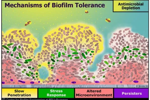
In addition to the increasing prevalence of antibiotic resistant bacteria,20-22 biofilm-based bacterial infections are notoriously resistant to standard antimicrobial treatment.4-6, 23, 24 Antibiotic treatments that are effective at killing planktonic bacteria are often ineffective against biofilms, with biofilm eradication often requiring a 1,000–10,000-fold increase in therapeutic dose.6, 15 Of importance, the common mechanisms of antibiotic resistance, including expression of efflux pumps, modified enzymes and target sites, and production of alternative metabolic pathways, are not necessarily responsible for the protection of these bacterial communities.22, 24 Biofilms instead exhibit several unique protective mechanisms that contribute to their overall robustness (Figure 1.2).4, 5, 24
6
measurements of antibiotic penetration in biofilms have exhibited a wide variation in permeation rates through such matrices, dependent on both the antibiotic structure and biofilm architecture.23, 24 Significant limitations in biofilm penetration have been reported for beta-lactams and aminoglycosides.23 In particular, positively-charged aminoglycosides are believed to bind to the negatively charged polymers within the biofilm, leading to antibiotic adsorption to the biofilm matrix and inhibited biofilm penetration.24 In addition to restricted penetration, antimicrobial agents may be deactivated through reaction with or binding to the biofilm EPS matrix.23, 25 For example, Nichols et al. demonstrated that the addition of alginate during biofilm formation reduced inhibition zones for tobramycin against Escherichia coli and S. aureus, suggesting the EPS matrix either interferes with antimicrobial action of tobramycin or inhibits its diffusion through the biofilm.26 In a similar study, Coquet et al. observed a decrease in antimicrobial action for both tobramycin and imipenem against alginate-embedded P. aeruginosa biofilms compared to planktonic bacteria.27 While the combination of tobramycin and imipenem was synergistic against planktonic cultures of P. aeruginosa, no synergy was displayed for the biofilm cultures.
7
conditions.6 These chemical gradients also lead to heterogeneity within the bacterial community with respect to growth states, with populations ranging from the continuously proliferating to the metabolically inactive.5, 23, 24 The depletion of nutrients may cause bacteria to enter a slow- or non-growing state where they are protected from killing. The varied rates of bacterial growth dramatically dictate the effectiveness of antibiotics, many of which only target rapidly growing bacteria.6, 23, 24
Quorum sensing (QS) is a method of cell-to-cell communication between bacterial cells that allows for cell density and/or population-based gene regulation.28, 29 Individual bacterial cells produce and release small QS signaling molecules to detect and relay information on the characteristics of the surrounding environment. The development of antibiotic and biocide tolerance or resistance phenotypes in biofilms is thought to be partially regulated by QS, although the exact mechanism is unknown.28, 30 The dense, confined biofilm environment enables the accumulation of QS signaling molecules, triggering QS-regulated gene expression and affecting the host immune response.28 For example, the release of various virulence factors by pathogenic bacteria is regulated by QS processes.2, 31 In the case of P. aeruginosa, production of the leukocidal toxin rhamnolipid B shields against invading neutrophils and other cellular components of the host response, contributing to the persistence of P. aeruginosa biofilms.2, 30
8
to resist killing by chemical disinfectants (e.g., chlorine bleach, glutaraldehyde, hydrogen peroxide) and antibiotics.23, 34 Further, this subpopulation of highly persistent bacteria is observed in newly formed biofilms that are too thin to provide an adequate barrier to antibiotic diffusion or metabolic gradients.34, 35 The presence of a persister subpopulation within a bacterial biofilm may promote the longevity and wide-spread resistance of bacteria to antimicrobial agents and disinfectants comprised of varying chemistries.
1.2 Current research into anti-biofilm therapeutics
The protective mechanisms exhibited by biofilms contributes to their ability to withstand treatment with antibiotics or antiseptics. Further, the increased occurrence of antibiotic-resistant bacteria coupled with a decline in new antibiotic research has necessitated the development of novel therapies capable of lessening or completely eliminating biofilm-based infections. This section describes the ongoing research aimed at either disrupting biofilm formation or killing established biofilms to reduce the bacterial burden in clinical infections.
1.2.1 Prevention of initial biofilm attachment
9
As microbial cell membranes tend to be hydrophobic and negatively-charged, antifouling properties are often imparted to surfaces through chemical modification with hydrophilic or negatively charged functional groups.48 Poly(ethylene glycol) (PEG) is one of the most highly investigated hydrophilic polymers for antifouling biomedical applications. The utility of PEG stems from reducing protein adsorption and bacterial attachment through hydration and steric effects.49, 50 Unfortunately, resisting protein adhesion alone does not necessarily correlate with reduced bacterial adhesion due to adhesion mechanisms that do not rely on proteins and the instability of PEG coatings in physiological solutions.51, 52 The use of negatively charged polymer coatings (e.g., heparin, albumin) has resulted in significant reductions in catheter-related infections.53, 54 For example, a recent study demonstrated reduced adhesion of Staphylococcus epidermidis with a heparin-like polyurethane containing negatively charged sulfate groups.55 Similarly, polycationic antimicrobial surfaces have been developed by tethering quaternary ammonium moieties to polymer coatings.56-58 While the QA-modified surfaces effectively killed adhered bacteria, these immobilized antimicrobial or anti-infective agents are often only toxic to the first wave of incoming bacteria, providing little antimicrobial action once layers of dead bacterial cells accumulate.11, 56
10
variation between adhesins allows for the development of antibodies that should be effective against a wide range of bacterial strains. Langermann et al. reported antibodies against a single FimH adhesin protein with specificity for >90% of E. coli strains expressing the FimH adhesin.61 This approach blocked the binding of E. coli to bladder cells in vitro. Vaccination of mice with the FimH adhesin vaccine reduced the in vivo colonization of bladder mucosa by E. coli by >99% and elicited a long-term immune response to FimH.62
Instead of studying individual adhesins, Schneewind and coworkers attempted to identify chemical reactions or binding steps that are shared by a large majority of surface proteins.29, 63 Sortases are membrane enzymes present in many Gram-positive strains that catalyze the covalent anchoring of surface proteins to the outer peptidoglycan layer, making them an excellent target for anti-adhesion agents with broad clinical applications.63-65 Several sortase inhibitors have been identified to date, including methane-thiosulfonate and mercurial p-hydroxymercuribenzoic acid, which block Cys184 residues in the active pocket of sortase A.66, 67 However, these compounds exhibit poor therapeutic value due to their high toxicities.29
1.2.2 Exopolysaccharide matrix disruption
11
aureus, P. aeruginosa, E. coli), iron (Fe3+) is crucial for bacterial growth and metabolic enzyme function.11 Gallium (Ga), alternatively, exhibits many similar features as Fe3+ (i.e., ionic radius) but does not perform the same functions critical for proper enzyme performance; as such, the uptake of Ga over Fe3+ by biological systems renders the enzymes non-functional.11, 70 Kaneko et al. found that while P. aeruginosa grown in Ga concentrations >2 µM displayed a dose-dependent decrease in growth rate, sub-inhibitory Ga concentrations (1 µM) allowed for uninhibited P. aeruginosa growth but prevented biofilm formation.71
One of the most studied components of the Gram-positive EPS matrix is the polysaccharide intercellular adhesion (PIA) protein synthesized by the icaADBC operon in staphylococci, which is required for staphylococcal biofilm development.36 The ica gene thus represents a viable potential target for the development of biofilm inhibitors. Sub-inhibitory concentrations of the common antibacterial agent povidone-iodine have exhibited anti-biofilm activity against S. epidermidis by reducing the transcription of the icaADBC operon through activation of the icaR transcriptional repressor.72 The inhibition of PIA synthesis and staphylococcal biofilm disruption have also been demonstrated using the organosulfur compound allicin against S. epidermidis and sulfhydryl compounds (i.e., dithiothreitol, beta-mercaptoethanol, cysteine) against S. aureus.36, 73, 74 As dimerization of specific protein domains in the presence of zinc (Zn2+) are required for biofilm formation by both S. epidermidis and S. aureus, Zn2+ chelation was similarly shown to prevent biofilm formation by both staphylococcal bacteria, including antibiotic-resistant strains.75
1.2.3 Quorum sensing inhibition
12
infection, QS inhibitors have been developed to reduce bacterial pathogenicity.28, 29 Quorum sensing inhibition is achieved through the binding of the inhibitor molecule to the QS receptor proteins, displacing the QS regulation center.36
Two of the leading classes of QS inhibitor candidates are furanones and RNA III inhibiting peptide (RIP).29 Furanones in particular have been investigated for their broad-spectrum QS inhibitory mechanisms and ability to inhibit the QS centers in both negative and Gram-positive pathogens. Additionally, furanones incapable of QS inhibition have been determined to increase biofilm susceptibility to antibiotics and antiseptics. Hentzer et al. treated P. aeruginosa
biofilms with several natural furanone compounds and found the furanones specifically targeted QS systems, inhibited virulence factor expression, and increased bacterial susceptibility to tobramycin and certain detergents.76 The QS inhibitor RIP has also been shown to suppress pathogenic virulence, biofilm formation, and antibiotic resistance in certain staphylococcal strains.77-80 Although RIP exhibited promising results as a QS inhibitor during studies with in vivo animal models, clinical applications have been limited due to concerns over in vivo stability and drug toxicity.29 These concerns notwithstanding, several in vitro studies have established the potential of QS inhibitors as surface-immobilized anti-biofilm agents, qualifying their ability to disrupt biofilm formation on surfaces.81, 82
1.2.4 Development of novel anti-biofilm agents
13
one such area.25, 29, 36 While antibiotic delivery vehicles demonstrate several advantages, including more efficient antibiotic delivery, in vivo drug retention, and bacterial killing, fostering bacterial resistance continues to be a major drawback of these systems.
Ionic silver (Ag+) has historically been used as an antimicrobial agent because it offers broad-spectrum activity at relatively low concentrations (0.05 ppm in PBS, ~50 ppm in biological fluids).83-86 The antimicrobial activity of silver ions stems from a number of biocidal mechanisms, including surface binding to and damaging of bacterial cell membranes, interfering with DNA replication, poisoning respiratory enzymes, and denaturing proteins (including DNA and RNA).83,
84, 87 Although wide-spread resistance to silver is unlikely due to its multi-mechanistic biocidal
activity, bacterial resistance to silver has been observed clinically.84, 86, 87 Several different types of silver-resistant clinical strains, mostly Gram-negative bacteria, have been recently isolated.84-86,
88, 89 Bacterial resistance to silver is observed in two predominant forms: 1) the binding of silver
by cells in the form of an intracellular complex; and, 2) the excretion of silver from microorganisms using cellular efflux pumps.86, 89 In addition to potential bacterial resistance, silver use is associated with several clinical disadvantages. The use of silver products often leads to potential irritation or discoloration of the surrounding tissue (argyria), especially when using silver nitrate.87 Silver-based products also poorly discriminate between healthy cells and pathogenic bacteria, resulting in toxicity to both keratinocytes and fibroblasts at bactericidal silver levels.87, 89
14
membranes makes them potentially effective against slow-growing or resistant bacteria embedded within biofilms; the development of resistance to this kind of mechanical disruption is rare.90, 91 Unfortunately, AMPs are both difficult and expensive to synthesize in large quantities, have low specificity, and are sensitive to protease digestion, limiting current therapeutic utility.29
Modification of AMPs has led to the development of selectively targeted antimicrobial peptides (STAMPs), which represent a novel strategy for species-specific biofilm control. A typical STAMP contains a species-specific targeting peptide connected to an antimicrobial peptide via a 2 to 20 amino acid peptide chain.29 This configuration allows for specific microbe targeting without reducing antibacterial activity. Eckert et al. first synthesized a P. aeruginosa-specific STAMP with increased bactericidal action and faster killing against P. aeruginosa than the non-targeted, general-killing peptide control.92 The development of STAMPs has been extended to selectively remove both Gram-positive and Gram-negative bacterial strains from multi-species planktonic cultures.92, 93 To date, however, the use of STAMPs alone has only resulted in inhibitory effects against P. aeruginosa and various cariogenic biofilms, though the co-administration of STAMPs with tobramycin has enhanced their biofilm eradication capabilities.94 1.3 Nitric oxide
15
This section will consider the physiological roles of endogenously produced NO, including its innate antimicrobial action.
1.3.1 Physiological roles of nitric oxide
Nitric oxide is produced endogenously through the oxidation of L-arginine to L-citrulline via nitric oxide synthase (NOS).9, 95, 96, 98 The three distinct isoforms of NOS are divided into two classes – constitutive (cNOS) and inducible (iNOS). The constitutive class encompasses the endothelial (eNOS) and neuronal (nNOS) isoforms with regulation by Ca2+ fluxes and binding to the messenger protein calmodulin.95, 96 iNOS is present in epithelial, endothelial, and inflammatory cells (e.g., macrophages and neutrophils) and upregulates upon cytokine expression and/or inflammatory stimuli.9, 95, 96 Although the three isoforms catalyze the same reaction to produce NO, they vary greatly with respect to regulation, concentration, and duration of NO production. The lowest levels of NO are produced by eNOS (pM), an enzyme located in the vascular endothelium, neurons, epithelial cells, and cardiac myocytes.98 The low, intermittent levels of NO generated by eNOS help maintain basal vascular tone and proper blood flow and pressure.95 Similarly, nNOS rapidly produces low, transient levels of NO (pM–nM) in neurons, skeletal muscle, and epithelial cells, allowing NO to function as a neurotransmitter in neuronal tissue.95, 98 As might be expected, iNOS has the highest capacity for NO generation, yielding significantly greater quantities of NO (µM) per mole of enzyme per minute than either of the cNOS isoforms.95, 98 The iNOS isoform is expressed in several cell types, particularly neutrophils and macrophages, as part of the innate immune response against invading pathogens and will be detailed further in Section 1.3.2.95, 96, 98
16
reacts with the iron center of guanylate cyclase, activating the production of cyclic guanosine monophosphate (cGMP).95 The upregulation of cGMP leads to smooth muscle relaxation and vasodilation. Nitric oxide production in the cardiovascular system also regulates vascular tone, myocardial contractility, endothelial-leukocyte interactions, and antithrombotic effects, while NO deficiencies due to endothelium injury can lead to cardiovascular conditions such as atherosclerosis, hypertension, coronary heart disease, and stroke.7, 95 Additionally, cNOS-derived NO acts as an intracellular messenger and neurotransmitter in the central nervous system through the stimulation of neuronal cGMP production.97, 99 The cGMP-dependent nervous system functions of NO include regulating the firing of neurons in various areas of the brain and spinal cord, the release of specific neurotransmitters (e.g., acetylcholine, serotonin), and membrane depolarization.99 The roles of endogenous NO span a wide range of functions in various biological systems, demonstrating its significance as a regulatory and signaling molecule.
1.3.2 Antimicrobial properties of nitric oxide
17
18
fungi, and parasites.8, 9, 105-107 The broad-spectrum antimicrobial action of NO is attributed to its multi-mechanistic killing pathways, exerting both nitrosative and oxidative stresses on pathogens through the formation of reactive nitrogen and oxygen intermediates (Figure 1.3).7, 8, 104 Reactive nitrogen intermediates (e.g., N2O3) apply nitrosative stress to bacterial cells, modifying DNA, proteins, and lipids.8 DNA targeting results in deamination, abasic sites, and strand breaks.9 While nitrosation of protein thiols is one of the most important protein targets of NO, nitrosative stress can occur at heme groups, iron-sulfur clusters, or amines in addition to reactive thiols.8, 9 Reaction of NO with superoxide (O2-) to form peroxynitrite (ONOO-) leads to oxidative DNA and protein damage as well as lipid peroxidation.8, 104, 108, 109 This multi-mechanistic action not only allows NO to exhibit broad-spectrum antimicrobial activity but also makes it unlikely to foster bacterial resistance.7 Indeed, Gram-positive, Gram-negative, and antibiotic-resistant bacteria that survived exposure to lethal exogenous NO doses after several passages (20 d) demonstrated minimal increases in inhibitory NO concentrations, rendering the development of NO release a viable option for designing novel antibacterial agents that do not foster bacterial resistance.10
19
less efficient than NO at scavenging superoxide, making it only partially effective at mitigating toxicity at high NO concentrations.111
1.4 Nitric oxide-releasing materials
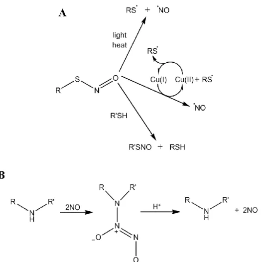
Exogenous NO donors have displayed substantial antibacterial and anti-biofilm activity against a broad range of microorganisms. Indeed, applying gaseous NO to infected dermal wounds was found to reduce the microbial burden and improve wound healing.112, 113 Both Gram-negative and Gram-positive bacteria, including antibiotic-resistant strains, have been found to be susceptible to gaseous NO.114 Unfortunately, the rapid reactivity of gaseous NO makes its use impractical in a clinical setting, limiting any potential therapeutic utility. A number of NO donors have been developed to allow for the controlled storage and delivery of NO, with the hope of expanding the range of potential clinical applications. Several classes of NO donors exist, including metal nitrosyls, organic nitrites and nitrates, oximes, S-nitrosothiols (RSNOs), and N -diazeniumdiolates.7, 115 To date, however, none have been translated to the clinic primarily due to challenging manufacturing issues.7 S-nitrosothiol and N-diazeniumdiolate NO donors represent the most extensively researched NO-release agents with the greatest potential as therapeutics, though still only in the development stages.115
1.4.1 Nitric oxide donors
20
ascorbate).118 The multiple decomposition pathways and triggers to liberate NO results in certain stability issue in vivo, complicating the progression of RSNO-based therapies to the clinic.115
N-Diazeniumdiolate NO donors form on secondary amines under high pressures of NO gas in basic conditions to yield zwitterionic dialkylamino diazeniumdiolate salts (Figure 1.4B).119-121 The resulting NO-releasing materials are generally stable in basic solutions or as solids at room temperature or below.120 In aqueous solutions at neutral or acidic pH, protonation of the secondary amine nitrogen initiates NO donor breakdown, yielding two moles of NO for every mole of NO donor.120, 122 As such, N-diazeniumdiolate moieties undergo spontaneous dissociation to yield biologically active NO under physiological conditions (i.e., pH 7.4), facilitating the therapeutic utility of this NO donor class. The rate of NO release is dependent on solution pH, with increasing solution acidity resulting in faster NO release.122 Moreover, the rate and duration of NO release can be tuned by varying the identity of the amine precursor, allowing for chemical control of the NO-release kinetics.121, 122 Similar to RSNO-based applications, no N -diazeniumdiolate-containing compounds have been clinically approved; however, a variety of animal studies have demonstrated the biomedical utility of N-diazeniumdiolate NO donors as a therapeutic source of NO.115 Due to their increased stability and spontaneous NO release under physiological conditions, this work will primarily focus on N-diazeniumdiolate NO donor storage and delivery chemistries.
1.4.2 Macromolecular nitric oxide-release scaffolds
21
22
quantities of NO (7.24 µmol/mg) due to the accessibility of secondary amines.123 Poor NO donor stability triggers rapid NO release (t1/2 = 1.8 s), however, resulting in NO liberation prior to association with bacteria and necessitating greater PROLI/NO doses for effective therapeutic action.124 In contrast, the small molecule N-diazeniumdiolate-modified diethylenetriamine (DETA/NO) exhibits slower NO donor breakdown, allowing for greater broad-spectrum antibacterial activity.125 Nevertheless, the antibacterial action of low molecular weight donors remains limited by uncontrolled NO release initiation and insufficient bacterial targeting, warranting the development of macromolecular scaffolds for NO delivery.
Modifying macromolecular scaffolds with NO-release capabilities has several advantages over small molecule NO donors, including more controllable NO payloads and release rates, modifiable surface chemistries, and reduced toxicity to mammalian cells. Several types of macromolecular scaffolds have been investigated for their NO-release capabilities, including liposomes,126-129 metal nanoparticles,130 metal organic frameworks,126, 131-133 chitosan,127, 134, 135 silica nanoparticles,104, 136-140 and dendrimers.141-143 The use of NO-releasing macromolecular vehicles improves both control over NO release and bacterial targeting/association. For example, the inhibitory concentration of S-nitrosated human serum albumin was decreased against bacteria by a factor of 5000 compared to two small molecule RSNO donors as a result of scaffold-bacteria association.144 Hetrick et al. also observed that NO-releasing silica eradicated planktonic P. aeruginosa cultures at NO doses 20x lower than those required from the small molecule NO donor PROLI/NO.104 The increase in bactericidal action was attributed to more efficient (targeted) NO delivery by the NO-releasing silica particles to the bacteria compared to PROLI/NO.
23
functionalization. Backlund and coworkers reported that NO-releasing silica nanoparticles with greater surface charge facilitated increased bacterial association, NO delivery efficiency, and bactericidal action over particles with lower zeta potential but similar NO totals and kinetics.136 Carpenter et al. evaluated the ability of NO-releasing silica nanoparticles to reduce planktonic P. aeruginosa bacterial viability as a function of particle size through the synthesis of 50, 100, and 200 nm silica nanoparticles, independent of total NO payload.137 Smaller (50 nm) particles were more effective at killing P. aeruginosa than larger (200 nm) particles, which was attributed to faster particle-bacteria association. In a similar study, Slomberg et al. found that 14 and 50 nm NO-releasing silica particles were more effective at killing planktonic P. aeruginosa than 150 nm particles.140 Interestingly, the 50 and 150 nm NO-releasing particles exhibited similar action against planktonic Gram-positive S. aureus cultures, with increased bactericidal action observed for the 14 nm particles, demonstrating a dependence of Gram designation on NO antibacterial activity. Lu and coworkers expanded these studies to investigate the effects of particle shape on bactericidal activity. Nitric oxide-releasing silica nanorods were synthesized with varied aspect ratios, maintaining the same particle volume and NO release while increasing the rod length.145 Greater antibacterial action against planktonic P. aeruginosa was observed with increasing aspect ratio due to more efficient NO delivery by the longer silica nanorods that associated with the bacteria longitudinally.
1.4.3 Effect of nitric oxide-releasing scaffold properties on biofilm eradication
24
nitroprusside, SNAP, GSNO) to disperse bacterial cells using P. aeruginosa, E. coli, S. epidermidis, and multi-species biofilms.147, 148 To fully harness the anti-biofilm capabilities of NO, the development of NO-releasing scaffolds has focused on understanding the physicochemical properties of the scaffold with respect to enhanced biofilm eradication.
Hetrick et al. reported broad-spectrum anti-biofilm activity for NO-releasing MAP3 silica nanoparticles against Gram-negative, Gram-positive, and pathogenic fungus biofilms.139 Further, the MAP3/NO silica nanoparticles demonstrated ~1000-fold greater efficacy against P. aeruginosa
25 1.5 Designing dual-action therapeutics
The use of antibiotic combinations has become common in clinical practice in efforts to increase the spectrum of antimicrobial activity, lower doses to circumvent toxicity, and minimize the likelihood of emerging bacterial resistance.149 Combination therapies can be classified into four modes of action that allow for enhanced antimicrobial activity (Figure 1.5).150 The first three modes combine a secondary adjuvant compound that augments activity of the primary drug by either 1) preventing its degradation or modification, 2) allowing for its accumulation and retention, or, 3) inhibiting the repair or tolerance mechanism of microbial cells to the primary drug. The fourth mode involves the combination of two or more mechanistically different biocides. Employing multiple compounds that work through different biocidal pathways should not only increase the bactericidal action and antibacterial sphere of influence, but also lessen the emergence of resistance due to the improbability of bacteria developing resistance to both biocidal mechanisms.150, 151 Further, combining two or more antibacterial agents can result in synergy, where the bactericidal efficacy of the combination is more effective than their individual sums.149, 150 As such, the design of dual-action therapeutics has become a viable option for the development of novel antibacterial agents.
26
27
bacteriostatic drugs bactericidal. For example, the bactericidal drug Synercid (King Pharmaceuticals; Bristol, Tennessee) is the combination of two static antibacterial compounds.152 More recent work has focused on the development of dual-action compounds on a single scaffold. For example, Oxaquin (BioVertis; Vienna, Austria) combines the therapeutic components of two different compounds in one molecule.150, 154, 155
The majority of dual-action antibacterial agents utilize a non-depleting, contact-based biocide as the base scaffold. Quaternary ammonium (QA) compounds are a popular non-depleting biocide due to their broad-spectrum efficacy and ability to kill bacteria without altering the QA structure, allowing for continued bactericidal activity.156-158 The antibacterial action of QA compounds is derived from the attractive electrostatic interactions between the cationic QA group and the negatively charged bacterial cell membrane, in turn disrupting natural chemical balances by replacing essential metal cations (Figure 1.6).156, 159 Adding alkyl chain groups to the QA moiety increases the biocidal action by promoting bacterial membrane penetration and disruption.156, 157, 159, 160 Although their simple structure has allowed for the facile incorporation of QA moieties into a variety of systems (e.g., polymers, films, particles), tethering QAs to polymers or particles limits their action to only the bacteria that come into contact with the QA-modified surface. As a result, combining QA moieties with releasable antibacterial agents should increase the biocidal sphere of influence in addition to improving the overall bactericidal action.
28
Hu et al. fabricated dual-action cellulose fibers by co-grafting N-halamine and QA salt monomers, allowing for the release of oxidative chlorine (Cl+) from QA-modified cellulose.162 The resulting dual-action fibers exhibited rapid and increased bactericidal action against Gram-negative and Gram-positive bacterial strains over the single-action QA-modified or Cl+-releasing cellulose alone.
The efficacy of combining tethered QA moieties with releasable silver is another area of active research. Multi-layer antibacterial coatings capable of both release- and contact-based killing were produced by incorporating QA moieties and silver in a layer-by-layer method.44, 163 For example, Grunlan et al. fabricated multi-layer films by dipping substrates into solutions containing either cetyltrimethylammonium bromide (QA moiety) or silver nitrite. The resulting dual-action films exhibited increased ZOIs against both E. coli and S. aureus versus the single-action silver-releasing films.44 Alternatively, the antibacterial films developed by Li et al. were composed of a functional reservoir for silver ion release capped with QA-modified nanoparticles. The bactericidal action of the dual-action coatings was greater than the single-action films, with the QA nanoparticles allowing for retained antibacterial action even after silver depletion.163 Similarly, Song et al. formed QA-modified polymer fibers doped with silver nanoparticles that were more bactericidal against E. coli and S. aureus than silver sulfadiazine.164
29
30
action particles were more effective at eradicating planktonic S. aureus than controls. Further, the bactericidal action of the NO-releasing, QA-modified particles against S. aureus increased with increasing alkyl chain length, establishing the benefit of combining biocidal moieties with NO release. Despite the improved antibacterial efficacy against Gram-positive bacteria, the NO-releasing, QA-modified silica particles demonstrated toxicity to L929 fibroblast cells at concentrations required to eradicate planktonic bacteria, necessitating the development of an alternative dual-action macromolecular scaffold with improved bactericidal action and negligible toxicity to mammalian cells.
1.6 Dendrimers as scaffolds for dual-action antibacterial agents
Dendrimers are a family of hyperbranched macromolecular scaffolds exhibiting unique multivalent architectures and modifiable exterior functional groups (Figure 1.7).165, 166 Generally small in size (<5 nm) and monodisperse, dendrimers have recently been investigated as dual-action antibacterial scaffolds.167-169 Increasing the dendrimer generation allows for synthetic control over size and the number of terminal functional groups.170 Much work has focused on the modification of the dendrimer exterior with antibacterial functional groups, allowing for both the specific targeting of bacterial cell membranes and the combination of multiple biocides on a single scaffold.167, 171 The ability of dendrimers to associate with, cross, or even disrupt bacterial cell membranes, combined with their small scaffold size, makes dendrimers an attractive scaffold for developing combination therapeutics.172-174
1.6.1 Antibacterial dendrimer scaffolds
31
poly(amidoamine) (PAMAM) and poly(propyleneimine) (PPI) dendrimers have been shown to elicit antibacterial activity against Gram-negative and Gram-positive bacteria.167, 168, 171-174 Xue et al. reported the ability of amino-terminated PAMAM dendrimers to inhibit the growth of S. aureus
and E. coli as a function of dendrimer generation.176 They found that the higher generations (generations 2 to 4) were more effective than the generation 1 (G1) scaffold. S. aureus bacteria that survived exposure to G2 PAMAM dendrimers after several passages (15 d) displayed minimal increases in inhibitory concentration, indicating a lack of induction of bacterial resistance by the dendrimer scaffold.
Amphiphilic compounds are known to perturb and disrupt bacterial cell membranes. Meyers et al. synthesized an anionic amphiphilic dendrimer scaffold that possessed efficient antibacterial activity against Gram-positive bacteria with minimal toxicity to eukaryotic cells.171 Unfortunately, studies evaluating the effect of alkyl chain length on amphiphile bactericidal action resulted in no improvement in antimicrobial action with increasing alkyl chain length. Similarly, Tulu et al. observed antibacterial action against Gram-negative and Gram-positive pathogens with both anionic- and cationic-terminated PAMAM dendrimers, with the cationic dendrimers generally demonstrating greater bactericidal efficacy.177
32
33
modifications, with the latter systems exhibiting negligible antibacterial action at the concentrations tested.180 The antibacterial activity of cationic carbosilane dendrimers containing QA moieties was also reported by Javier de la Mata and coworkers.181-183 Increasing the number of QA moieties at the exterior of the carbosilane dendrimer scaffold improved the bactericidal action against both Gram-negative and Gram-positive bacteria over amine- and single QA-functionalized dendrimers.183 Despite establishing the potential of QA-modified dendrimers for use as antibacterial agents, these initial investigations lacked a mechanistic understanding of bactericidal efficacy as a function of bacterial Gram designation and QA alkyl chain length.
1.6.2 Nitric oxide-releasing dendrimers
Previous work in the Schoenfisch lab demonstrated the ability to modify both PPI and PAMAM dendrimers with N-diazeniumdiolate and S-nitrosothiol NO donors.142, 143 Stasko et al. first reported the modification of PPI dendrimers with N-diazeniumdiolate moieties at primary amine sites.143 Due to the instability of primary amine N-diazeniumdiolates, the dendrimers were further functionalized to impart secondary amines on the scaffold prior to NO donor formation. Functionalizing the dendrimer scaffold altered the NO-release kinetics, with the more hydrophobic modifications resulting in longer NO-release half-lives than their hydrophilic counterparts.143 Increasing the dendrimer generation from G3 to G5 within the same exterior modification slightly extended the NO-release half-life, although the statistical significance is unclear.
34
the dendrimer generation had an indiscriminate effect on NO-release kinetics. While the more hydrophilic modifications demonstrated an increase in NO-release half-life at higher generations, little variance in the NO-release kinetics was observed for the hydrophobic groups; however, the statistical significance of these variations is again unclear.141 Alternatively, Lu et al. modified G1 and G3 PAMAM scaffolds with a ratio of methyl and decene alkyl chains. The resulting amphiphilic dendrimers were then tuned to exhibit similar NO-release totals and half-lives regardless of the hydrophobicity ratio or dendrimer generation (though dendrimers containing only hydrophilic methyl groups still maintained slightly faster NO-release kinetics).184 Based on these studies, the NO-release kinetics were observed to be more dependent on the hydrophobicity of the modification than the dendrimer generation.
Nitric oxide-releasing dendrimers have displayed antibacterial action against both Gram-negative and Gram-positive bacteria, including antibiotic-resistant strains. For example, Sun et al. reported the bactericidal action of G2 and G5 PPI dendrimers against planktonic cultures of P. aeruginosa and S. aureus as a function of dendrimer generation, exterior modification, and NO-release capabilities.185 As expected, the more hydrophobic functionalities (e.g., benzene rings) were more effective at killing planktonic cultures than other modifications (e.g., PEG, methyl groups) due to increased association with the hydrophobic bacterial cell membrane. The addition of NO-release generally improved the antibacterial action of the dendrimer scaffolds against both bacterial strains. Additionally, the higher dendrimer generations exhibited greater bactericidal action than the lower generations, corresponding with more efficient NO delivery.