Salvinorin A: A potent naturally occurring
nonnitrogenous
opioid selective agonist
Bryan L. Roth*†‡§¶, Karen Baner*, Richard Westkaemper㥋, Daniel Siebert**, Kenner C. Rice††, SeAnna Steinberg*, Paul Ernsberger*‡‡, and Richard B. Rothman§§
*National Institute of Mental Health Psychoactive Drug Screening Program, and Departments of†Biochemistry,‡Psychiatry,§Neurosciences, and ‡‡Pharmacology and Nutrition, Case Western Reserve University Medical School, Cleveland, OH 44106;§§Clinical Psychopharmacology Section, Intramural Research Program, National Institute on Drug Abuse, National Institutes of Health, Baltimore, MD 21224;㛳Department of Medicinal Chemistry, Medical College of Virginia, Richmond, VA 23298; **TheSalvia divinorumResearch and Information Center, Malibu, CA 90263; and††Laboratory of Medicinal Chemistry, National Institute of Diabetes and Digestive and Kidney Diseases, National Institutes of Health, Bethesda, MD 20892
Edited by Erminio Costa, University of Illinois, Chicago, IL, and approved July 9, 2002 (received for review April 18, 2002)
Salvia divinorum, whose main active ingredient is the neoclero-dane diterpene Salvinorin A, is a hallucinogenic plant in the mint family that has been used in traditional spiritual practices for its psychoactive properties by the Mazatecs of Oaxaca, Mexico. More recently,S. divinorumextracts and Salvinorin A have become more widely used in the U.S. as legal hallucinogens. We discovered that Salvinorin A potently and selectively inhibited 3H-bremazocine
binding to clonedopioid receptors. Salvinorin A had no signifi-cant activity against a battery of 50 receptors, transporters, and ion channels and showed a distinctive profile compared with the prototypic hallucinogen lysergic acid diethylamide. Functional studies demonstrated that Salvinorin A is a potentopioid agonist at cloned opioid receptors expressed in human embryonic kidney-293 cells and at native opioid receptors expressed in guinea pig brain. Importantly, Salvinorin A had no actions at the 5-HT2Aserotonin receptor, the principal molecular target
respon-sible for the actions of classical hallucinogens. Salvinorin A thus represents, to our knowledge, the first naturally occurring nonni-trogenous opioid-receptor subtype-selective agonist. Because Salvinorin A is a psychotomimetic selective foropioid receptors, opioid-selective antagonists may represent novel psychothera-peutic compounds for diseases manifested by perceptual distor-tions (e.g., schizophrenia, dementia, and bipolar disorders). Addi-tionally, these results suggest that opioid receptors play a prominent role in the modulation of human perception.
S
alvia divinorum, a member of the mint family, is a psycho-active plant that has been used in traditional spiritual practices by the Mazatec people of Oaxaca, Mexico for many centuries (1).S. divinorumalso grows in California and has been used as a legal hallucinogen for several years (2). Traditionally, S. divinorumis ingested as a quid or smoked for its psychoactive properties (1) and has been reported to have potent hallucina-tory actions (1, 3). The main active ingredient ofS. divinorumis Salvinorin A (Fig. 1), a novel neoclerodane diterpene of known absolute configuration (4) whose structure was determined by single-crystal x-ray analysis in two independent studies (5, 6). Salvinorin A is structurally distinct from the naturally occurring hallucinogensN,N-dimethyltryptamine, psilocybin, and mesca-line and synthetic hallucinogens such as lysergic acid diethylam-ide (LSD), 4-bromo-2,5-dimethoxyphenylisopropylamine (DOB), and ketamine. Salvinorin A has been reported to be the most potent naturally occurring hallucinogen, with an effective dose in humans in the 200- to 1,000-g range when smoked (1, 3). Salvinorin A thus rivals the synthetic hallucinogens LSD and DOB in potency. Salvinorin A has been reported to induce an intense hallucinatory experience in humans, with a typical duration of action being several minutes to an hour or so (1).Several prior investigations attempted unsuccessfully to iden-tify the molecular and cellular targets responsible for the actions of Salvinorin A (1, 3) by using mainly nonhuman molecular targets. Since then, it has become widely recognized that the
pharmacological properties of rodent and human molecular targets are frequently distinct (7), and that tissue-based radio-ligand binding assays frequently yield inaccurate estimates of drug potency and selectivity. Accordingly, we reexamined the molecular pharmacological profile of the novel diterpene Salvi-norin A at a large number of cloned human G protein-coupled receptors (GPCRs), channels, and transporters. We report here that Salvinorin A is a potent and selective opioid receptor (KOR) agonist and represents, to our knowledge, the first nonalkaloid opioid subtype-selective drug. We suggest that because the KOR has long been recognized as a target for psychotomimetic agents, KOR antagonists may represent a novel class of psychotherapeutic compounds. Our results also suggest that the KOR兾dynorphin peptide system functions to modulate human perception.
Materials and Methods
Materials.Two sources of Salvinorin A were used for the studies described here: Biosearch and theSalvia divinorum Research and Information Center, Malibu, CA; both samples were iden-tical by thin-layer chromatography and mass spectroscopy and showed the expected molecular ion in the mass spectrum. In addition, the Biosearch sample showed the reported melting point (6), and the Varian 300 MHz NMR spectrum was identical with that reported. The coding region of the KOR was cloned via PCR-amplification of ‘‘Quick-Clone’’ cDNA (CLONTECH) and subcloned into the eukaryotic expression vector pIRESNEO viaNotI adaptors to yield pIRESNEO-KOR. The entire insert was verified by automated double-stranded DNA sequencing (Cleveland Genomics, Cleveland). A stable human embryonic kidney-293 cell line expressing the KOR was also constructed (KOR-293) and was used for radioligand-binding and functional assays. GF-62 cells, a stable cell line expressing the 5-HT2A
receptor (8), was used for functional studies of 5-HT2Areceptors.
All other receptors were obtained as previously described (9, 10) as part of the National Institute of Mental Health Psychoactive Drug Screening Program (NIMH-PDSP) resource.
Frozen guinea pig brains and rat brains were purchased from Harlan Bioproducts for Science (Indianapolis). [D
-Ala-2-MePhe4,Gly-ol5]enkephalin (DAMGO),D-Phe-Cys-Tyr-D
-Trp-Arg-Thr-Pen-Thr-NH2 (CTAP), and H-Tyr-Tic-Phe-Phe-OH
(TIPP) were obtained from Multiple Peptide Systems (San Diego) through arrangement with Paul Hillery of the Research Technology Branch, National Institute on Drug Abuse. SNC-80 was obtained from K.C.R. (⫺)-Nor-binaltorphine 2HCl (Nor-BNI) and (⫹)-U69593 were obtained from Research
Biochemi-This paper was submitted directly (Track II) to the PNAS office.
Abbreviations: KOR,opioid receptor; MOR,opioid receptor; DOR,␦opioid receptor; LSD, lysergic acid diethylamide; NIMH-PDSP, National Institute of Mental Health Psycho-active Drug Screening Program; GPCR, G protein-coupled receptor.
cals (Natick, MA). [35S]Guanosine 5⬘-(␥-thio)-triphosphate
([35S]-GTP[␥S], 45 TBq兾mmol) was purchased from DuPont兾
NEN. BSA, naloxone, GDP, and GTP[␥S] were purchased from Sigma.
Radioligand-Binding Assays.Radioligand-binding assays at human
cloned GPCRs, ion channels, and transporters were performed as previously detailed (9, 10) by using the resources of the NIMH-PDSP. Detailed on-line protocols are available for all assays at the NIMH-PDSP web site (http:兾兾pdsp.cwru.edu). opioid radioligand-binding assaysin situin guinea pig brain and opioid receptor (MOR)- and␦opioid receptor (DOR)-binding assays in rat brain were performed as previously detailed (11). Initial screening assays were performed by using 10M Salvi-norin A or 10M LSD by using quadruplicate determinations and the percent inhibition of specific binding determined. Where
10M test compound inhibited⬎50% of specific binding,Ki
determinations were performed by using six concentrations of unlabeled ligand spanning a 10,000-fold dose range.Kis were
calculated by usingGRAPHPAD PRIZMand represent the mean⫾
SEM of quadruplicate determinations.
Functional Assays.Phosphoinositide hydrolysis assays at 5-HT2A
receptors were performed as previously described (8, 12). opioid agonist-dependent inhibition of adenylate cyclase was performed by using the KOR-cell line. Briefly, cells were split into polylysine-coated 24-well plates and then incubated over-night in serum-free medium. The next day, medium was replaced with Hanks’ F12 medium containing 100M isobutylmethyl-xanthine and 100M forskolin together with various concen-trations of test agent. After incubation at 37°C for 15 min, the reaction was terminated and cAMP content determined as
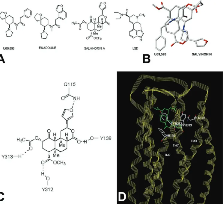
Fig. 1. Molecular modeling predicts Salvinorin A is a structurally novelopioid ligand.Ashows the structure of Salvinorin A, enadoline, U69593 and LSD whereasBshows a superimposition of the structures of Salvinorin A and U69593.Cshows potential residues on the KOR identified by molecular modeling, which might interact with Salvinorin A, andDshows a model of Salvinorin A’s interactions with the KOR (see supporting information for further details).
described previously (13). All data reported here represent the results of at least four separate experiments with EC50andEmax
values calculated by using GRAPHPAD PRIZM (Graph PAD,
San Diego).
The35S-GTP[␥S]-binding assay proceeded with modifications
of the methods described previously (14, 15). Guinea pig caudate membranes, prepared as described previously (16) (10–20g of protein in 300l of 50 mM Tris䡠HCl, pH 7.4, with 1.67 mM DTT and 0.15% BSA) were added to polystyrene 96-well plates filled with 200l of a reaction buffer containing 50 mM Tris䡠HCl, pH 7.4, 100 mM NaCl, 10 mM MgCl2, 1 mM EDTA, 100M GDP,
0.1% BSA, 0.05–0.01 nM35S-GTP[␥S], and varying
concentra-tions of drugs. The reaction mixture was incubated for 3 h at 22°C (equilibrium). The reaction was terminated by the addition of 0.5 ml of ice-cold Tris䡠HCl, pH 7.4 (4°C) followed by rapid vacuum filtration through Whatman GF兾B filters previously soaked in ice-cold Tris䡠HCl, pH 7.4 (4°C). The filters were washed twice with 0.5 ml of ice-cold distilled H2O (4°C). Bound radioactivity
was counted at an efficiency of 98% by liquid scintillation spectroscopy. Nonspecific binding was determined in the pres-ence of 10M GTP[␥S].
Molecular Modeling.Molecular modeling investigations were
con-ducted by using theSYBLmolecular modeling package (Ver. 6.7,
2001, Tripos Associates, St. Louis). Molecular mechanics min-imizations of receptor models and complexes were performed after the addition of hydrogen atoms by using the Tripos force field with Gasteiger–Hu¨ckel charges (distance-dependent di-electric constant, nonbonded cutoff⫽8 Å) without constraints and were terminated at an energy gradient of 0.05 kcal兾mol兾Å, essentially as previously described (17–19) TheUNITYprogram withinSYBLwas used to perform the three-dimensional database searches. Full details of the modeling methods and results are published as supporting information (Tables 3–5) on the PNAS web site, www.pnas.org.
Results
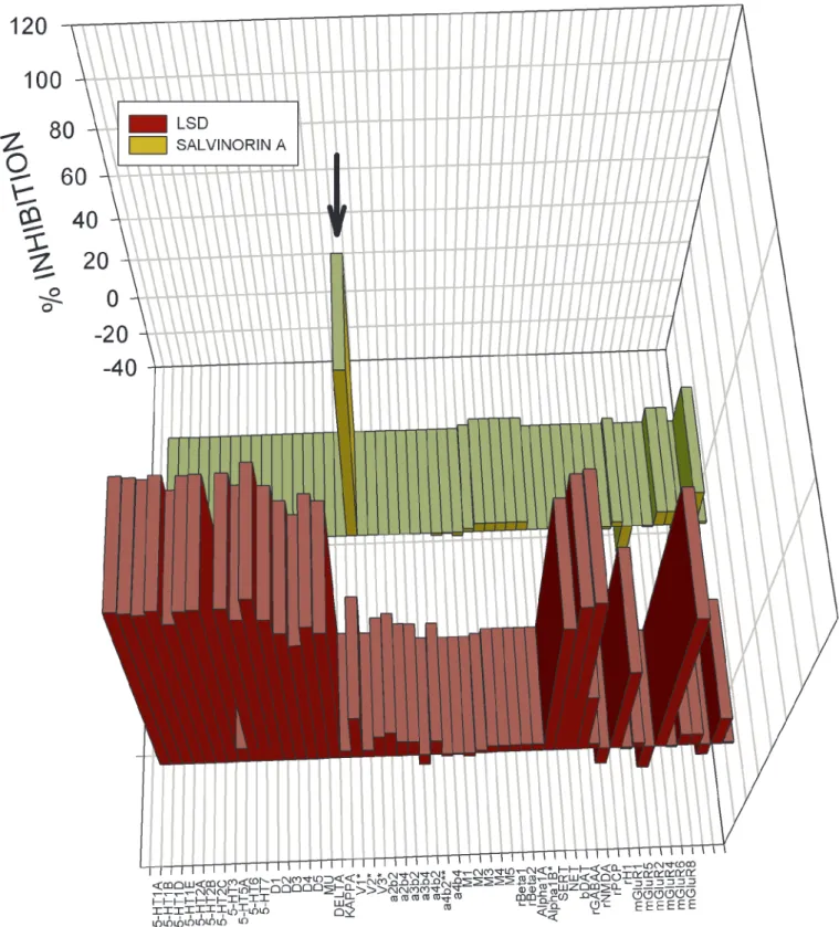
Salvinorin A Selectively Inhibits KOR Binding.To identify Salvinorin A’s molecular target, we screened Salvinorin A (10 M) at a large panel of mainly cloned human GPCRs, transporters, and ligand-gated ion channels by using the resources of the NIMH-PDSP. For comparison, we screened the same molecular targets with the prototypic hallucinogen LSD, also at 10M. As shown in Fig. 1 and in supporting information, Salvinorin A inhibited only [3H]-bremazocine-labeled KORs and did not significantly
inhibit binding to cloned human(MOR) or␦opioid (DOR) receptors or any of the 48 other molecular targets screened.Ki
determinations (Table 1) showed that Salvinorin A was a potent agonist of KOR and guinea pig (gp)KOR. Additionally, Salvi-norin A hadKivalues⬎5,000 nM at the gpMORs and gpDORs
(Table 1). These results indicate that Salvinorin A is, to our knowledge, the first naturally occurringopioid selective ligand. By comparison, LSD potently inhibits the binding of a large number of biogenic amine receptors (Fig. 1) withKis⬍50 nM for
several GPCRs (data not shown). Interestingly, Salvinorin A had no detectable affinity for the 5-HT2Aserotonin receptor and did
not activate 5-HT2Areceptors (not shown), which represent the
main molecular target responsible for the classical hallucinogens such as LSD,N,N⬘-dimethyltryptamine, psilocybin, mescaline, and 4-bromo-2,5-dimethoxyphenylisopropylamine (20, 21).
Salvinorin A Represents a Structurally Novel KOR Ligand.Because Salvinorin A represents a structurally novel hallucinogen, we next performed molecular modeling studies to provide insights into how this compound might interact with KORs. A previously reported model of the KOR complexed with the KOR-selective agonist U69593 was used as a starting point (22). This model has the advantage that it was derived from a set of distance con-straints between potential hydrogen bond-forming pairs unique to the opioid receptor sequences themselves. The result thus does not depend directly on any direct experimental structural data for rhodopsins. Although this model was constructed before the publication of the crystal structure of rhodopsin (23), it is remarkable that the overall configurations are quite similar (rms deviation ⫽ 4 ⌬ by fitting the helix C-␣ atoms of identical residues in both sequences). The U69593 KOR complex places the arylacetamide portion of the ligand in a position analogous to the tyramine moiety with the carbonyl hydrogen bonded to Y139 (22). The only structural similarity between U69593 and Salvinorin A (Fig. 2A) is the presence of an aromatic ring and the amide and ester carbonyl groups separated by a short linkage. Because of this similarity, and the nearly complete lack of similarity of salvinorin and any known KOR ligand, the salvi-norin crystal structure (5) was initially docked by superimposi-tion of aromatic centroids and the carbonyl atoms of salvinorin with those of bound U69593. The role of the carbonyl function-ality for arylacetamide ligands as a hydrogen bond acceptor has been demonstrated experimentally (24) and indirectly supports the proposed role of Y139 and its interaction with the lactone carbonyl of salvinorin. Multiple sterically allowed complexes were generated by using a systematic conformational search about all rotatable Y139 bonds, a dummy bond between a Y139 OH hydrogen atom, and salvinorin carbonyl, following a previ-ously described method (18). Candidate complexes were evalu-ated interactively for steric fit and hydrogen bond donating properties of the receptor cavity visualized as a Connolly channel plot color coded for hydrogen-bonding potential.
Only one family of complexes allowed simultaneous hydrogen bond formation between the receptor side chains and ligand features shown in Fig. 2C(see Table 3 for additional modeling results and details of modeling procedures). In this orientation, the furan substituent of Salvinorin A pointed toward TM1 and TM2, the 4-methoxycarbonyl toward TM5 and TM6, with the A and C rings toward the extra- and intracellular sides, respectively (Fig. 2Dand supporting information). Not unexpectedly, there is very little atom-by-atom correspondence between bound U69593 and Salvinorin A, although both occupy a similar space (Fig. 2B). Docking of salvinorin into hydrogen bond potential-coded Connolly channels defining the binding sites of the MOR and DOR models (22) indicates that salvinorin is sterically compatible with each in slightly different binding modes but could not as readily accommodate the four-point hydrogen bond donor兾acceptor scheme (Fig. 2D) seen with the KOR receptor (e.g., the KOR models could accommodate the furan oxygen and 4-methoxycarbonyl functionality but not the 2-acetoxy group). Residues potentially forming the salvinorin-binding site of the KOR receptor model are listed in Table 4. The identities of 11 of these are conserved in both the MOR and DOR, whereas the remaining seven are variable. The variable residues cause sig-nificant alterations in the steric and electronic characteristics of MOR and DOR in the regions analogous to the salvinorin-binding site of the KOR. The substantial differences in the region of the salvinorin-binding site between the KOR and MOR兾DOR receptors are consistent with the observed KOR selectivity of salvinorin.
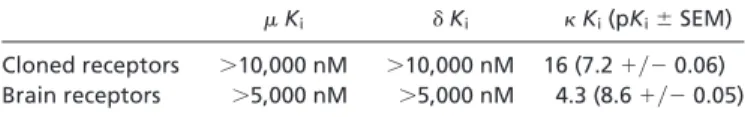
Table 1. Salvinorin A is a potent and selectiveopioid ligand
Ki ␦Ki Ki(pKi⫾SEM)
Cloned receptors ⬎10,000 nM ⬎10,000 nM 16 (7.2⫹兾⫺0.06) Brain receptors ⬎5,000 nM ⬎5,000 nM 4.3 (8.6⫹兾⫺0.05)
The proposed KOR salvinorin-binding site model is also consistent with what little is known about the structural features of salvinorin required for psychotropic activity. For example, the
one-position carbonyl of salvinorin is not able to form specific donor兾acceptor contacts with residues in the receptor model, partially because of its sterically hindered environment, and is
Fig. 2. Large-scale screening of human cloned GPCRs reveals Salvinorin A is selective for KOR. Shown is the mean percent inhibition of radioligand binding or functional activity (metabotropic glutamate receptors only) to 50 receptors and transporters for LSD (yellow bars) and Salvinorin A (red bars) tested at 10M. With the exception of the rat1 and2 adrenergic and bovine dopamine transporter (DAT) all of the assays were performed with cloned human receptors heterologously expressed (seeMaterials and Methodsand supporting information on the PNAS web site for details). As can be seen (arrow), Salvinorin A inhibited only KOR binding at 10M. See Table 5 for details. SERT, serotonin transporter; NET, norepinephrine transporter; DAT, dopamine transporter; rGABAA, rat GABA-A receptor.
not essential for psychotropic activity (25). The 2-acetoxy group of salvinorin does make specific donor兾acceptor contacts in the model and is required for activity (5). Interestingly, a three-dimensional search of the National Cancer Society Database using the pharmacophore features and geometries derived from salvinorin docked with the KOR model produced splendidin (26) and deoxydeoxygedunin (27) (not shown). Splendidin was orig-inally isolated fromSalvia splendens, a species distinct fromS. divinorumand from which salvinorin is derived.S. splendenshas been reported to have psychotropic activity.
Salvinorin A Is a Potent KOR Agonist at Recombinant KORs and KORs
Expressed in Situ. We next examined the agonist兾antagonist
properties of Salvinorin A by using two model systems: KOR
stably expressed in human embryonic kidney-293 cells and gpKOR expressedin situ in guinea pig brain. As shown in Fig. 3, Salvinorin A was a potent KOR agonist with an EC50 for
inhibition of adenylate cyclase of 1.05 nM as compared with an EC50 for the KOR agonist U69593 of 1.2 nM (Table 2).
Salvinorin A was also a potent agonist at gpKOR expressedin situ with an EC50 for [35S]GTP[␥S] binding of 235 nM with
U69593 having an EC50of 377 nM (Table 2). Taken together,
these results indicate that Salvinorin A represents, to our knowledge, the first nonnitrogenous KOR-selective agonist.
Discussion
The main finding of this paper is that Salvinorin A, the active ingredient of the hallucinogenic plantS. divinorum, is a potent and selective KOR agonist. Salvinorin A is a novel nonalkaloid diterpene that has no structural resemblance to any known hallucinogens but does have modest structural homology to enadoline, a selective KOR agonist (Fig. 1AandC). Salvinorin A thus represents a class of hallucinogens with potent actions at KORs. Because KOR agonists have long been known to have psychotomimetic actions (28), these results imply that the actions of Salvinorin A in humans are mediated, at least in part, via activation of KORs. Additionally, these results imply that KOR-selective antagonists could conceivably represent treatments for diseases in which hallucinations are prominent, including schizo-phrenia, depression with psychotic features, and the hallucinosis associated with certain dementias (Alzheimer’s, Huntington’s, and Pick diseases) and certain types of drug abuse (e.g., am-phetamine and cocaine psychosis) (29, 30). Previous studies evaluating naltrexone, which is a nonselective opioid antagonist, for the treatment of schizophrenia have yielded equivocal results (31, 32).
Dynorphin was discovered in 1979 by Goldsteinet al.(33) and was demonstrated to be an extraordinarily potent endogenous peptide with selectivity for the KOR (34), a GPCR in the opioid receptor family (35). Before the cloning of the KOR, a large amount of behavioral (36, 37), developmental (38, 39), and biochemical (40, 41) data suggested the existence of distinct KORs. Although dynorphin and related peptides represent, in some cases, potent and relatively selective endogenous ligands for the KOR, other types of naturally occurring ligands have heretofore not been identified. The discovery that Salvinorin A is a potent naturally occurring nonalkaloid agonist for the KOR is thus unexpected.
Fig. 3. Salvinorin A is a potent KOR agonist.Ashows that Salvinorin A potently inhibits 3H-bremazocine binding to cloned KORs, whereasBshows the ability of Salvinorin A to inhibit forskolin-stimulated adenylate cyclase in KOR-393 cells. Data represent the mean⫾SD of triplicate determinations from a representative experiment that has been replicated three times. For the inhibition of forskolin-stimulated cyclase activity, an EC50value of 1⫾0.5 nM was calculated for Salvinorin A, compared with an EC50value of 1.2⫾0.6 nM for U69593.
Table 2. Salvinorin A is a potent-opioid agonist: [35S]GTP-␥-S studies using guinea pig brain
caudate membranes
Drug Unblocked CTAP, 200 nM TIPP, 20 nM Nor-BNI, 0.2 nM
Percent stimulation
DAMGO 414⫾47 11,124⫾2,126 494⫾56 355⫾61
0.96⫾0.02 0.99⫾0.09 0.97⫾0.02 0.92⫾0.03
SNC80 758⫾131 987⫾120 9,565⫾4,115 855⫾119
0.91⫾0.04 0.94⫾0.03 0.90⫾0.19 0.86⫾0.3
U69,593 377⫾39 540⫾57 442⫾76 1,554⫾168
1.70⫾0.04 1.70⫾0.04 1.60⫾0.06 1.60⫾0.05
Percent of maximal stimulation produced by 10M U69,593
Salvinorin A 235⫾26 204⫾20 259⫾40 643⫾128
0.79⫾0.04 0.83⫾0.03 0.81⫾0.06 0.89⫾0.10
It is now well established that the activation of KORs induces a large number of behavioral effects that include analgesia, sedation, and perceptual distortions. In the past, studies on the precise role of KORs in humans were hampered by the lack of selective agonists, although studies with compounds such as cyclazocine and ketocyclazocine suggested that KOR agonists were psychotomimetic (28). More recently, human studies with the highly selective KOR agonist enadoline (42) indicated that KOR activation induced visual distortions, feelings of unreality, and depersonalization. These effects of enadoline are reminis-cent of those previously reported for Salvinorin A (2, 3). Taken together, these results suggest that the KOR兾dynorphinergic system functions to modulate human perception and cognition, as might be inferred from detailed anatomical studies of dynor-phin peptide distribution studies (43–45).
One of the implications of these results is that KORs or KOR signaling may also be important in the pathogenesis of diseases characterized by perceptual distortions. The most obvious eases implicated are schizophrenia, dementia, and bipolar dis-orders, because all are characterized by hallucinations and delusions. Prior studies evaluating KORs in schizophrenia have
yielded conflicting results (46–48), whereas one study examining affective disorder was negative (47). On the other hand, two well-controlled studies have demonstrated an up-regulation of KORs in Alzheimer’s disease (49, 50), whereas MORs and DORs were down-regulated (50) or unchanged (49).
In conclusion, we report the discovery that Salvinorin A is a potent selective KOR agonist. Salvinorin A thus represents a unique structural class of nonnitrogenous opioid subtype-selective agonists. Additionally, these results suggest that KORs play a prominent role in the regulation of human perception and suggest that KOR antagonists could represent a novel drug class with specific activity in diseases in which alterations in percep-tion are predominant. Finally, these results imply that the KOR兾dynorphinergic system functions to modulate human per-ception and cognition.
We gratefully acknowledge Christina M. Dersch, Beth Popadok, and Sandra Hufesein for superb technical assistance. This work was sup-ported in part by a Research Scientist Development Award KO2MH01366 (B.L.R.) and by the NIMH-PDSP NO2MH80004 (B.L.R.).
1. Valdes, L. J., III (1994)J. Psychoactive Drugs26,277–283.
2. Giroud, C., Felber, F., Augsburger, M., Horisberger, B., Rivier, L. & Mangin, P. (2000)Forensic Sci. Int.112,143–150.
3. Siebert, D. J. (1994)J. Ethnopharmacol.43,53–56.
4. Koreeda, M., Brown, L. & Valdes, I. L. (1990)Chem. Lett.2015–2018. 5. Valdes, L. J., Butler, W. M., Hatfield, G. M., Paul, A. G. & Koreeda, M. (1984)
J. Org. Chem.49,4716–7720.
6. Ortega, A., Blount, J. F. & Manchand, P. S. (1982)J. Chem. Soc. Perkins Trans. 1, 2505–2508.
7. Adham, N., Tamm, J. A., Salon, J. A., Vaysse, P. J., Weinshank, R. L. & Branchek, T. A. (1994)Neuropharmacology33,387–391.
8. Roth, B. L., Palvimaki, E., Berry, S., Khan, N., Sachs, N., Uluer, A. & Choudhary, M. (1995)J. Pharmacol. Exp. Ther.275,1638–1646.
9. Glennon, R. A., Lee, M., Rangisetty, J. B., Dukat, M., Roth, B. L., Savage, J. E., McBride, A., Rauser, L., Hufeisen, S. & Lee, D. K. (2000)J. Med. Chem.43,
1011–1018.
10. Rothman, R. B., Baumann, M. H., Savage, J. E., Rauser, L., McBride, A., Hufeisen, S. J. & Roth, B. L. (2000)Circulation102,2836–2841.
11. Ananthan, S., Kezar, H. S., III, Carter, R. L., Saini, S. K., Rice, K. C., Wells, J. L., Davis, P., Xu, H., Dersch, C. M., Bilsky, E. J.,et al.(1999)J. Med. Chem.
42,3527–3538.
12. Roth, B. L., Shoham, M., Choudhary, M. & Khan, N. (1997)Mol. Pharmacol.
52,259–266.
13. Gray, J. A., Sheffler, D. J., Bhatnagar, A., Woods, J. A., Hufeisen, S. J., Benovic, J. L. & Roth, B. L. (2001)Mol. Pharmacol.60,1020–1030. 14. Sim, L. J., Selley, D. E., Xiao, R. & Childers, S. R. (1996)Eur. J. Pharmacol.
307,97–105.
15. Partilla, J. S., Carroll, F. I., Thomas, J. B., Rice, K. C., Zimmerman, D. M. & Rothman, R. B. (1999)Analgesia4,27–32.
16. Thomas, J. B., Mascarella, S. W., Burgess, J. P., Xu, H., McCullough, K. B., Rothman, R. B., Flippen-Anderson, J. L., George, C. F., Cantrell, B. E., Zimmerman, D. M. & Carroll, F. I. (1998) Bioorg. Med. Chem. Lett.8,
3149–3152.
17. Shapiro, D. A., Kristiansen, K., Weiner, D. M., Kroeze, W. K. & Roth, B. L. (2002)J. Biol. Chem.277,11441–11449.
18. Westkaemper, R. B., Runyon, S. P., Savage, J. E., Roth, B. L. & Glennon, R. A. (2001)Bioorg. Med. Chem. Lett.11,563–566.
19. Choudhary, M. S., Sachs, N., Uluer, A., Glennon, R. A., Westkaemper, R. B. & Roth, B. L. (1995)Mol. Pharmacol.47,450–457.
20. Glennon, R. A., Titler, M. & McKenney, J. D. (1984)Life Sci.35,2505–2511. 21. Roth, B. L., Willins, D. L., Kristiansen, K. & Kroeze, W. K. (1998)Pharmacol.
Ther.79,231–257.
22. Pogozheva, I. D., Lomize, A. L. & Mosberg, H. I. (1998)Biophys. J.75,612–634. 23. Palczewski, K., Kumasaka, T., Hori, T., Behnke, C. A., Motoshima, H., Fox, B. A., Le Trong, I., Teller, D. C., Okada, T., Stenkamp, R. E.,et al.(2000)
Science289,739–745.
24. Lavecchia, A., Greco, G., Novellino, E., Vittorio, F. & Ronsisvalle, G. (2000)
J. Med. Chem.43,2124–2134.
25. Valdes, I. L., Chang, H. M., Visger, D. C. & Koreeda, M. (2001)Org. Lett.3,
3935–3937.
26. Savona, G., Paternostro, M. P. & Piozzi, F. (1979)J. Chem. Soc. Perkins Trans. 1, 533–534.
27. Bevan, C. W. L., Halsall, T. G., Nwaji, M. N. & Taylor, D. A. H. (1962)J. Chem. Soc. Perkins Trans. 1, 768–771.
28. Pfeiffer, A., Brantl, V., Herz, A. & Emrich, H. M. (1986)Science233,774–776. 29. Rothman, R. B., Gorelick, D. A., Heishman, S. J., Eichmiller, P. R., Hill, B. H.,
Norbeck, J. & Liberto, J. G. (2000)J. Subst. Abuse Treat.18,277–281. 30. Rothman, R. B. (1994)Analgesia1,27–49.
31. Sernyak, M. J., Glazer, W. M., Heninger, G. R., Charney, D. S., Woods, S. W., Petrakis, I. L., Krystal, J. H. & Price, L. H. (1998)J. Clin. Psychopharmacol.
18,248–251.
32. Marchesi, G. F., Santone, G., Cotani, P., Giordano, A. & Chelli, F. (1995)Prog. Neuropsychopharmacol. Biol. Psychiatr.19,1239–1249.
33. Goldstein, A., Tachibana, S., Lowney, L. I., Hunkapiller, M. & Hood, L. (1979)
Proc. Natl. Acad. Sci. USA76,6666–6670.
34. Chavkin, C., James, I. F. & Goldstein, A. (1982)Science215,413–415. 35. Xie, G. X., Meng, F., Mansour, A., Thompson, R. C., Hoversten, M. T.,
Goldstein, A., Watson, S. J. & Akil, H. (1994)Proc. Natl. Acad. Sci. USA91,
3779–3783.
36. Martin, W. R., Eades, C. G., Thompson, J. A., Huppler, R. E. & Gilbert, P. E. (1976)J. Pharmacol. Exp. Ther.197,517–532.
37. Martin, W. R. (1979)Br. J. Clin. Pharmacol.7,273S–279S.
38. Spain, J. W., Bennett, D. B., Roth, B. L. & Coscia, C. J. (1983)Life Sci.33,
235–239.
39. Spain, J. W., Roth, B. L. & Coscia, C. J. (1985)J. Neurosci.5,584–588. 40. Chang, K. J., Hazum, E. & Cuatrecasas, P. (1981)Proc. Natl. Acad. Sci. USA
78,4141–4145.
41. Kosterlitz, H. W., Paterson, S. J. & Robson, L. E. (1981)Br. J. Pharmacol.73,
939–949.
42. Walsh, S. L., Strain, E. C., Abreu, M. E. & Bigelow, G. E. (2001) Psycho-pharmacology (Berlin)157,151–162.
43. Chavkin, C., Bakhit, C., Weber, E. & Bloom, F. E. (1983)Proc. Natl. Acad. Sci. USA80,7669–7673.
44. McGinty, J. F., van der Kooy, D. & Bloom, F. E. (1984) J. Neurosci.4,
1104–1117.
45. McGinty, J. F., Henriksen, S. J., Goldstein, A., Terenius, L. & Bloom, F. E. (1983)Proc. Natl. Acad. Sci. USA80,589–593.
46. Owen, F., Bourne, R. C., Poulter, M., Crow, T. J., Paterson, S. J. & Kosterlitz, H. W. (1985)Br. J. Psychiatr.146,507–509.
47. Peckys, D. & Hurd, Y. L. (2001)Brain Res. Bull.55,619–624.
48. Izenwasser, S., Staley, J. K., Cohn, S. & Mash, D. C. (1999)Life Sci.65,
857–862.
49. Barg, J., Belcheva, M., Rowinski, J., Ho, A., Burke, W. J., Chung, H. D., Schmidt, C. A. & Coscia, C. J. (1993)Brain Res.632,209–215.
50. Mathieu-Kia, A. M., Fan, L. Q., Kreek, M. J., Simon, E. J. & Hiller, J. M. (2001)
Brain Res.893,121–134.
![Table 2. Salvinorin A is a potent -opioid agonist: [ 35 S]GTP-␥-S studies using guinea pig brain caudate membranes](https://thumb-us.123doks.com/thumbv2/123dok_us/8115737.2152102/5.945.201.743.822.1008/table-salvinorin-potent-opioid-agonist-studies-caudate-membranes.webp)
