Copyright © 2004, American Society for Microbiology. All Rights Reserved.
Complementation in Cells Cotransfected with a Mixture of Wild-Type
and Mutant Human Immunodeficiency Virus (HIV) Influences the
Replication Capacities and Phenotypes of Mutant Variants
in a Single-Cycle HIV Resistance Assay
Hongmei Mo,* Liangjun Lu, Ron Pithawalla, Dale J. Kempf, and Akhteruzzaman Molla
Global Pharmaceutical Research and Development, Abbott Laboratories, Abbott Park, IllinoisReceived 30 October 2003/Returned for modification 5 May 2004/Accepted 5 June 2004
The impact of cotransfection of mixtures of mutant and wild type (WT) virus on the observed phenotype and replication capacity (RC) in a single-cycle human immunodeficiency virus (HIV) phenotypic assay has been investigated by cotransfecting mutant HIV clones expressing the firefly luciferase expression gene with a WT
clone expressingRenillaluciferase. Four mutant constructs with different genotypes displayed <1% RC when
transfected alone. Cotransfection of as little as 9% of the WT clone resulted in an 18- to 33-fold increase in the
RC of the mutant clones. In addition, the 50% inhibitory concentration (IC50) of lopinavir against seven mutant
clones decreased by up to 97% after incremental cotransfection of 9 to 50% of the WT clone. The enhancement
of RC and decrease in IC50for mutant variants following cotransfection with the WT variant appear to be due
to complementation rather than genetic recombination. These findings suggest that the RC and susceptibility of plasma isolates from patients who are off therapy or not adherent to treatment, in which WT virus may expand to significant levels, should be interpreted with caution.
Human immunodeficiency virus (HIV) resistance testing methods have been shown to improve the success of antiret-roviral therapy in treatment-experienced patients (3, 10, 11, 25, 36). As such, resistance testing is now recommended to help guide the choice of regimens after first- and multiple-drug treatment failure by three consensus panels (the International AIDS Society-USA, the EuroGuideline Group, and the U.S. Department of Health and Human Services) (10, 12, 34). In addition, consideration of resistance testing in primary HIV infection is also recommended (12, 34). Two types of tests can be beneficial: genotypic assays, which document the presence of mutations known to confer decreased drug susceptibility (29), and phenotypic assays, which determine the concentra-tion of an antiretroviral agent that reduces HIV replicaconcentra-tion by 50% (IC50) in tissue culture (14, 16, 18, 24). Conventional
phenotypic assays are labor-intensive, time-consuming, and in-efficient because of the requirement for virus isolation from peripheral blood mononuclear cells (18). To overcome this problem, recombinant phenotypic assays, based on direct am-plification of the patient’s gene of interest (protease [PR] gene and a portion of the reverse transcriptase [RT] gene) from viral RNA in plasma, have been developed (14, 24). These recombinant virus assays are rapid, reproducible, and adapt-able to large-scale application through use of robotics. To capture and preserve the PR and RT sequence heterogeneity of the plasma virus, the target cells were cotransfected with either the “pool” of DNA generated from patient plasma to-gether with a linearized HIV genomic vector or with the pool of the resistance testing vectors generated by cloning the pool
of PCR-generated DNA into a modified HIV vector that lacks the analogous sequence (14, 24). In both cases, a pool of recombinant viruses were generated from the transfected cells and used for drug susceptibility determination. Then, these population-based approaches measure the drug susceptibility of the viral population, compared to a standard wild-type (WT) virus, represented as the incremental factor of change in IC50
(FC).
Recent modifications of the single-cycle phenotypic assay, employing a standardized transfection of HIV proviral DNA, have also allowed the estimation of the replication capacities (RCs) of patient viruses (20, 24, 27; R. Haubrich, T. Wrin, and N. Hellmann, Abstr. XI Int. HIV Drug Resist. Workshop, abstr. 121, 2002). Both the FC and RC have been shown to correlate with clinical response (3, 10, 11, 25, 36; Haubrich et al., Abstr. XI Int. HIV Drug Resist. Workshop; N. Hellmann, T. Wrin, and M. Bates, Abstr. XI Int. HIV Drug Resist. Work-shop, abstr. 63, 2002). However, plasma samples from treat-ment-experienced patients might contain mixtures of HIV vari-ants with substantially different FCs and RCs, particularly if therapy has been discontinued for a sufficient period of time to allow archival WT strains to expand within the quasispecies (6, 17, 28). Under conditions of the single-cycle assay, cotransfec-tion of different viral variants into the same cell might provide the opportunity for genetic recombination and/or complemen-tation. Since viruses with drug resistance mutations often ex-hibit reduced RCs compared to standard WT strains (21), the impact of mixed species on RC and FC is uncertain. In one study (24), mixing a mutant strain with 25, 50, and 75% of WT virus reduced the FC of nelfinavir by 70, 88, and 93%, respec-tively. In each case, the percent decrease in observed FC was greater than that for the cotransfected WT strain, suggesting an interaction between mutant and WT viruses in the cotrans-fection. The effect of this potential interaction (complementa-* Corresponding author. Mailing address: Department R47D,
Building AP52N, Abbott Laboratories, 200 Abbott Park Rd., Abbott Park, IL 60064-6217. Phone: (847) 937-5933. Fax: (847) 938-2756. E-mail: [email protected].
4169
on May 15, 2020 by guest
http://jcm.asm.org/
tion or recombination) between WT and mutant on the output (FC and RC) of resistance testing assays has not been thor-oughly analyzed. The objective of the present study was to quantitate the effect of different proportions of WT virus on the apparent drug susceptibility and RC of mutant virus when both are present in mixtures by using WT and mutant clones that express different reporter genes.
MATERIALS AND METHODS
Plasma sample.Plasma samples were obtained from PR inhibitor (PI)-expe-rienced individuals receiving lopinavir (LPV)-ritonavir therapy either at baseline or at the time of viral rebound.
Construction of mutant and WT HIV clones.Proviral molecular clones pNL4-3-Fluc and pNL4-3-Rluc were kindly provided by J. He and N. Landau at Indiana University and The Salk Institute, respectively. pNL4-3-Fluc was constructed to contain a firefly luciferase expression gene in thenefregion and a frameshift in the envelope (13). A unique XmaI restriction cut site was introduced into pNL4-3-Fluc downstream of the PR coding region to construct the shuttle vector designated pNL4-3-Fluc-x. The pNL4-3-Fluc-x construct was used as the vector for construction of a variety of mutants.
Viral RNA was isolated from 200l of plasma with QIAamp RNA extraction kits (QIAGEN, Hilden, Germany) according to the manufacture’s instructions. Reverse transcription was performed with Superscript II (Gibco/BRL, Gaithers-burg, Md.). A fragment spanning the PR and the p7/p1 and p1/p6 cleavage sites was amplified with the PlatinumTaqDNA polymerase high-fidelity system (In-vitrogen, Carlsbad, Calif.) with forward and reverse primers containing ApaI and XmaI sites, respectively. After digestion with ApaI and XmaI, the PCR products were inserted into the pNL4-3-Fluc-x vector, in which the ApaI-XmaI fragment had been removed. Ligation reaction products were transformed into competent
Escherichia coli(Invitrogen). DNA plasmids purified from each individual colony were sequenced to determine their genotypes. The resulting constructs were designated mutant clones.
Drug susceptibility and RC assays.Drug susceptibility was determined by a single-cycle assay. To generate recombinant pseudotyped viruses, human embry-onic kidney 293 cells were cotransfected with either WT pNL4-3-Fluc-x or a mutant pNL4-3-Fluc-x clone along with an expression vector carrying the vesic-ular stomatitis virus envelope gene with Lipofectamine PLUS reagent (Invitro-gen). The transfection conditions were optimized to achieve⬎90% transfection efficiency according to the manufacturer’s instruction. Three hours after trans-fection, cells were trypsinized and seeded into 96-well plates containing serial dilutions of LPV. The culture supernatants harvested at 2 days posttransfection were used to infect fresh 293 cells at a concentration of 104cells/well. To
investigate the effect on drug susceptibility of viral mixtures, each mutant clone (expressing firefly luciferase) was mixed with WT pNL4-3-Rluc (expressing Re-nillaluciferase) at ratios of 1:0.1 (9%), 1:0.5 (33%), and 1:1 (50%). The mixtures of mutant and WT DNA were used for cotransfection as described above. Firefly andRenillaluciferase activities for each sample were sequentially measured with the Dual-Luciferase reporter assay system according to the manufacturer’s in-structions (Promega Corporation, Madison, Wis.) in order to minimize experi-mental variations from differences in pipetting volumes, cell lysis efficiency, amount of cells, and assay efficiency. In each experiment, mutant pNL4-3-Fluc-x and WT pNL4-3-Rluc as well as pNL4-3-Fluc-x were transfected alone to serve as controls. Because of the single-replication-cycle format of this assay, the level of luciferase activity in transfected cells is proportional to the amount of HIV DNA transfected; therefore, the luciferase activities from transfected or cotrans-fected cells were measured for each transfection in order to normalize the transfection efficiency. The percent inhibition was calculated from the formula [1 ⫺(luciferase activity in the presence of drug/luciferase activity in the absence of drug)]⫻100. The IC50was determined by nonlinear regression curve fitting with
the Prism program. As controls, the LPV IC50 for the WT was determined
following cotransfection with the mutant clones and transfection of the WT pNL4-3-Rluc clone as well as transfection of WT pNL4-3-Fluc-x alone. The FC is the IC50for the mutant virus/the IC50for WT pNL4-3-Fluc-x.
The RC for each mutant was calculated by comparing the firefly luciferase activity generated by the mutants to that generated by WT pNL4-3-Fluc-x, after adjusting for minor differences in transfection efficiencies. The RC values were expressed as percentages of that for the WT and reflect the levels of replication for mutant viruses compared to that for the WT control.
Sequence analysis.The ApaI-SmaI fragment containing the p7/p1 and p1/p6 cleavage sites as well as the PR gene was amplified by RT-PCR from supernatant
harvested from cotransfection of mutant and WT constructs. The amplified products were purified and then blunt end ligated into the TA cloning vector (Invitrogen). Miniprep plasmid DNA from individual colonies was purified and then sequenced with an ABI-373 DNA sequencer (Applied Biosystems, Foster City, Calif.).
RESULTS
The impaired RC of mutants is restored by cotransfected
WT DNA.To assess the effect of cotransfection of WT HIV on
the RC of mutant strains, we compared the RCs of 11 mutant clones after transfection alone or after cotransfection with WT pNL4-3-Rluc at ratios of mutant to WT of 10:1 (9% of WT), 2:1 (33% of WT), and 1:1 (50% of WT). WT pNL4-3-Rluc and WT pNL4-3-Fluc-x were included as controls. The genotypes of the 11 mutant clones are shown in Table 1. Each mutant clone contains one or two primary mutations and a number of secondary PI resistance mutations. Prior to the cotransfection experiments, the transfection efficiencies were optimized to achieve greater than 90% efficiency. Luciferase activity, mea-sured 48 h posttransfection, correlated well with the levels of p24 production from transfected cells (data not shown). Under optimized transfection conditions, the difference in transfec-tion efficiency between different reactransfec-tions was minor (⬍30%; data not shown). For 95% of the determinations, the values of RC from replicate assays differed by less than twofold. The values of standard deviations of RC were⬍30% of the means for 80% of the determinations.
Mutant clones 1 to 4 displayed low RCs (⬍1% of WT) when transfected alone. In contrast, cotransfection with as little as 9% of the WT clone increased the RCs of these clones by 18-to 33-fold (Table 2). Cotransfection with 33 and 50% of WT further enhanced the RCs of those four mutant clones to at least 10% and up to 76% of WT, respectively (incremental increase of 68- to 690-fold). In contrast, the other seven mu-tants (mutant clones 5 to 11) displayed only modestly de-creased RCs compared to the WT clone (15 to 68%) when transfected alone. The RCs of those seven mutants did not change significantly upon cotransfection with up to 50% of WT (one- to fourfold). Similarly, there was no significant change in the RC of WT virus after cotransfection with up to 67%
mu-TABLE 1. Genotype of the mutant clones
Mutant
clone Mutations in PR
1 ...L10I, L24I, E35D, M36I, M46L, I54V, L63P, I64V, V82A 2 ...L10F, L24I, E35D, M36V, N37S, R41K, M46I, I54V, D60E,
Q61E, I62V, L63P, I64V, V82A
3 ...L10F, I15T, E35D, M36V, N37S, R41K, I54V, D60E, Q61E, I62V, L63P, I64V, V82A
4 ...L10V, V32I, N37S, M46I, I47V, I62V, L63P, V77I, P79S, Q92K, I93L, C95F
5 ...L10F, V32I, E35D, M36I, M46L, I54V, L63P, I64V, V82A 6 ...L10I, E35D, N37D, L63P, A71V, T74P, I84V, L90M, I93L 7 ...L10I, E35D, N37D, M46I, I54V, L63P, A71V, T74P, I84V,
L90M, I93L
8 ...L10I, V32I, M46I, I47A, I62V, L63P, V77I, Q92K, I93L, C95F 9 ...L10I, G48V, I54V, L63P, A71V, I72M, V77I, V82A, L90M, I93L 10 ...L10I, V32F, G48V, I54V, V56K, L63P, A71V, I72M, V77I,
V82A, L90M, I93L
11 ...L10V, I15V, G16E, K20R, E35D, M36I, R41K, M46I, I50V, I54V, K55R, R57K, Q61G, I64L, A71V, I72R, V82A, L89I, L90M, Q92K
on May 15, 2020 by guest
http://jcm.asm.org/
[image:2.603.300.545.74.261.2]tants, as indicated by Renilla luciferase activity (data not shown).
The apparent phenotypes of mutants are decreased by
co-transfection with WT DNA.We also investigated the effect of
cotransfection of WT HIV on the phenotypic susceptibility (FC) of mutant clones. Prior to this study, validation of this phenotypic assay was performed. The susceptibility of WT pNL4-3-Fluc to LPV was determined in four independent ex-periments, and each experiment includes triplicate wells. The IC50values for LPV derived from four independent
experi-ments were 5.0-, 5.5-, 7.4-, and 7.8-fold, respectively. We also tested other PIs (ritonavir, indinavir, nelfinavir, and saquina-vir) in this assay; the IC50 values obtained with this assay
correlated well with the results published in the literature (data not shown). The variations among different experiments for at least 95% of the determinations were less than threefold.
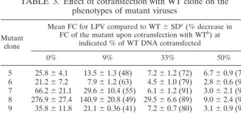
Since mutant clones 1 to 4 displayed insufficient RC for phenotypic evaluation, these experiments were limited to mu-tant clones 5 to 11. The reductions in LPV susceptibility of the seven mutants after transfection alone ranged from 21- to 277-fold, compared to the WT standard (Table 3). Upon
co-transfection with 9% WT DNA, the FCs for six of seven mu-tants decreased by approximately one-half (range, 41 to 63%). Cotransfection of 33 and 50% WT DNA substantially de-creased the FCs of all mutants (72 to 97%). In all cases, the percent decrease in FC for the mutant was greater than the percentage for the cotransfected WT strain, suggesting that FC values for mutant strains are underestimated when mixtures of mutant and WT DNA are cotransfected. In contrast, the sus-ceptibility of the WT clone was not significantly affected by cotransfection with up to 50 to 67% of the each of four mutant clones (Table 4). In each case, a twofold or less change in IC50
after cotransfection with the mutant was observed.
The interaction between WT and mutant occurs in
cotrans-fected cells. To investigate the mechanism of the observed
interaction between mutant and WT viruses in the single-cycle assay, we performed coculture, coinfection, and cotransfection experiments comparing the WT and two mutant clones with poor RCs (clones 3 and 4), as well as two with higher RCs (clones 5 and 6). In contrast to the results of cotransfection experiments, coculture of cells containing individually trans-fected mutants and WT at a ratio of 1:1 did not affect the RCs of the mutants, as indicated by firefly luciferase activity (Fig. 1). Similarly, upon coinfection of cells with a 1:1 ratio of mutants and WT viruses harvested from individually transfected cells the RCs of the mutant clones did not change significantly from those observed upon infection without WT virus (Fig. 1). These results indicate that the complementation of the mutant RC by WT HIV occurs primarily in cotransfected cells when the mutant and WT DNA coexist in the same cells.
No evidence of recombination in cotransfected cells.To
ad-dress whether the enhancement of RCs and decrease in FCs of the mutant clones following cotransfection with the WT clone are due to genetic recombination, sequences of 16 to 20 indi-vidual clones from the supernatants harvested from the co-transfection of mutant clones 4 and 6 with WT were deter-mined. As shown in Table 5, the ratios of mutant to WT sequences corresponded to the relative amounts of mutant and WT clones used for cotransfection (1:1, 2:1, and 10:1, respec-tively). No recombination was observed, suggesting that the interaction of mutant and WT viruses in the above single-cycle replication assays is not due to recombination.
DISCUSSION
[image:3.603.298.543.89.185.2]In this study, we analyzed the effect of mixtures of viral strains on the apparent RCs and FCs of HIV molecular clones. We observed that even relatively small amounts of WT virus TABLE 2. Effect of cotransfection with WT virus on
the RCs of mutant viruses
Mutant clone
Mean RC (%)⫾SDa(fold increase in RCb) at indicated % of WT DNA cotransfected
0% 9% 33% 50%
1 0.07⫾0.008 1.6⫾0.5 (22) 10.9⫾1.4 (155) 22.1⫾6.3 (315) 2 0.08⫾0.05 1.7⫾0.7 (21) 43.7⫾10.4 (546) 55.2⫾4.2 (690) 3 0.16⫾0.6 5.3⫾0.7 (33) 48.2⫾2.8 (301) 64.3⫾11.2 (402) 4 0.7⫾0.4 12.3⫾4.7 (18) 47.7⫾4.1 (68) 75.9⫾8.1 (108) 5 14.7⫾3.0 19.8⫾3.6 (1) 46.8⫾5.8 (3) 57.0⫾5.5 (4) 6 67.6⫾4.9 83.5⫾18.0 (1) 146.8⫾2.4 (2) 160⫾5.5 (2) 7 32.5⫾8.5 32.9⫾7.3 (1) 34.3⫾7.6 (1) 50.2⫾1.6 (2) 8 32.7⫾0.3 41.8⫾3.4 (1) 52.5⫾1.1 (2) 56.0⫾10.1 (2) 9 47.9⫾4.8 47.2⫾4.8 (1) 57.3⫾2.4 (1) 97.4⫾5.5 (2) 10 20.4⫾5.6 21.5⫾1.9 (1) 27.3⫾7.7 (1) 22.6⫾0.3 (1) 11 38.6⫾13.2 31.6⫾1.8 (1) 44.1⫾2.6 (1) 40.5⫾5.1 (1)
aThe relative RC was determined by single-cycle assay as described in Mate-rials and Methods. RC percentages are calculated from the formula: (mean luciferase activity of the test samples/mean luciferase activity of WT pNL4-3-Fluc-x from six replicates for each experiment)⫻100. Values were derived from two or three separate experiments.
bValues are RCs of mutants in mixture/RCs of mutant transfected alone.
TABLE 3. Effect of cotransfection with WT clone on the phenotypes of mutant viruses
Mutant clone
Mean FC for LPV compared to WT⫾SDa(% decrease in FC of the mutant upon cotransfection with WTb) at
indicated % of WT DNA cotransfected
0% 9% 33% 50%
5 25.8⫾4.1 13.5⫾1.3 (48) 7.2⫾1.2 (72) 6.7⫾0.9 (74) 6 21.2⫾7.2 7.9⫾1.2 (63) 4.5⫾1.0 (79) 2.8⫾0.6 (97) 7 66.2⫾21.1 29.6⫾10.4 (55) 6.1⫾1.2 (91) 3.0⫾2.1 (95) 8 276.9⫾27.4 140.9⫾20.8 (49) 29.5⫾6.6 (89) 9.0⫾2.4 (97) 9 35.8⫾11.8 21.1⫾0.36 (41) 7.2⫾0.7 (80) 3.1⫾0.9 (91) 10 54.5⫾6.6 24.4⫾3.8 (55) 5.0⫾1.7 (91) 3.0⫾0.4 (95) 11 150.1⫾17.1 132.6⫾37.0 (11) 31.3⫾2.8 (79) 16.7⫾3.5 (89)
aValues were determined from the formula IC
50of mutant either alone or in
mixture/IC50of WT pNL4- 3-Fluc. The mean changes in IC50were derived from
[image:3.603.45.283.89.228.2]two or three independent experiments, and each experiment contains triplicates. bValues are calculated from the formula (FC of mutant in mixture/FC of mutant alone)⫻100.
TABLE 4. Effect of cotransfection of mutant clones on the LPV susceptibility of WT virus
Mutant clone cotransfected
IC50for WT pNL4-3-Rluc of
LPV (M) at indicated % of mutant DNA cotransfected
50% 67%
4 0.011 0.021
8 0.013 0.013
9 0.014 0.024
10 0.017 0.025
pNL4-3-Fluc-x (WT) 0.014 0.013
on May 15, 2020 by guest
http://jcm.asm.org/
[image:3.603.44.285.570.683.2]within a viral population could significantly enhance the ap-parent RCs and decrease the FCs of mutant strains. Cotrans-fection of the mutant strain with as little as 9% WT clone resulted in a significant increase in the RCs of all four mutant strains that had low RCs when transfected alone. Cotransfec-tion with 33 and 50% WT strain further increased the RC to a level close to that for the WT. In contrast, we observed that, when mutant clones with only modestly reduced RCs were cotransfected with WT virus, the RC did not change signifi-cantly. Furthermore, the RC of the WT virus was not affected by cotransfection with mutants with low RC.
Similarly, cotransfection of as little as 9% WT clone de-creased the FCs of mutants by up to 63%. Similar results were obtained in a previous study in which the apparent FC of mutant virus decreased by 70 to 93% upon mixing with 25 to 75% WT virus (24). The agreement between these two studies suggests that (i) the assay used in this study is reliable and (ii) the observed effects of virus mixtures on the RC and FC in the present study are likely to be observed in any recombinant phenotypic assay employing transfection of mixed populations. The present results indicate that cotransfected WT virus can complement the replication defect of mutant strains, presum-ably through an intracellular interaction. The construction of WT and mutant clones expressing different luciferase reporters allows quantitation of the effect of this complementation. Since no recombinant clones were observed upon sequence analysis of the viruses harvested from the cotransfection, genetic re-combination is unlikely to be responsible for the effect. Lack of evidence of recombination is in agreement with the nature of single-cycle assays and the short period of viral replication (4 days total). Our results also indicate that complementation occurs only following cotransfection, not during coculture or coinfection, suggesting that the mechanism of the
[image:4.603.128.458.69.266.2]complemen-tation requires the presence of both mutant and WT DNA in the same cell. Under coculture or coinfection conditions, the probability of coexistence of WT and mutant strains within the same cell is low (8, 26, 30). The fact that the complementation between WT and mutant occurs only in cotransfected cells indicates that this event may have no significant impact on the RCs and drug susceptibilities of mutants in vivo because it requires superinfection, which could occur in vivo but is not common. The reduced RC of PI-resistant virus is likely in many cases to be attributable to reduced efficiency in the cleav-age of the Gag and Gag-Pol polyproteins by a mutant HIV PR, through decreased substrate binding and cleavage and/or im-paired dimerization (4, 20, 27, 35). Indeed, some of the muta-tions selected during PI therapy apparently contribute to re-sistance by enhancing proteolytic efficiency and RC (21, 22, 23). Furthermore, mutations in the p1/p7 and NC/p1 substrate sites can also partially alleviate the reduced RC of PI-resistant virus (5, 20). Our results suggest that, following cotransfection of WT and mutant HIV DNA, independent transcription and FIG. 1. Relative replication capacity was determined by measuring the single-cycle growth of recombinant viruses containing a luciferase reporter gene. Mutant alone, WT Rluc and mutant DNAs individually transfected into 293 cells. Equivalent amounts of WT pNL4-3-Fluc-x or mutant viruses harvested from the individual transfected cells were used to infect fresh 293 cells. Coinfection was performed by infecting fresh 293 cells with a 1:1 mixture of mutants and WT viruses that were harvested from individually transfected cells 48 h posttransfection. Coculture was done by mixing cells containing individually transfected mutants and WT at a ratio of 1:1 3 h after transfection and then coculturing for 48 h. Cotransfection was performed by transfecting a 1:1 mixture of WT and mutant DNA. Luciferase activity was quantified 48 h postinfection, and relative virus growth was normalized to that of its parental WT pNL4-3-Fluc-x. The results were derived from a single experiment with six replicates.
TABLE 5. Frequencies of mutant and WT sequences detected in supernatant from the cotransfection
Mutant clone
cotransfected cotransfected% of WT
No. of positive clones of (% of total)
Mutant WT Total
4 50 9 (45) 11 (55) 20
33 14 (73) 5 (27) 19
9 20 (100) 0 (0) 20
6 50 8 (45) 10 (55) 18
33 11 (68) 5 (32) 16
9 16 (84) 3 (16) 19
on May 15, 2020 by guest
http://jcm.asm.org/
[image:4.603.300.541.625.725.2]translation produce both mutant and WT Gag-Pol polypro-teins. The coexistence of WT and mutant PR may complement the defective catalytic efficiency of mutant PR. Gag and Pol proteins processed by WT PR would be expected to nonselec-tively package HIV RNA derived from either pNL4-3-Rluc (WT) or the mutant clone, leading to an artificial increase in the number of infectious “mutant” particles (those containing mutant RNA and leading to the expression of firefly lucif-erase). A similar mechanism is likely to produce the decrease in the FCs of the mutants. The reduced susceptibility of PI-resistant virus is generally attributable to the loss in affinity for the binding of a PI to the mutant HIV PR active site (4, 7, 32). In cells cotransfected with WT and mutant strains, a significant amount of total (WT plus mutant) PR activity is inhibited at drug concentrations substantially lower than the IC50of the
pure mutant strain (WT PR is preferentially inhibited). If pro-teolytic processing is rate-determining for replication (15, 19, 31), the lowered total PR activity (due to smaller amounts of active PR and reduced processing efficiency of mutant PR, particularly for WT Gag and Gag-Pol) would be expected to lower the observed IC50of the mutant (firefly luciferase
activ-ity). In contrast, the presence of mutant PR in the cotrans-fected cells would presumably have little impact on the RC of the WT strain because sufficient WT PR is present to complete processing. Furthermore, if the WT PR is responsible for the majority of processing, the effect of cotransfecting mutant PR is expected to have minimal impact on the FC of the WT strain. Further studies are needed for fully understanding the mech-anism of the above observations.
These observations have implications for the interpretation of RC and susceptibility during the clinical management of patients by phenotypic resistance testing assays. Since the co-transfection of even relatively small amounts of WT virus (that may not be detectable by population sequencing methods) may significantly increase RC and reduce FC, these parameters should be interpreted with caution in the following groups of patients: (i) patients who were previously heavily treated and are currently off therapy or not adherent to treatment, in which WT virus or mutant variants with better fitness may expand to significant levels in the population (9, 17), and (ii) treatment-naive patients whose resistant mutants are just emerging and are in competition with WT virus (1, 33). The present study was limited to the investigation of mixing WT and mutant strains; the effect of mixing divergent mutants has not been assessed. The present study is also limited by the use of indi-vidual clones and is not amenable to the more-complex viral mixtures that are likely to be present in vivo (9, 17). Further-more, the present study is limited to the investigation of the effect of cotransfection of a mixture on the phenotypic suscep-tibility of mutants to LPV. It is speculated that this apparent decrease in resistance may be also seen with other PIs. How-ever, it is not clear if this would be the case for RT inhibitors (RTIs). The effect of cotransfection of mixtures on the suscep-tibility to other PIs and RTIs should be further studied. None-theless, our results suggest that complementation between vi-ral populations can occur with samples from patient plasma. Since the effect of complementation is not assessable to cur-rent commercial HIV resistance testing methodology, the in-terpretation of testing results should be always performed in the context of other clinical data and treatment history.
In summary, because of the unique cotransfection step in-herent to single-cycle HIV resistance assays, even relatively small amounts of WT virus within a viral population can sig-nificantly impact the apparent RCs and phenotypes of mutant strains. This effect appears to be due to complementation rather than genetic recombination. The RCs and susceptibili-ties of plasma isolates should be interpreted with caution for patients who are off therapy or not adherent to treatment.
ACKNOWLEDGMENTS
We are grateful to Johnny He and Nathaniel R. Landau for provid-ing pNL4-3-Fluc and pNL4-3-Rluc constructs and to Scott C Brun, William Kohlbrenner, and Shing Chang for supporting this study.
REFERENCES
1. Bi, X., H. Gatanaga, S. Ida, K. Tsuchiya, S. Matsuoka-Aizawa, S. Kimura, and S. Oka.2003. Emergence of protease inhibitor resistance-associated mutations in plasma HIV-1 precedes that in proviruses of peripheral blood mononuclear cells by more than a year. J. Acquir. Immune Defic. Syndr.
34:1–6.
2. Call, S. A., M. S. Saag, A. O. Westfall, J. L. Raper, S. V. Pham, J. M. Tolson, N. S. Hellmann, G. A. Cloud, and V. A. Johnson.2001. Phenotypic drug susceptibility testing predicts long-term virologic suppression better than treatment history in patients with human immunodeficiency virus infection. J. Infect. Dis.183:401–408.
3. Cohen, C. J., S. Hunt, M. Sension, C. Farthing, M. Conant, S. Jacobson, J. Nadler, W. Verbiest, K. Hertogs, M. Ames, A. R. Rinehart, and N. M. Graham.2002. A randomized trial assessing the impact of phenotypic resis-tance testing on antiretroviral therapy. AIDS16:579–588.
4. Croteau, G., L. Doyon, D. Thibeault, G. McKercher, L. Pilote, and D. Lamarre.1997. Impaired fitness of human immunodeficiency virus type 1 variants with high-level resistance to protease inhibitors. J. Virol.71:1089– 1096.
5. Doyon, L., G. Croteau, D. Thibeault, F. Poulin, L. Pilote, and D. Lamarre.
1996. Second locus involved in human immunodeficiency virus type 1 resis-tance to protease inhibitors. J. Virol.70:3763–3769.
6. Frost, S. D., M. Nijhuis, R. Schuurman, C. A. Boucher, and A. J. Brown.
2000. Evolution of lamivudine resistance in human immunodeficiency virus type 1-infected individuals: the relative roles of drift and selection. J. Virol.
74:6262–6268.
7. Goldblum, A.1990. Modulation of the affinity of aspartic proteases by the mutated residues in active site models. FEBS Lett.261:241–244. 8. Gonzales, M. J., E. Delwart, S. Y. Rhee, R. Tsui, A. R. Zolopa, J. Taylor, and
R. W. Shafer.2003. Lack of detectable human immunodeficiency virus type 1 superinfection during 1072 person-years of observation. J. Infect. Dis.
188:397–405.
9. Hance, A. J., V. Lemiale, J. Izopet, D. Lecossier, V. Joly, P. Massip, F. Mammano, D. Descamps, F. Brun-Vezinet, and F. Clavel.2001. Changes in human immunodeficiency virus type 1 populations after treatment interrup-tion in patients failing antiretroviral therapy. J. Virol.75:6410–6417. 10. Hanna, G. J., and R. T. D’Aquila.2001. Clinical use of genotypic and
phenotypic drug resistance testing to monitor antiretroviral chemotherapy. Clin. Infect. Dis.32:774–782.
11. Harrigan, P. R., K. Hertogs, W. Verbiest, R. Pauwels, B. Larder, S. Kemp, S. Bloor, B. Yip, R. Hogg, C. Alexander, and J. S. Montaner.1999. Baseline HIV drug resistance profile predicts response to ritonavir-saquinavir pro-tease inhibitor therapy in a community setting. AIDS13:1863–1871. 12. Haubrich, R., and L. Demeter.2001. International perspectives on
antiret-roviral resistance. Clinical utility of resistance testing: retrospective and prospective data supporting use and current recommendations. J. Acquir. Immune Defic. Syndr.26(Suppl. 1):S51–S59.
13. He, J., and N. R. Landau.1995. Use of a novel human immunodeficiency virus type 1 reporter virus expressing human placental alkaline phosphatase to detect an alternative viral receptor. J. Virol.69:4587–4592.
14. Hertogs, K., M. P. de Bethune, V. Miller, T. Ivens, P. Schel, A. Van Cau-wenberge, C. Van Den Eynde, V. Van Gerwen, H. Azijn, M. Van Houtte, F. Peeters, S. Staszewski, M. Conant, S. Bloor, S. Kemp, B. Larder, and R. Pauwels.1998. A rapid method for simultaneous detection of phenotypic resistance to inhibitors of protease and reverse transcriptase in recombinant human immunodeficiency virus type 1 isolates from patients treated with antiretroviral drugs. Antimicrob. Agents Chemother.42:269–276. 15. Huang, M., J. M. Orenstein, M. A. Martin, and E. O. Freed.1995. p6Gag is
required for particle production from full-length human immunodeficiency virus type 1 molecular clones expressing protease. J. Virol.69:6810–6818. 16. Iga, M., Z. Matsuda, A. Okayama, W. Sugiura, S. Hashida, K. Morishita, Y.
Nagai, and H. Tsubouchi.2002. Rapid phenotypic assay for human immu-nodeficiency virus type 1 protease using in vitro translation. J. Virol. Meth-ods106:25–37.
on May 15, 2020 by guest
http://jcm.asm.org/
17. Izopet, J., C. Souyris, A. Hance, K. Sandres-Saune, M. Alvarez, C. Pasquier, F. Clavel, J. Puel, and P. Massip.2002. Evolution of human immunodefi-ciency virus type 1 populations after resumption of therapy following treat-ment interruption and shift in resistance genotype. J. Infect. Dis.185:1506– 1510.
18. Japour, A. J., D. L. Mayers, V. A. Johnson, D. R. Kuritzkes, L. A. Beckett, J. M. Arduino, J. Lane, R. J. Black, P. S. Reichelderfer, R. T. D’Aquila, C. S. Crumpacker, the RV-43 Study Group, and the AIDS Clinical Trials Group Virology Committee Resistance Working Group.1993. Standardized periph-eral blood mononuclear cell culture assay for determination of drug suscep-tibilities of clinical human immunodeficiency virus type 1 isolates. Antimi-crob. Agents Chemother.37:1095–1101.
19. Jardine, D. K., D. P. Tyssen, and C. J. Birch.2000. Effect of protease inhibitors on HIV-1 maturation and infectivity. Antiviral Res.45:59–68. 20. Maguire, M. F., R. Guinea, P. Griffin, S. Macmanus, R. C. Elston, J.
Wol-fram, N. Richards, M. H. Hanlon, D. J. Porter, T. Wrin, N. Parkin, M. Tisdale, E. Furfine, C. Petropoulos, B. W. Snowden, and J. P. Kleim.2002. Changes in human immunodeficiency virus type 1 Gag at positions L449 and P453 are linked to I50V protease mutants in vivo and cause reduction of sensitivity to amprenavir and improved viral fitness in vitro. J. Virol.76:
7398–7406.
21. Mammano, F., V. Trouplin, V. Zennou, and F. Clavel.2000. Retracing the evolutionary pathways of human immunodeficiency virus type 1 resistance to protease inhibitors: virus fitness in the absence and in the presence of drug. J. Virol.74:8524–8531.
22. Menzo, S., A. Monachetti, C. Balotta, S. Corvasce, S. Rusconi, S. Paolucci, F. Baldanti, P. Bagnarelli, and M. Clementi.2003. Processivity and drug-dependence of HIV-1 protease: determinants of viral fitness in variants resistant to protease inhibitors. AIDS17:663–671.
23. Nijhuis, M., R. Schuurman, D. de Jong, J. Erickson, E. Gustchina, J. Albert, P. Schipper, S. Gulnik, and C. A. Boucher.1999. Increased fitness of drug resistant HIV-1 protease as a result of acquisition of compensatory muta-tions during suboptimal therapy. AIDS13:2349–2359.
24. Petropoulos, C. J., N. T. Parkin, K. L. Limoli, Y. S. Lie, T. Wrin, W. Huang, H. Tian, D. Smith, G. A. Winslow, D. J. Capon, and J. M. Whitcomb.2000. A novel phenotypic drug susceptibility assay for human immunodeficiency virus type 1. Antimicrob. Agents Chemother.44:920–928.
25. Piketty, C., E. Race, P. Castiel, L. Belec, G. Peytavin, A. Si-Mohamed, G. Gonzalez-Canali, L. Weiss, F. Clavel, and M. D. Kazatchkine.1999. Efficacy of a five-drug combination including ritonavir, saquinavir and efavirenz in
patients who failed on a conventional triple-drug regimen: phenotypic resis-tance to protease inhibitors predicts outcome of therapy. AIDS13:F71–F77. 26. Potash, M. J., and D. J. Volsky.1998. Viral interference in HIV-1 infected
cells. Rev. Med. Virol.8:203–211.
27. Prado, J. G., T. Wrin, J. Beauchaine, L. Ruiz, C. J. Petropoulos, S. D. Frost, B. Clotet, R. T. D’Aquila, and J. Martinez-Picado.2002. Amprenavir-resis-tant HIV-1 exhibits lopinavir cross-resistance and reduced replication capac-ity. AIDS16:1009–1017.
28. Rayner, M. M., B. Cordova, and D. A. Jackson.1997. Population dynamics studies of wild-type and drug-resistant mutant HIV in mixed infections. Virology236:85–94.
29. Shafer, R. W.2002. Genotypic testing for human immunodeficiency virus type 1 drug resistance. Clin. Microbiol. Rev.15:247–277.
30. Taddeo, B., M. Federico, F. Titti, G. B. Rossi, and P. Verani.1993. Homol-ogous superinfection of both producer and nonproducer HIV-infected cells is blocked at a late retrotranscription step. Virology194:441–452. 31. Tessmer, U., and H. G. Krausslich.1998. Cleavage of human
immunodefi-ciency virus type 1 proteinase from the N-terminally adjacent p6* protein is essential for efficient Gag polyprotein processing and viral infectivity. J. Vi-rol.72:3459–3463.
32. Todd, M. J., I. Luque, A. Velazquez-Campoy, and E. Freire.2000. Thermo-dynamic basis of resistance to HIV-1 protease inhibition: calorimetric anal-ysis of the V82F/I84V active site resistant mutant. Biochemistry39:11876– 11883.
33. Vasudevachari, M. B., Y. M. Zhang, H. Imamichi, T. Imamichi, J. Falloon, and N. P. Salzman.1996. Emergence of protease inhibitor resistance muta-tions in human immunodeficiency virus type 1 isolates from patients and rapid screening procedure for their detection. Antimicrob. Agents Che-mother.40:2535–2541.
34. Wilson, J. W.2003. Update on antiretroviral drug resistance testing: com-bining laboratory technology with patient care. AIDS (Reading)13:25–30, 35–38.
35. Xie, D., S. Gulnik, E. Gustchina, B. Yu, W. Shao, W. Qoronfleh, A. Nathan, and J. W. Erickson.1999. Drug resistance mutations can effect dimer sta-bility of HIV-1 protease at neutral pH. Protein Sci.8:1702–1707. 36. Zolopa, A. R., R. W. Shafer, A. Warford, J. G. Montoya, P. Hsu, D.
Katzen-stein, T. C. Merigan, and B. Efron.1999. HIV-1 genotypic resistance pat-terns predict response to saquinavir-ritonavir therapy in patients in whom previous protease inhibitor therapy had failed. Ann. Intern. Med.131:813– 821.