Initial Study on Mitral Valve Detection from
Echocardiography Sequences
Lina Farhana Mahadi Department of Electrical and
Electronic Engineering, University Tun Hussein Onn Malaysia
(UTHM)
Batu Pahat, Johor, Malaysia [email protected]
Mohd Thariq Zaluwi Department of Cardiology (Invasive
and Non Invasive Cardiac Lab), Pantai Hospital Klang, A Branch of Pantai Medical Centre
Sdn.Bhd. Selangor Darul Ehsan [email protected]
Nabilah Ibrahim Department of Electrical and
Electronic Engineering, University Tun Hussein Onn Malaysia
(UTHM)
Batu Pahat, Johor, Malaysia [email protected]
Muhammad Haniff S.M Johan Department of Engineering,
Pantai Hospital Klang, A Branch of Pantai Medical Centre
Sdn.Bhd. Selangor Darul Ehsan [email protected]
Shahnoor Shanta Department of Power Electrical
Engineering
University Tun Hussein Onn Malaysia (UTHM)
Batu Pahat, Johor, Malaysia [email protected]
Hideyuki Hasegawa Graduate School of Science and Engineering, Toyama University,
Toyama Prefecture, Japan [email protected]
Abstract—This paper provides the explanation of the concepts of similarity measuring technique to be implemented in image registration process. Template matching has been used for many applications in image processing. This paper discussed about the implementation of template matching for automatically detection of the mitral valve image. An experiment is carried out which covers the patient scanning who suffers from mitral valve disease. Here, the normalized cross correlation is employed to conduct the template matching. The performance of the method is validated by comparing the value of correlation coefficient produces by different images.
Keywords—Template matching, Mitral valve, Image processing, Normalized cross correlation.
I. INTRODUCTION
The two chambers (atrium and ventricle) of the left side of the heart are separated by mitral valve. Mitral valve prolapse is the condition when the leaflets of the mitral valve bulge (prolapse) into the left atrium as the heart contract [1]. Mitral valve regurgitation or incompetence is a heart disorder when the mitral valve does not properly closed when the heart pumps the bloods out, thus, the blood leaking into the atrium from the ventricle causes by mitral valve prolapse [2]. Therefore, during the contraction, it allows blood to flow in two directions. Supposedly, the blood will flow from the ventricle through the aortic valve, however, in this case some of the blood will flow back into the atrium. Thus, the blood volume and pressure in the area will increase due to the leakage.
The evaluation of patients with mitral valve disease is one of the most challenging and promising clinical application of echocardiography. The purposes of this research is to deal with echocardiographic assessment of the mitral valve. Ultrasound machines are non-invasive, low cost and save time, but it depends mostly on the operators skills and experiences to capture clear images. Image processing is vital which involve altering the features of an
image in order to either better its descriptive information for human interpretation or render it more suitable for autonomous machine recognition.
Image registration is a process to find corresponding points in two images or more either by automatic or manual procedure and align them into a single image. Template matching is one of the methods used to find the area of a sub image (template) that match to the reference image. This paper implements the idea of the template matching on mitral valve image by using normalized cross-correlation method.
II. THEORY AND RELATED WORK A. Mitral Valve
The mitral valve, otherwise called the bicuspid valve or left atrioventricular valve, is a valve with two folds in the heart, which lies between the two chambers (left atrium and the left ventricle). The mitral valve and the tricuspid valve are known collectively as the atrioventricular valves because they lie between the atria and the ventricles of the heart. Fig. 1 (a) shows the view of opened heart where white arrow indicates of normal blood flow and mitral valve labelled at the center right.
In typical conditions, blood courses through an open mitral valve amid diastole with compression of the left atrium, and the mitral valve closes amid systole with constriction of the left ventricle. The valve opens and closes as a result of pressure contrasts, opening when there is more prominent pressure in the left atrium than ventricle, and shutting when there is more noteworthy pressure in the ventricle than atrium. In anomalous conditions, blood may stream in reverse through the valve (mitral regurgitation) or the mitral valve might be limited (mitral stenosis). Rheumatic heart disease often affects the mitral valve; the valve may also prolapse with age, and be affected by infective endocarditis.
The mitral annulus as shown in Fig. 1 (b) is a fibrous ring that is attached to the mitral valve leaflets. Unlike prosthetic valve (artificial), it is not continuous. The mitral This work is supported by Graduate Research Grant (GPPS: no of
annulus is saddle shaped and changes in shape throughout the cardiac cycle [3]. The annulus contracts and reduces its surface area during systole to help provide complete closure of the leaflets. Otherwise, if the leaflets does not close tightly it can result to functional mitral regurgitation [4]. The normal diameter of the mitral annulus is 2.7 to 3.5 centimeters, and the circumference is 8 to 9 centimeters. Microscopically, there is no evidence of an annular structure anteriorly, where the mitral valve leaflet is contiguous with the posterior aortic root [5].
(a) (b) Fig. 1. (a) View of the open heart (b) Mitral annulus
B. Template Matching
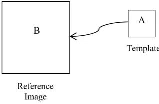
The idea of the template matching is to compare and match the rectangular blocks of pixels from a set of two ×
[image:2.595.57.287.173.293.2]images of the template and the selected image. The template must be smaller that the reference image for this to work. In this process, the template is being moved pixel by pixel on the selected image to obtain the most matched pixels. Fig. 2 shows the template matching .
Fig.2. Template matching .
In this section, also known as a sliding dot product or sliding inner-product, the normalized cross correlation method has been used for template matching. In signal processing, cross correlation is a measure of similarity of two waveforms as a function of a time-lag applied to one of them. The cross correlation mathematically can be expressed by
, ∑ ∑ , , . (3)
It is denoted by ⊗ where kernel being cross-correlation with the input signal , to generate an output . It also can be simply expressed by
, ∑ , (4) and the normalized correlation can be expressed by
_ , ∑
∑ ∑ , (5) where the value obtained is proportional to the signals similarity. Note that the numerator is indicates the cross correlation of the image and the template, and cannot be used as similarity measure because it will produce false matching results. Thus, the denominator is used to normalize the cross correlation to obtain the right match.
III. RESEARCH METHODOLOGY A. Sampling and Image Acquisition
Ultrasound images were obtained by the S5-1 sector probe, connected to ultrasonic diagnosis equipment Philips iE33, which located in Medical Imaging Laboratory Pantai Hospital Klang. A subject who suffers from mitral valve disease was selected in this study. The images were scanned from the left parasternal window. The parasternal long axis (PLAX) view as shown in Fig. 3 is obtained by placing the transducer in the three to four left intercostal spaces close to the sternum with the beam oriented toward the patient’s right shoulder. This orientation is slicing through the heart on a long axis from base to apex [6]. Based on the image, it can be seen that mitral valve (MV) is the boundary between the left atrium (LA) and left ventricle (LV). After collecting the video file from the ultrasound imaging system, the video file was extracted into frame image format by using MATLAB.
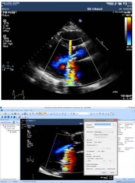
The ultrasound image can only be read by DICOM software specialize for medical image purpose. Therefore, in order for the image can be processed in MATLAB software, the image must first be converted into video file (Avi video). Fig. 4 shows the Dicom interface for reading the ultrasound image and the steps to export the file to video format. Here the size of the video should be similar to the original one and no compression selected in order to preserve the originality of the image obtained.
Fig. 3. The parasternal long axis (PLAX) view. LA, RA, LV, and RV stand for left/right atrium, and left/right ventricle, respectively, while MV and Ao is mitral valve and aorta.
Template
Reference Image
[image:2.595.76.236.439.543.2] [image:2.595.339.513.520.641.2]Fig. 4. Dicom interface for reading the ultrasound image and video conversion format.
B. Steps of Template Matching
This method has been implemented in MATLAB software. The algorithms used for template matching codes are as follows:
1. Create a template: It was created based on the original image. The rgb2gray conversion has been used to convert the colour image to grayscale. The template must be smaller than the image compared, thus, cropping is necessary. After that, the template (cropped image) must be saved under a different name.
2. Image selection: The image has to be selected and rgb2gray conversion is used to convert the colour image to grayscale. The size of the image based on row and column can be checked through the workspace.
3. Normalized cross correlation: The normxcorr2 function is being used in the Matlab software. Based on the function, the cross-correlation in the spatial or the frequency domain, depending on the size of images is calculated. Next, calculate local sums by precomputing running sums, where the local sums are used to normalize the cross-correlation to get correlation coefficients [7,8].
4. Match box: A coloured box that indicates the match template with image is created around the maximum match for easier visualization.
IV. IMPLEMENTATION OF NORMALIZED CROSS CORRELATION This section presented the result of the image registration by template matching. The experiment was conducted by using PLAX view, focusing on mitral valve
with the grayscale image of size 600 × 800. The experiment was conducted on two male patients both suffering with mitral valve disease. Patient 1 in Fig. 5 has the artificial mitral valve thus the valve does not move much throughout the complete cardiac cycle. Meanwhile, patient 2 suffering from flail mitral valve where the valve has a ruptured chordae thus it cannot close completely as it should, which allowing the flow back of the blood to the heart chamber in the wrong direction. In the experiment, the template extracted from patient 2 consist of mitral valve open (cardiac diastole) and mitral valve close (cardiac systole).
[image:3.595.316.538.527.717.2]Fig. 5-9 shows the result of every different template matching using a same size of different grayscale image with different size of template by normalizes cross correlation method. Each figure consists of four separated image the original 600 × 800 grayscale image, varies sizesqa of template image and the surface plot.The coloured box in the matching image indicates the matching position of the template that extracted from the reference image. The peaks on the cross correlation "surface" are the positions of the best matches in the image of the template. Surface plots are used for visualizing matrices that are too large to display in numerical form and for graphing functions of two variables.
Fig. 8 and Fig. 9 shows the result of matching image obtained and surface plot by using a false template. The template used in this process in Fig. 8 is from patient 2 and the reference image is from patient 1. Meanwhile, Fig. 9 shows the correlation when applied the false template of mitral valve open with the reference image of mitral valve closed obtained from patient 2. The system still produced the most likely match position of the template to be in the reference. The difference can be seen in the surface plot when being compared to the other first three trials that uses the true template with the true reference image. The first three clearly shows a distinctive peak compared to those applied to the false template, where the surface shows a fuzzy plot indicates the uncertainty of the match position.
Fig.6. a) Original image size 600 × 800 b) Template of size 144 × 122 c) Matching image d) Surface plot
Fig.7. a) Original image size 600 × 800 b) Template of size 114 × 112 c) Matching image d) Surface plot
Fig.8. a) Original image size 600 × 800 b) Template of size 114 × 112 c) Matching image d) Surface plot
Fig.9. a) Original image size 600 × 800 b) Template of size 144 × 122 c) Matching image d) Surface plot
V. DISCUSSION
Cardiac diseases are a major health concern worldwide. In particular, useful information about the cardiac function can be extracted by analyzing of echocardiography sequences. However, echocardiographic images are difficult to analyze and depends mostly on the operator’s skills and lifetime experiences to obtain a clear and better image. Unclear images may result in false diagnosis due to the tendency of existing speckle noise. In this paper, an initial study on mitral valve detection is proposed. In average normal person, mitral valve will open and close due to the pressure contrast of the blood flow. The failure of the mitral valve to properly close during the pumping out of blood, resulting in the leaking of blood to the atrium.
ACKNOWLEDGMENT
The authors would like to express their gratefulness for the support and generosity from Universiti Tun Hussein Onn Malaysia (UTHM), Microelectronics and Nanotechnology – Shamsuddin Research Centre (MiNT-SRC), and Pantai Hospital Klang, Medical Imaging Laboratory for having the space doing the experiment and analysis also data collecting for this research.
REFERENCES
[1] Shah PM, ""Current concepts in mitral valve prolapse-diagnosis and management,"" Journal of Cardiology, vol. vol. 56, no. no. 2, pp. pp. 125-133, 2010.
[2] Retrived from Webmed. (2018).
https://www.webmd.com/heart/mitral-valve-prolapse-symptoms-causes-and-treatment#1
[3] Feroze Mahmood et al., ""Mitral Annulus: An Intraoperative Echocardiographic Perspective". ," Journal of Cardiothoracic and Vascular Anesthesia, vol. 27 (6), pp. 1355–1363.
[4] JK Perloff and WC Roberts, ""The mitral apparatus. Functional anatomy of mitral regurgitation". Circulation," in Cardiovascular Pathology, Jagdish Butany L. Maximilian Buja, Ed., 1972, vol. 46 (2), pp. 227–39.
[5] Catherine M. Otto and Robert Bonow., "Valvular Heart Disease: A Companion to Braunwald's Heart Disease. ," Elsevier Health Sciences., September 2009.
[6] Retrieved from Servier Medical Art, www.servier.com on May 2008.
[7] Lewis, J. P., "Fast Normalized Cross-Correlation," Industrial Light & Magic, May 15-19, 1995, p. 120-123.