Vol.45, No. 1 JOURNALOFVIROLOGY, Jan. 1983,p.343-353
0022-538X/83/010343-11$02.00/0
CopyrightC1983,AmericanSocietyfor Microbiology
Genetic
Analysis of Temperature-Sensitive
Mutants
Which
Define
the
Genes for the Major Herpes Simplex Virus Type
2
DNA-Binding Protein
and a New Late
Function
RICHARD A. F. DIXON,tDENNIS J. SABOURIN, AND PRISCILLA A.SCHAFFER*
Laboratory of Tumor Virus Genetics, Sidney Farber Cancer Institute, and Departmentof Microbiology and
Molecular Genetics, Harvard Medical School, Boston, Massachusetts02115
Received 2 August 1982/Accepted4October 1982
Eleven temperature-sensitive mutantsofherpes simplex virus type 2 (HSV-2) exhibit overlapping patterns of complementation that define four functional
groups.Recombinationtestsconfirmedtheassignment ofmutants to complemen-tation groups 1 through 4 and permitted the four groups to be ordered in an
unambiguous lineararray. Combined recombinationand marker rescue tests (A.
E. Spang, P. J. Godowski, and D.M. Knipe, J.Virol. 45:332-342, 1983)indicate thatthemutations lie inatight clusternearthecenterofULtotheleft ofthegene
for DNA polymerase in the order 4-3-2-1-polymerase. The seven mutants that
make up groups 1 and 2 fail to complement each other and mutants in HSV-1
complementation
group1-1, thegroupthoughttodefine the structuralgeneforthemajor HSV-1 DNA-bindingprotein withamolecularweight of 130,000. At38°C,
mutants in groups 1 and 2
synthesize
little or no viral DNA, and unlike cells infected with the wild-type virus, mutant-infected cells exhibit no detectablenuclearantigen reactive with monoclonalorpolypeptide-specific antibodyto the
major HSV-2 DNA-bindingprotein.Thefourmutantsthat make up groups 3 and 4
donotcomplement eachother,nordotheycomplementmutantsingroup 2.They do, however, complementmutantsingroup 1 aswellasrepresentativemutantsof HSV-1 complementation group 1-1. At 38°C, mutants in groups 3 and 4 are
phenotypically
DNA',
and nuclei of mutant-infected cells contain the HSV-2 DNA-binding protein. Thus, the four functional groups appear to define twoclosely linked genes, one
encoding
an early viral functionaffecting
both viralDNA synthesis and
expression
of the DNA-bindingprotein
with a molecular weight of 130,000 (groups 1 and 2), and the other encoding a previously unidentified late viral function (groups 3 and 4). The former gene presumablyrepresents the structuralgenefor themajor HSV-2 DNA-bindingprotein.
Functional homology between the genes of herpes simplex virus types 1 and 2 (HSV-1, HSV-2)wasfirst demonstrated by the failureof
phenotypically
similartemperature-sensitive (ts)mutants of the two virus types to complement
(8,
29). That the orderof
genes on thechromo-somes of the two viruses is at least roughly similar was demonstrated by the
viability
of intertypicrecombinants andby the colinearity ofHSV-1 and HSV-2 DNA sequences specifying polypeptides of
approximately
the same size andkinetic class (12, 15-17). Although studies of intertypic recombinants suggest extensive
func-tionalhomologyandgeneticcolinearitybetween
HSV-1 and HSV-2DNAs,comparative
comple-tPresent address: Department ofMolecularBiologyand Genetics, The Johns Hopkins University School of Medicine, Baltimore,MD 21205.
mentation andmapping studies of phenotypical-ly similar mutants of each virus type provide
more definitive proof of such homology. To
date,
comparison
of thefunctional defects oftsand other
mutants
of HSV-1 and HSV-2 by intertypic complementation, combined with ef-forts to locate the mutationsby
physical
map-ping procedures,
have demonstrated functionalhomology andgeneticcolinearity for three viral genes:
thymidine
kinase(11), DNA polymerase (3), and alkaline DNase (18, 21). We reportherein the results of a study defining a third
homologous gene: the gene for the major viral
DNA-binding protein.
Thefollowing observations prompted this
in-vestigation. A collaborative studyof29 ts mu-tants of HSV-2 identified 20
complementation
groups (25). A total of5 ofthe 29 mutants, aswell as 6 mutants not included in the original
343
on November 10, 2019 by guest
http://jvi.asm.org/
344 DIXON, SABOURIN, AND SCHAFFER
study, exhibit overlapping patterns of comple-mentation among themselves, complement
mu-tantsinothergroupsefficiently, and differ
mark-edly in their viral DNA phenotypes. Importantly, several of the 11 HSV-2 mutants
fail to complement mutants in HSV-1 comple-mentationgroup1-1, which defines thegenefor the major HSV-1 DNA-binding protein with a
molecular weight of 130,000 (130K) (5, 29a). Herein we describe complementation tests,
recombination analysis, and selected phenotypic properties of the series of HSV-2ts mutants. In relatedpapers,Spangetal. (27) and Welleretal. (29a) describe fine-structure physical mapping studies of the HSV-2 mutants and the series of analogous HSV-1 mutants, respectively. Taken together, these three studies and the earlier study of Conley et al. (5) define a fourth HSV
genewhich is both functionally homologous and
colinearon thegenomes of HSV-1 and HSV-2: thegene for the major HSV DNA-binding
pro-tein. In addition, we describe a previously
un-identified late viral genewhich maps tothe left ofthe HSV-2 DNA-binding proteinnearposition
0.36onthe viral genome.
MATERIALS AND METHODS
Cells. Humanembryonic lung (HEL)cells and
Afri-cangreen monkey kidney (Vero) cellswereused for
virus growthandfor complementationand
recombina-tiontests. HEL cellswereused fordeterminingviral
DNA phenotypes, virus assays were conducted in
Verocells, and immunofluorescence tests were
con-ducted inprimary Syrianhamsterembryocells
(Lake-view Syrian hamsters, Lakeview Hamster Colony,
Newfield, N.J.). Cells were propagated in Dulbecco
modified Eagle minimal essentialmedium containing
10% newborncalfserum,0.03%glutamine, and 0.25%
NaHCO3. Cells weremaintainedin thesamemedium
containing5% newborn calfserum.
Virus andvirus assays. The ts mutants of HSV-2,
strains 186, IPB2, and UW268, were isolated after
mutagenesis with5-bromodeoxyuridine, UV light,
N-methyl-N'-nitro-N-nitrosoguanidine, andnitrous acid.
The properties of these mutants and of the HSV-1
mutant used in intertypic complementation tests,
tsJ12,arepresented in Table 1.
Virus stockswere prepared in HELorVero cells,
and plaque assays were performed in Vero cells as
previously described (25). Permissive and
nonpermis-sivetemperatureswere34 and38°C, respectively.The
plating efficiencies of mutants [(PFU/ml at
38'C)/(PFU/ml at34°C)] ranged from 10-6to10-4.
Genetic methods.Complementationand
recombina-tiontestswereconductedasdescribed previously(24,
26). Complementationgroupsofmutants,where
previ-ously determined, areshown in Table 1.
Viral DNA phenotypes. Viral DNA phenotypes of mutantsweredetermined asdescribed by Aronetal.
(1).
Immunofluorescence tests. Indirect immunofluores-cence testswere conductedby the method of Porter
(19)asmodified by Flanneryetal.(9).
Monoclonal antibody to the 130K HSV-2
DNA-binding proteinwaskindly providedasmouseascitic
fluids by AnthonyMinson(UniversityofCambridge,
England). This antibody is immunoglobulin G
anti-body; purified130Kproteincompetesfor the antibody
in radioimmunoassays; and theantibody precipitates
the purified proteinandproducesthesamedistribution
offluorescence in HSV-2-infected cells as
polypep-tide-specific antiserum directed against the entire
130K protein(A. Minson, personal communication).
Polypeptide-specific antiserum to the 130Kprotein
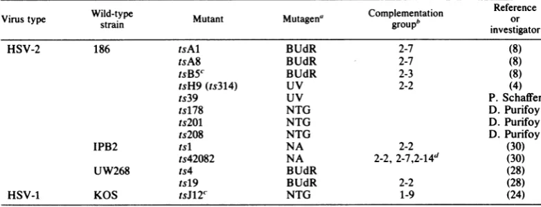
TABLE 1. Propertiesof HSV-1 and HSV-2 ts mutantsused in thisstudy
Wild-type Complementation Reference
Virus type strain Mutant Mutagena
groupb
orinvestigator
HSV-2 186 tsAl BUdR 2-7 (8)
tsA8 BUdR 2-7 (8)
tsB5c BUdR 2-3 (8)
tsH9(ts314) UV 2-2 (4)
ts39 UV P. Schaffer
ts178 NTG D.Purifoy
ts2WM NTG D.Purifoy
ts208 NTG D.Purifoy
IPB2 tsl NA 2-2 (30)
ts42082 NA 2-2,
2-7,2-14d
(30)UW268 ts4 BUdR (28)
tsl9 BUdR 2-2 (28)
HSV-1 KOS tsJ12'' NTG 1-9 (24)
a Abbreviations: BUdR,5-bromodeoxyuridine; UV,UVlight; NTG,N-methyl-N'-nitro-N-nitrosoguanidine;
NA, nitrous acid.
bDeterminedpreviouslywhere shown(8, 25).
cMutant tsB5specifiesathermolabileDNApolymerase(22);mutanttsJ12 of HSV-1 strain KOS is defectivein
theexpression ofglycoproteingAgB (14).
dMutantts42082failedtocomplementmutants in threegroups(25).
on November 10, 2019 by guest
http://jvi.asm.org/
[image:2.489.55.446.464.614.2]HSV-2 ts MUTANTS 345 waspreparedasdescribed by Courtney and
Benyesh-Melnick (6). The patterns of 130K protein-specific
fluorescence produced by this antiserum in cells in-fected with and transformed by HSV-2 have been describedpreviously (9).
Fluorescein isothiocyanate-labeled rabbit anti-mouse immunoglobulin G (Cappel Laboratories, Cochranville, Pa.)wasusedtodetectmonoclonal and polypeptide-specific antibodyonfixed monolayers.
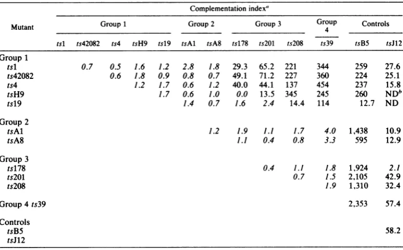
RESULTS
Complementation. The results of previous complementation tests placed HSV-2 mutants
tsH9, tsl, and tsl9 into one complementation group(2-2) and mutanttsA8 into anothergroup
(2-7) (25). Mutant ts42082 failedtocomplement members of both groups, and because of this
overlap it couldnotbeassignedunequivocallyto
either. Theunusualoverlappingpatternsof com-plementation exhibited not only by ts42082 but alsoby six other HSV-2tsmutants notincluded in the original study (ts4, ts39, ts178, ts201,
ts208, and tsAl) prompted a reexamination of
the functional relatedness ofthese mutants by complementation analysis.
The results ofquantitative complementation
testsamongthe 11 mutantsare shown in Table
2. The HSV-2 mutant tsB5, which specifies
thermolabile DNA polymerase activity and is a
member ofcomplementationgroup2-3 (22, 25), and tsJW2, a mutant defective in glycoprotein gAgB andamemberofgroup1-9(14, 25), were
included as controls. By using an index of10 rather than 2 (24)tosignify positive complemen-tation, the 11 mutants were classified into four
groups numbered 1, 2, 3, and 4 (Table 2).
Members ofagivengroupfailed tocomplement each other; however, members of different
groups exhibited overlapping patterns of
com-plementation. Thus,mutantsingroup1failedto
complement mutants ingroup2. Moreover,
ex-ceptfor pairs tsH9 + ts178, tsl9 + ts178, and
tsl9+ts201, the fivemutantsingroup1
comple-mented mutantsingroups3 and4efficiently. In
contrast,the group2 mutantsfailed to
comple-ment mutants in groups 3 and 4. Likewise,
mutants in group 3 failed to complement the group4mutant.Notably, allmutantsingroups1 through 4 (except for the pairs tsH9 + tsJ12 and tsl9 + tsJW2, which were not tested, and ts178 + tsJ12, which yielded an index of2.1) complemented control mutants tsB5 of HSV-2
and tsJ12 of HSV-1 efficiently. The fact that
mutantscomplemented tsB5 and tsJ12 indicates that theyareverylikely defective ingenesother than polymerase (tsB5) or glycoprotein gAgB
(tsJ12).
[image:3.489.46.439.400.644.2]Recombination. The observation that the 11
TABLE 2. Complementationamong tsmutantsof HSV-2
Complementationindex'
Mutant Group1 Group2 Group3 Group Controls
tsl ts42082 ts4 tsH9 tsl9 tsAl tsA8 ts178 ts2WM ts208 ts39 tsB5 tsJ12
Group1
tsl 0.7 0.5 1.6 1.2 2.8 1.8 29.3 65.2 221 344 259 27.6
ts42082 0.6 1.8 0.9 0.8 0.7 49.1 71.2 227 360 224 25.1
ts4 1.2 1.7 0.6 1.2 40.0 44.1 137 454 237 15.8
tsH9 1.7 0.6 1.0 0.0 13.5 345 245 260 NDb
tsl9 1.4 0.7 1.6 2.4 14.4 114 12.7 ND
Group2
tsAl 1.2 1.9 1.1 1.7 4.0 1,438 10.9
tsA8 1.1 0.4 0.8 3.3 595 12.9
Group3
ts178 0.4 1.1 1.8 1,924 2.1
ts2WM 0.7 1.5 2,105 42.9
ts208 1.9 1,310 32.4
Group4ts39 2,353 57.4
Controls
tsB5 58.2
tsJ12
aComplementation tests were conducted as previously
described
(24). Complementation indices <10 areitalicized.
bND,Notdone.
VOL.45, 1983
on November 10, 2019 by guest
http://jvi.asm.org/
TABLE 3. Recombination among11 ts mutantsof HSV-2 Recombinationfrequency'
Mutant Test Group1 Group2 Group3 Group Control
no. __ 4
tsl ts42082 ts4 tsH9 tsl9 tsAl tsA8 ts178 ts201 ts208 ts39 tsB5
Group1
tsl 1
2 3
0.002 1.0
0.1 0.8
ts42082 1
2
3
ts4
0.1
0.4
1 2 3
0.1 0.03
0.001 0.01
0.3 0.7
0.04 0.04 2.0
0.1 1.1 0.7 9.1 2.5 6.8 6.0
0.8 1.2 1.8 4.8 4.3 9.6 5.0
0.4 2.4 0.6 2.0 1.3 5.3 5.0
0.6 1.0 1.6 ,5.8 4.8 9.5 5.9
0.5 0.7
0.6 3.7
1.7 4.5
4.0
3.6 7.4
4.5 9.0
8.0
11.0 8.0
0.001 1.3 1.5 2.6 2.5 2.8 7.9 1.4
2.2 2.1 0.8 5.1 4.0 8.0 1.3
0.001 0.7 1.1
0.0 0.7 2.3
1.9
0.3 2.2
2.0 6.5
19.2 15.2
0.9 0.5 0.6 4.0 17.5
0.8 2.1 1.7 7.3 17.1
0.0 1.3 2.0 14.5
0.0 0.8 4.8 23.0
1.4 0.6 25.8
0.0 4.5 24.9
2.0 24.8
4.5 25.9
47.6 44.1
aRecombination testswereconductedasdescribed previously (26).
mutants could be classified into fourfunctional groupsbasedonoverlappingpatternsof
comple-mentation raised thequestionwhether the
muta-tions ineachgroupcould be ordered by
recom-bination. Totestthispossibility, recombination
analysis was conducted by two-factor crosses.
Thepolymerasemutant,tsB5,wasagain includ-edas anoutside marker since the physical map
location of its mutation (0.40 to 0.42) has been
determined (27). The HSV-1 mutant tsJ12 was
notincludedinthese crossesbecause intertypic
recombination frequencies are usually low and maynotreflect the order ofmarkers faithfully(7).
Unfortunately, no equivalent mutant (i.e., no
mutantdefective in glycoprotein gAgB) hasyet
been identified in the HSV-2 system. Three
separate recombination tests were performed.
Tests 1and 2wereconducted in HELcells,and
test3 was conducted in Verocells. The results
of these tests arepresentedin Table 3, and the
3
3
1
2 3
1 2 tsH9
tsl9
Group 2 tsAl
tsA8
Group 3 tsl78
Ws20
ts208
Group4
ts39
1 2
1 2 1 2
1
2
on November 10, 2019 by guest
http://jvi.asm.org/
HSV-2 ts MUTANTS
4
TEST NO.
'39
-
-1-2
3
39
.__.
3 2 I
208 r
:201(178) 'AlAS (178)
'4
142082 B5I.
201 'I " " " 42082
208 178 " )ASAI ) "' 4 1
I. . . . .. . BS
201 208 (178), AIAS
.. . .. ..
42082
H9
(178) '4119
..- _5
-J I~~~~~~~~~~~~~~~~~~~~~
L- -____-, L- __ __ -JL-____j I-_ __
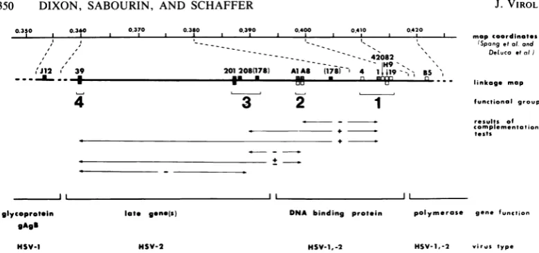
_--FIG. 1. Linkagemapsgeneratedby two-factorcrossesamong12tsmutantsof HSV-2.Mutations in members ofthe four functionalgroups areenclosedby dashed lines and numbered attop.
linkagemapsderived from these dataare shown
in Fig. 1.
Although recombination frequencies between markers variedamongthethreetests(Table 3), both the order of markers and the relative
dis-tancesbetweenmarkerswerethesamewhen the
three maps were compared on the same scale
(Fig. 1).
The classification ofmutants into fourgroups
by complementation analysis was confirmed by
recombination analysis (Table 3, Fig. 1). Thus,
withoneexception, mutations ofmutantsin the
same group were more closely linked to each otherthan tothe mutations ofmutantsin other
groups, and the mutations of members of the
fourgroups were ordered unambiguously in all
three tests with respect to the outside marker, the mutation of tsB5. The exception to this
pattern was ts178, for which the mutation
mapped eitherto the right of ts201 and ts208 in
group3orbetweengroups1 and 2 intests1 and 3 (Fig. 1). It will be recalled that mutant ts178 also produced ambiguous results in complemen-tationtests (Table 2).
Phenotypic properties of mutants. (i) Viral DNA phenotypes. The results of complementa-tion and recombinacomplementa-tion analysis indicated that the fourgroups ofmutants may represent four distinct and separablefunctions. Comparison of the viral DNA phenotypes of the 11 mutants
[image:5.489.53.443.58.187.2]furthersupportsthishypothesis (Table 4). Thus, viral DNA synthesis at 38°C ranged from <1 to TABLE 4. Phenotypic properties of13HSV-2 ts mutants
Fluorescentstainingb
Viral DNA 340C 380C
Virus synthesisat38°C
(% wild-type)' Nucleus Cytoplasm Nucleus Cytoplasm
MC PS MC PS MC PS MC PS
Wild-type 100 + + + + + + - +
tsB5 5
Group1
tsl9 <1
tsH9 <1 + + + + - - + +
ts4 <1
ts42082 <1
tsl 5
Group2
tsA8 10 + + + + - - - +
tsAl 9 + + + + - - + +
Group3
ts208 80
ts201 52 + + +
ts178 50
Group4
ts39 119 + + +
'Measuredasdescribed byAronet al.(1).
bStainingwith monoclonal antibody (MC) and polypeptide-specific antibody(PS) to the 130Kprotein. +,
Brightfluorescence; ±, moderatetofaintfluorescence; -, nospecific staining.
347
VOL. 45, 1983
l
on November 10, 2019 by guest
http://jvi.asm.org/
[image:5.489.47.442.404.661.2]5% of wild-type levels in mutants of group 1, from 9 to10%in mutantsofgroup 2,and from50 to 80% in mutants of group 3. The group 4 mutant synthesized wild-type levels of viral DNA. All mutants were equally effective in
shutting off host cell DNA synthesis; each mu-tantreduced levelsof cellularDNAsynthesisto 10 to 30% of uninfected cell levels (data not shown).
(ii)Immunofluorescence tests. Intertypic
com-plementationtestshaveshown thatonemember
ofgroup 1 (tsH9) and both members ofgroup 2 (tsAl and tsA8) do notcomplement a series of
sixtsmutantsin HSV-1 complementationgroup 1-1 (29a). Mutants in group 1-1 aredefective in
viral DNA synthesis, and their mutations map
between coordinates 0.385 and 0.413;
further-more, mutant-infected cells fail to express the
130K DNA-binding protein at 39°C in immuno-fluorescence tests with monoclonal antibody to
the HSV-1 DNA-binding protein. Because our dataindicatethat mutants tsH9, tsAl,and tsA8, aswell asother members ofgroups 1 and2,are
defective in the equivalent HSV-2 gene, we tested mutants representing all four groups for their ability to express the 130K DNA-binding proteinat38°C. In these tests we used
monoclo-nal antibody to the HSV-2 130K protein. The resultsofthese tests aresummarized in Table4, and photomicrographs of typical fluorescence
reactions are shown in Fig. 2. As in cells
infect-ed with wild-type virus (Fig. 2A), the protein
wasdetected inthenucleiand to alesserextent
in thecytoplasm of mutant-infected cellsat34°C (Fig. 2C, E, and G). At 38°C, the antigen was
detected in nuclei but not in the cytoplasm of cells infected with the
wild-type
virus (Fig. 2B)orwith mutants in groups 3 and 4 (e.g., ts201; Fig.2H). In contrast, theantigenwasnot
detect-edinthenuclei ofcellsinfectedwith one mutant ingroup 1 (tsH9) orwithboth mutantsingroup 2
(tsAl and tsA8). Cytoplasmic fluorescencewas,
however, detected in cells infected with tsH9
(Fig. 2F)andtsAl, butnot incells infected with
tsA8 (Fig. 2D), at 38°C.
Because the monoclonal antibody could, by
definition, detect only a single epitope of the 130K protein and because mutant forms of the 130K protein may be sufficiently altered
struc-turally to render the protein undetectable with this monoclonal antibody, we also tested cells
infected with selected mutants and with the
wild-type virus, using antiserum prepared
againstthepurified 130Kprotein (Table4). Such
polypeptide-specific
antiserum should bereac-tivewith the spectrum ofantigenicdeterminants onthe 130Kprotein. Like the monoclonal
anti-body, the polypeptide-specific antiserum was reactive with both the nuclei and thecytoplasm of cells infected withwild-type andwith mutant
viruses at 34°C. Moreover, unlike the monoclo-nal antibody, the polypeptide-specific antiserum
produced positive fluorescencein thecytoplasm
of cells infected with wild-type virus at38°C. Importantly, no nuclear fluorescence was ob-servedin cells infected with tsH9,tsAl, ortsA8
at 38°C. The failure to detect nuclear fluores-cence with polypeptide-specific antiserum strongly suggests that the protein is, infact, not present in the nucleus and notsimply that it may be present in an altered antigenic state, asone
might conclude from tests with monoclonal anti-body.
Thus, with one exception, the patterns of fluorescence observed in mutant-infected cells wereidentical with both antibody preparations. The exception was that no cytoplasmic fluores-cence was detectable in tsA8-infected cells at 38°C with monoclonal antibody, but moderate cytoplasmic fluorescence was observed with the
polypeptide-specificantiserum.
Takentogether, the results of immunofluores-cence tests indicate that mutants ingroups 1 and 2 are defective in a functionaffecting the trans-port of the 130Kproteintothe nuclei of infected cells at38°C,whereasmutantsingroups 3 and 4 exhibit no such defect. These observations thus provide additional support for the functional
grouping suggested by the other genetic and
phenotypicparameters described above.
DISCUSSION
Eleven ts mutants define four functional groups. The combined results of
complementa-tion, recombination, and phenotypic analysis
indicate that the 11 HSV-2 ts mutants can be
assignedtofour functional groups (Fig. 3). The unusual overlapping patterns of
comple-mentation exhibited by these mutants (Table 1) provided the initial evidence for the existence of
four groups. One possible explanation for the
overlap is thatthe mutants possess multiple ts
mutations. This explanation appears unlikely, however, because recombination frequencies were (i)higher than one would expect in crosses between double mutants and (ii) sufficiently ad-ditive to construct linkage maps. Moreover, all mutants reverted with low but consistent fre-quency (10-6to
10-4).
Importantly, the grouping by
complementa-tionwassupportedbyrecombinational and
phe-notypic data. Generally speaking, recombina-tionfrequencies betweenmutantsingroups that did not complement were low, demonstrating
close linkage, whereas recombination
frequen-cies werehigher betweenmutantsingroups that didcomplement.
An interesting finding from recombination studies concerns theunexpectedly high
on November 10, 2019 by guest
http://jvi.asm.org/
FIG. 2. Photomicrographs of cells infected with wild-type and ts mutant viruses, stained with monoclonal
antibodyto the majorHSV-2 DNA-binding protein of 130K. Shown are cells infected withwild-typevirus at34°C
(A) and38°C(B); with tsA8, group 2, at34°C(C) and38°C(D); with tsH9, group 1, at34°C(E) and38°C(F); and withts201, group 3, at34°C(G) and38°C (H).
349
on November 10, 2019 by guest
http://jvi.asm.org/
350 DIXON, SABOURIN, AND SCHAFFER
0.350 0.3t0 0.370 0.380 0.390 0.400 0.410 0420
mapcoordinotes
---. (Spongetat or
- -~--- --- ---- 42082 \Dehucoeta
201208(178) AlA8 1718r 4 1| i9g-= BS
*. uu u ''' linkoge map
'I
I
'Jl12 X' 39 _ .-i .. #
4
3
2
1
Ind
a1 1
functional group
results of Complementotion
tests
i
DNA binding protein
HSV-1,-2
polymerose gene function
HSV-1,-2 virus type
FIG. 3. Genetic andphenotypic properties of 11 ts mutantsofHSV-2. The heavy line isthelinkage map
generatedbytwo-factorcrosses(Fig. 1).Thetopline indicates thephysicalmapcoordinates ofmutationsshown
on the linkage map. The physical map location of the HSV-1 mutation of tsJ12 was determined by DeLuca
(personal communication).ThemutanttsJ12 isamember ofcomplementationgroup1-9(25),which defines the
genefor HSV-1glycoprotein gAgB (14).Thephysicalmaplocations of mutationsinmembers ofgroups1through
4and of tsB5weredeterminedbySpangetal.(27).ThemutanttsB5 isamember ofcomplementationgroup2-3
(25),whichdefinesthegenefor HSV-2 viral DNApolymerase(2, 22).The viralDNAphenotypesofmutants at
38°Careindicatedbyboxes above thelinkagemapline: -M,>20% ofwild-typelevels of viralDNA; JIlL,s20%
ofwild-type levelsof viral DNA; n ,nodetectableviral DNA.Expressionof the 130KDNA-binding proteinin
nuclei of mutant-infected cellsat 38°C is indicated by boxes belowthe line: T, brightnuclearfluorescence;
, no specificfluorescence. Results ofcomplementation tests between mutants ingroups 1 through 4 are
shownbeneath thelinkagemap,wherecomplementationindices >10(+),between2and 10(±),and<2(-)are
distinguished. Theidentity of viralgenesdefined by thets mutants studied and thevirus type inwhich these
geneshave been identified(HSV-1, HSV-2, orboth)areillustrated atthe bottom.
cy of recombination between tsB5 and ts39, whose mutations representtheopposite termini of the linkage map. Indeed, recombination
be-tween tsB5 and ts39 approached 50%, whereas the actual physical distance between the two
mutations is less than 10% of the viral genome
(Fig. 3 and accompanying text). A frequency approaching 50% suggests the presence ofone
ormore recombinational hotspots between the
two markers. Whether such a hot spot
consti-tutestheputative origin of viral DNA synthesis thought toreside between 0.35 and 0.45 (10, 13, 23)-the physical map location of the 11
muta-tions involved in this study-is an intriguing
possibility.
Although the results of recombination tests
were sufficiently additive to order the four
groupsofmutationsunambiguously withrespect to oneanother and to tsB5, the order of
muta-tions withineachgroupis less certain. The order
shown in Fig. 3 represents the average of the
threepositions for each mutation shown in Fig. 1 andshould be considered suggestive rather than definitive.
The fact that mutants in the same functional
group have similar phenotypic properties with regard to both the levelof viral DNA synthesis and the ability to detect the 130K protein in nuclei of mutant-infected cells provides
addi-tional support for the classification of the 11
mutantsinto four functionalgroups. The failure
todetectthe 130K protein in the nuclei of cells infected withmutantstsH9, tsAl, and tsA8 is of special interest. Results obtained with polypep-tide-specific antiserum suggestthat the protein isnot,infact,presentinnuclei. It is possible,on
the other hand, that the antibody preparations used in thesetestswerenotsensitive enoughto
detect small quantities of 130K protein. Indeed, Spang et al. (27) have presented evidence to
suggest that the protein is associated with the
nuclear fraction of cells infected with selected
mutants in groups 1 and 2. Clearly, additional
experimentation will benecessarytoresolve this problem. Inanyevent,itis clear thatmutantsin
groups 1 and 2 possess a defect in nuclear
expression of the 130Kproteinnotcharacteristic ofmutantsingroups 3 and 4 whenexpression is assessed with polypeptide-specific antisera in immunofluorescence tests.
The results of the studies described in this
reportalso addressamorefundamentalproblem
with the genetic analysis of HSV. Based upon
the criterion that complementation indices .2 reflect the ability oftwo mutantstocomplement (24), 29 ts mutants of HSV-2 were shown to
define 20 nonoverlapping complementation
groups(25).The recent studies of S. K. Weller,
glycoprotein gAgS
HSV-1
late gene(s)
HSV-2
on November 10, 2019 by guest
http://jvi.asm.org/
[image:8.489.56.446.61.244.2]HSV-2 ts MUTANTS 351
W. R. Sacks, D. M. Coen, and P. A. Schaffer (submitted for publication) now indicate that
complementation indices between 2 and 10 are
ambiguous and may represent either
intracis-tronic or intercistroniccomplementation. These findings are supported by the results of our
studies with mutants in groups 1 and 2. These
resultshave, therefore, promptedusto reclassi-fymutantsincomplementationgroup 2-2 and 2-7 to a common group, group 2-2. For the same reasons, the results of complementation tests
reported herein, as well as the results of all future tests, will utilize a value of 10 to signify
positive complementation. Itis anticipated that the problem of intracistronic versus
intercis-tronic complementation will ultimately be re-solved by combined studies
involving
physical mapping, transcriptional mapping, and in vitrotranslation ofcarefully mapped transcripts.
Order, orientation, and physical map location of mutations in the four groups. The order, orientation, and physical map location of the
mutationspresented inFig.3 are based upon the
following observations.
(i) Mutant tsB5 synthesizes a thermolabile
DNApolymerase(22) and doesnotcomplement the HSV-2 DNA polymerase mutant of strain
HSG52, ts6, a member of
complementation
group 2-3 (2). Both the tsB5 and ts6 mutationshave been mapped to a 3.2-kilobase
region
be-tweencoordinates0.40 and 0.42onthephysical
map ofHSV-2(3, 27).
(ii) HSV-2 mutations ofmembers of comple-mentation group 2-2 as well as their HSV-1
counterparts in group 1-1 (i.e., those in the
majorHSV-1 DNA-bindingprotein) map imme-diatelytotheleftof mutations inthepolymerase
gene (27,29a).
(iii) In the HSV-1 system, the nearest
recog-nized markertothe left of mutations in members
of group 1-1 is the geneforglycoprotein gAgB.
The HSV-1tsJ12mutant, alatemutantdefective
ingAgB expression, maps betweencoordinates
0.357 and 0.360(N. DeLuca,personal communi-cation); analogous HSV-2 mutations in glyco-proteingAgB have not yet been identified. Be-cause three of four mutants in groups 3 and 4
complement mutant tsJ12
efficiently (Table
2)andbecause ts39(group 4), ts201 (group3),and ts178 (group 3) do not exhibit alteredexpression
ofgAgB (B. Pancake, personalcommunication)
astsJ12 does(14), it isunlikelythatanyofthese mutantsisdefective in thegenefor gAgB. If this is indeed the case, then the DNA-positive
mu-tantsingroups 3 and4mustrepresenta newlate
gene(orgenes)
lying
between thegenesfor the 130K protein andglycoprotein gAgB (Fig. 3).(iv) Spang et al. (27) have recently demon-strated that mutationsrepresenting groups 2, 3, and4map withinaregion withmap coordinates
0.360 to 0.380. This region lies immediately to the right of the sequence in which the tsJ12 mutation lies (0.357 to 0.360) and to the left of a sequence from 0.380 to 0.400 containing the
mutationin tsl (group 1). The physicalmapping data ofSpang et al. thus support the order (right to left) tsB5 (polymerase), groups 1 (2, 3, 4), tsJ12 (glycoprotein gAgB) (Fig. 3).
(v) Fine mapping of viral transcripts found in
abundance before viral DNA synthesis has shown that two speciescorrespond to the genes forglycoprotein gAgB and the 130K protein(L.
Holland,personalcommunication). Moreover, a protein of 128K was translated in vitro by Con-ley et al. (5) from transcripts mapping in or near
the region encodingthe 130K protein.
Fourfunctional groups: How many genes? The most reasonable interpretation of the genetic andphenotypic data summarized in Fig. 3 is that the 11 mutants represent two (or perhaps three) viral genes.
One gene encodes the130K protein. This gene is represented by mutants in groups 1 (ts4, tsl,
ts42082, tsH9, and tsl9) and 2 (tsAl and tsA8).
Thisinterpretation is basedupon (i) thefailureof mutants in both groups to complement each other; (ii) the common defect in viral DNA synthesis and the failure to express the 130K
polypeptide in the nucleus characteristic of these mutants at 38°C; (iii) the observation that the mutations of HSV-1 ts mutants with similar
phenotypic properties map to a corresponding region ofthe HSV-1 genome (29a); (iv) the fact that at least three mutants ingroup 1 (tsl, ts19,
and tsH9) exhibit altered distribution of the 130K protein in nuclei and cytoplasm as
deter-mined by cellularfractionation and sodium
do-decylsulfate-polyacrylamide gel electrophoresis
(27); and (v) the observation that the purified
130K protein from tsH9-infected cells exhibits
reduced DNA-unwinding activity compared
with purified 130K protein from cells infected with wild-type virus (20). The hypothesis that mutants in groups 1 and 2 define a single gene implies that the 130K protein has at least two
functions; mutants in groups 1 and 2 are distin-guishable by complementation with mutants in other genes (i.e., mutants in group 1
comple-ment mutants in groups 3 and 4 efficiently,
whereas mutants in group 2 do not). What
specifically thesetwofunctions are is currently
notknown.
Baseduponphysical mappingstudies(27),the ts mutations of members of all fourgroupsmap within a 6.4-kilobase region between coordi-nates 0.36 and 0.4. This amount of DNA is
sufficient to encode aprotein ofapproximately
210K. Subtracting thecodingsequences for the 130K protein from this sum (the coding se-quences for the 130K protein map to the right
VOL.45, 1983
on November 10, 2019 by guest
http://jvi.asm.org/
352 DIXON, SABOURIN, AND SCHAFFER
handend ofthe region), the remainingDNA is sufficienttoencode aprotein(s) of approximate-ly 80K.Thetemplate for thisprotein(s) would lie in the left-hand end ofthe region, tothe left of thegenefor the130Kproteinand to theright of
the gene for glycoprotein gAgB. Unfortunately, insufficient phenotypic and physical mapping dataareavailabletodetermine whethermutants ingroups 3and4defineone or two genes. In any event, thesemutationsdefine one or more
previ-ously undescribed late HSV-2 functions.
As illustrated in
Fig.
3, the results of this investigation together withthefindings
ofSpang
et al. (27) serve todefine the gene for the major HSV-2 DNA-bindingprotein-a
gene that is both functionallyhomologous
andstructurally
colinear with the gene for the HSV-1 DNA
binding protein
(29a).Additionally,
thesestudies have identified apreviously undescribed HSV-2 lategene(s) for whichacounterpart hasrecently
been identified in the HSV-1 system (B.
Pan-cake, personal
communication).
ACKNOWLEDGMENTS
We thank N. DeLuca and D. Knipe for communicating
unpublishedresults and D. Coen, B.Pancake,and S.Weller for useful comments on the manuscript. We also thank M. Datzformanuscriptpreparation.
This investigationwassupported byPublic HealthService grants CA20260 and CA21082 from the National Cancer Institute and grantMV-77from the AmericanCancerSociety. D.J.S. was supported by training grant CA09031 from the National Cancer Institute.
LITERATURE CITED
1. Aron, G. M.,D.J. M.Purifoy,and P. A.Schaffer. 1975. DNAsynthesis and DNA polymeraseactivityofherpes simplex virus type 1 temperature-sensitive mutants. J. Virol. 16:498-507.
2. Chartrand, P., C. S. Crumpacker, P. A. Schaffer, and N. M. Wilkie.1980. Physical andgenetic analysis of the herpes simplex virus DNA polymerase locus. Virology 103:311-326.
3. Chartrand, P., N. D. Stow, M. C. Timbury, andN. M. Wilkie. 1979. Physical mapping of paar mutations of herpes simplex virus type 1 and type 2 by intertypic markerrescue.J. Virol. 31:265-276.
4. Chu, C.-T.,andP. A.Schaffer.1975.Qualitative comple-mentation tests fortemperature-sensitivemutantsof her-pessimplex virus. J. Virol. 16:1131-1136.
5. Conley,A.J.,D.M.Knipe,P. C.Jones,and B. Roizman. 1981. Moleculargenetics ofherpes simplex virus. VII. Characterization ofatemperature-sensitive mutant pro-duced by in vitro mutagenesis and defective in DNA
synthesis andaccumulation ofypolypeptides. J. Virol. 37:191-206.
6. Courtney,R.J.,andM.Benyesh-Melnick.1974.Isolation andcharacterization ofalargemolecularweight polypep-tide ofherpessimplexvirustype-1.Virology62:539-551. 7. Esparza, J., M. Benyesh-Melnick, and P. A. Schaffer. 1976.Intertypic complementationandrecombination be-tween temperature-sensitive mutants ofherpes simplex virustypes1and 2.Virology 70:372-384.
8. Esparza, J., D. J. M. Purifoy, P. A. Schaffer, and M.
Benyesh-Melnick. 1974. Isolation, complementation and preliminary phenotypic characterization of temperature-sensitivemutantsofherpessimplexvirustype2.Virology
57:554-565.
9. Flannery,V.L.,R.J.Courtney,andP. A.Schaffer.1977. Expressionofan early,nonstructural antigenofherpes
simplex virus in cells transformed in vitro by herpes
simplexvirus. J. Virol. 21:284-291.
10. Frenkel, N.,H.Locker,and D. A.Vlazny.1980.Studies of defective herpessimplex viruses.Ann. N.Y.Acad. Sci. 354:347-370.
11. Halliburton, I. W.,L. S.Morse,B. Roizman,andK. E.
Quinn. 1980. Mappingof the thymidine kinase genesof type1and type 2herpessimplexvirusesusingintertypic recombinants. J. Gen. Virol. 49:235-253.
12. Halliburton, I.W., R. E.Randall,R. A. Killington,and D. H. Watson. 1977. Some properties of recombinants between type1and type 2herpessimplexvirus. J. Gen. Virol. 36:471-484.
13. Kaerner,H.C., I.B.Maichle,andC.H.Schroder.1979.
Originoftwodifferent classes of defective HSV-1
Ange-lotti DNA. Nucleic Acids Res. 6:1467-1478.
14. Little, S. P., J. T. Jofre, R. J. Courtney, and P. A. Schaffer.1981. Avirion-associatedglycoproteinessential
for infectivityofherpes simplexvirus type1. Virology
115:149-160.
15. Marsden, H. S., N. D. Stow, V. G. Preston, M. C. Timbury, and N. M. Wilkie. 1978. Physical mapping of herpes simplex virus-induced polypeptides. J. Virol. 28:624-642.
16. Morse, L. S., T. G. Buchman, B. Roizman, and P. A. Schaffer. 1977. Anatomyofherpessimplex virus DNA. IX. Apparentexclusion ofsomeparental DNA
arrange-mentsin the generationofintertypic (HSV-1 x HSV-2)
recombinants. J. Virol. 24:231-248.
17. Morse,L.S.,L.Pereira,B.Roizman,and P. A.Schaffer. 1978.Anatomyofherpessimplexvirus(HSV)DNA. X.
Mapping of viral genes by analysisofpolypeptides and
functionsspecified byHSV-1 x HSV-2 recombinants. J. Virol. 26:389-410.
18. Moss, H.,P.Chartrand,M. C.Timbury,andJ.Hay.1979. Mutantofherpessimplexvirus type 2 with temperature-sensitive lesions affecting virion thermostability and DNaseactivity: identification of the lethal mutation and
physical mappingof thenuc-lesion. J. Virol. 32:140-146.
19. Porter, D. D., I. Wimberly, and M. Benyesh-Melnick. 1969. Prevalence of antibodies to EB virus and other herpesviruses.J.Am. Med. Assoc.208:1675-1679. 20. Powell, K. L., E. Littler, and D.J.M. Purifoy. 1981.
Nonstructuralproteinsofherpessimplexvirus.II.Major
virus-specific DNA-binding protein.J. Virol. 39:894-902. 21. Preston,C.M.,and M.G.Cordingley.1982. mRNA- and DNA-directed synthesis ofherpes simplexvirus-coded exonucleaseinXenopuslaevis oocytes. J. Virol. 43:386-394.
22. Purifoy,D.J. M.,and M. Benyesh-Melnick. 1975. DNA
polymeraseinductionbyDNA-negative
temperature-sen-sitivemutantsofherpes simplex virustype2. Virology 68:374-386.
23. Reyes, G. R., R.LaFemina, S. D. Hayward, and G. S.
Hayward. 1980. Morphological transformation byDNA
fragments of human herpesviruses: evidence for two
distincttransforming regionsinherpessimplexvirustypes
1and2and lack of correlation with biochemical transfer
ofthethymidinekinasegene.ColdSpringHarborSymp.
Quant.Biol.44:629-641.
24. Schaffer,P.A.,G. M.Aron,N.Biswal,and M. Benyesh-Melnick. 1973.Temperature-sensitive mutantsofherpes
simplex virus type 1: isolation, complementation and
partialcharacterization. Virology52:57-71.
25. Schaffer,P.A.,V.C.Carter, andM. C.Timbury.1978. Collaborativecomplementation studyof temperature-sen-sitivemutantsofherpessimplexvirus types 1 and2. J. Virol.27:490-504.
26. Schaffer,P.A.,M.J.Tevethia,and M.Benyesh-Melnick. 1974.Recombinationbetweentemperature-sensitive
mu-tantsofherpessimplexvirustype1.Virology58:219-228. 27. Spang, A. E., P.J. Godowski, and D. M. Knipe. 1983.
on November 10, 2019 by guest
http://jvi.asm.org/
HSV-2 ts MUTANTS 353 Characterization ofherpes simplex virus 2
temperature-sensitivemutantswhose lesionsmapinor nearthecoding
sequences for themajor DNA-binding protein. J. Virol. 45:332-342.
28. Takahashi, M., and K. Yamanishi. 1974. Transformation of hamsterembryo and human embryo cells by
tempera-ture-sensitive mutants of herpes simplex virus type 2. Virology 61:306-311.
29. Timbury, M. C., and J. H. Subak-Sharpe. 1973. Genetic interactions between temperature-sensitive mutants of
types 1 and 2 herpes simplex viruses. J. Gen. Virol. 18:347-357.
29a.Weller, S. K., K. J. Lee, D. J. Sabourin, and P. A. Schaffer. 1983. Genetic analysisof temperature-sensitive
mutants which define the gene for the major herpes simplex virus type 1 DNA-binding protein. J. Virol. 45:354-366.
30. Zygraich,N., and C. Huygelen. 1973. Invivobehaviourof
atemperature-sensitive (ts)mutantofherpesvirus hominis
type2. Arch. GesamteVirusforsch.43:103-111.
VOL.45, 1983