This is a repository copy of Cortical Functioning in Children with Developmental
Coordination Disorder: A Motor Overflow Study.
White Rose Research Online URL for this paper:
http://eprints.whiterose.ac.uk/87876/
Version: Accepted Version
Article:
Licari, M, Billington, J orcid.org/0000-0003-0995-8875, Reid, S et al. (7 more authors)
(2015) Cortical Functioning in Children with Developmental Coordination Disorder: A Motor
Overflow Study. Experimental Brain Research, 233 (6). pp. 1703-1710. ISSN 0014-4819
https://doi.org/10.1007/s00221-015-4243-7
[email protected] https://eprints.whiterose.ac.uk/
Reuse
Items deposited in White Rose Research Online are protected by copyright, with all rights reserved unless indicated otherwise. They may be downloaded and/or printed for private study, or other acts as permitted by national copyright laws. The publisher or other rights holders may allow further reproduction and re-use of the full text version. This is indicated by the licence information on the White Rose Research Online record for the item.
Takedown
If you consider content in White Rose Research Online to be in breach of UK law, please notify us by
Cortical Functioning in Children with Developmental Coordination Disorder: A Motor
Overflow Study
Melissa K. Licari1, Jac Billington2, Siobhan L. Reid1, John P. Wann3, Catherine M. Elliott4, Anne M. Winsor5, Erin Robins6, Ashleigh L. Thornton1, Randall Jones5 & Michael Bynevelt5,6
1School of Sport Science, Exercise & Health, The University of Western Australia 2Institute of Psychological Sciences. University of Leeds.
3
Department of Psychology, Royal Holloway, University of London
4School of Paediatrics & Child Health, The University of Western Australia 5
Neurological Intervention & Imaging Service of Western Australia, Sir Charles Gairdner Hospital
6Department of Diagnostic Imaging, Princess Margaret Hospital for Children
Correspondence:
Asst. Prof. Melissa Licari
School of Sport Science, Exercise & Health
The University of Western Australia
35 Stirling Highway
Crawley WA 6009
Ph: +61 8 6488 7282
Email: [email protected]
Abstract
This study examined brain activation in children with Developmental Coordination Disorder (DCD) to reveal
areas that may contribute to poor movement execution and/or abundant motor overflow. Using functional
magnetic resonance imaging (fMRI), 13 boys with DCD (mean age = 9.6yrs ± 0.8) and 13 typically developing
controls (mean age = 9.3yrs ± 0.6) were scanned performing two tasks (finger sequencing and hand clenching)
with their dominant hand, while a four-finger motion sensor recorded contralateral motor overflow on their
non-dominant hand. Despite displaying increased motor overflow on both functional tasks during scanning, there
were no obvious activation deficits in the DCD group to explain the abundant motor overflow seen. However,
sequencing task, an area which plays an integral role in executive and spatially oriented processing. Decreased
activation was also seen in the left inferior frontal gyrus, an area typically active during the observation and
imitation of hand movements. Finally, increased activation in the right postcentral gyrus was seen in children
with DCD, which may reflect increased reliance on somatosensory information during the execution of complex
fine motor tasks.
Keywords: Developmental Coordination Disorder, functional magnetic resonance imaging, brain function,
Introduction
Affecting up to 6% of school-aged children, Developmental Coordination Disorder (DCD) is a condition
characterized by the inability to execute movement skills at an age-appropriate level (American Psychiatric
Association 2013). Children display a wide variety of movement problems, including fine and gross motor
clumsiness, difficulty with balance and postural control, and the presence of neurodevelopmental immaturities
such as choreiform and mirror movements(World Health Organisation 2012). The coordination issues are
relatively mild when compared to other movement conditions, but significant enough to cause activity
limitations and participation restrictions, as well as negatively impacting social and emotional development
(Cairney et al. 2010).
The underlying etiology of the movement difficulties associated with DCD is largely unknown, but has
long been suspected to reflect subtle deficits in neurological functioning. To date, there have been limited
neuroimaging studies completed to examine suspected deficits in neurological functioning in this population. On
a response inhibition task, Querne et al. (2008) examined the functional connectivity of the attentional network
of nine children with DCD aged 8-13 years. Even though children with DCD performed the task as well as
controls and were found to engage similar cortical regions, activation patterns of the cortical networks were
different. Specifically, the connectivity between the middle frontal and anterior cingulate cortex to inferior
parietal cortex varied, potentially resulting in the engagement of other inhibitory mechanisms to sustain a good
level of inhibitory control. Other studies examining neurological functioning of children with DCD have looked
at performance on visuomotor tasks. Kashiwagi et al. (2009) found decreased activation in regions associated
with visuomotor control in 12 boys with DCD aged 9-12 years. Finally, Zwicker, Missiuna, Harris and Boyd
(2010) found extensive activation differences in the frontal, parietal and temporal regions of seven children with
DCD aged 8-12 years, with children with DCD activating a different neural network to complete a fine motor
task compared to typically developing children.
While the aforementioned studies have provided great insight into potential mechanisms underlying
DCD, there is certainly a need for ongoing research in this area. Due to the heterogeneous nature of DCD, future
research examining children who present with specific motor symptoms, and not purely on a diagnosis of DCD,
are likely to provide greater insight into cortical areas contributing to particular motor symptoms. One symptom
with the potential to reveal distinct differences in cortical activation patterns, particularly in relation to suspected
deficits in inhibitory control, is motor overflow. Motor overflow, one of a cluster of symptoms classified as a
actively involved in the performance of a task (Licari et al. 2006). Even though motor overflow is considered a
normal developmental phenomenon in children (Largo et al. 2003), abundance or persistence is likely to
substantially interfere with the aesthetics of motor performance(Kuhtz-Buschbeck et al. 2000) and potentially
delay motor development. Neurological soft signs were recently suggested to not be indicative for the diagnosis
of DCD (Blank et al. 2012), but evidence from our previous research suggests otherwise (Licari et al. 2006;
Licari and Larkin 2008), highlighting the need for further work to examine such signs in this population.
Motor overflow is displayed on a variety of different tasks and is classified according to the anatomical
location of extraneous movements presented. One type of motor overflow with the potential to be highly suited
to the neuroimaging environment, causing activation in homologous muscles on the side opposite to the primary
action, is contralateral motor overflow. Tasks commonly used to evoke contralateral motor overflow are
repetitive sequential finger movement tasks (Licari et al. 2006; Licari and Larkin 2008; Largo et al. 2001;
MacNeil et al. 2011), with such tasks employed in previous functional magnetic resonance imaging (fMRI)
research to investigate other clinical populations (Mostofsky et al. 2006). To date, no studies have been
conducted to investigate cortical activation patterns contributing to increased motor overflow in children with
DCD. This was the primary aim of the present study. In addition, this study sought to examine whether children
with DCD display activation profiles similar to previous fMRI studies investigating other complex visuomotor
tasks. Children with DCD were recruited into this study on the provision that they presented with abundant
motor overflow on clinical examination.
Materials and Methods
Participants
A total of 26 right-handed boys aged 8-10 years were recruited for this study, 13 from State Child Development
Centre in Western Australia with a diagnosis of DCD and 13 group-aged matched typically developing controls.
All children were assessed using the MABC-2 (Henderson et al. 2007) to ensure motor performance was below
that expected for chronological age in the DCD group (<5th percentile) and within the normative range for the control group (>15th percentile). Handedness was screened using the Edinburgh Handedness Inventory (Oldfield 1971). Children with DCD were also assessed using selected items from the Zurich Neuromotor Assessment
(Largo et al. 2001) to ensure they displayed moderate to severe contralateral motor overflow. These items
included finger tapping, finger sequencing and the pegboard. Finally, all children were screened using the
attention deficits that may impact motor overflow severity or alter neurological function.
Two weeks prior to scanning, all participants completed fMRI training to familiarise them with the
scanning environment (noise, confined space, head coil), scanning procedure and behavioural tasks to be
performed. Parental consent and child assent was obtained throughout each phase of the study. Ethics approval
was obtained from the Human Research Ethics Committees at the University of Western Australia
(RA/4/1/2572) and Princess Margaret Hospital for Children (1804).
Behavioral Tasks
Participants completed two active tasks on their dominant right hand. The first was a sequential finger
sequencing task which involved participants touching each finger onto their thumb one at a time (Figure 1a).
Children with DCD have been shown to present with abundant contralateral motor overflow on this task (Licari
et al. 2006; Licari and Larkin 2008)and it is a task easily adapted to the scanning environment. The second task
was a repetitive hand clenching task which involved participants opening and closing their hand (Figure 1b).
This task was selected because it involved movement of the same digits and activated similar cortical regions
during piloting. Children with DCD were also assessed performing this task prior to scanning and minimal
contralateral motor overflow was observed, making it a suitable contrast task for this study.
INSERT FIGURE 1 ABOUT HERE
Imaging Parameters
Imaging was conducted on a 3T Philips Achieva TX scanner using an 8 channel head coil. High resolution
anatomical images were acquired first (T1-weighted 3D FFE 160 slices 1x1x1 mm), followed by two functional
studies (T2-weighted gradient echo, TR/TE = 3000/35ms, flip angle 90o, 24 axial slices with a thickness of 4mm, interslice gap = 0mm). A randomized block design was employed, with participants completing a task
continuously for 27 seconds followed by a 12 second period of rest. Each task was completed 8 times in total.
Visual and auditory stimuli (metronome ticks at 0.8Hz) were used to prompt and coordinate each task. Total
scan time was 16 minutes.
An individually made thermo-plastic splint was worn by participants on the active dominant hand
during scanning to isolate movement in the digits. A custom built four-finger motion sensor glove (Mag Design
non-dominant inactive hand to record any displacement of digits during performance of functional tasks (sampling
rate 100Hz).
INSERT FIGURE 2 ABOUT HERE
Statistical Analysis
MRI data were analyzed using Brainvoyager QX software (version 2.1, Brain Innovations, Maastricht,
Netherlands). Preprocessing of the data included both three-dimensional motion correction (mean displacement
DCD = 1.08mm, Control = 0.92mm) and temporal filtering to remove head movement and signal drift
respectively. All images were smoothed using an 8mm full half width maximum Gaussian kernel to improve
registration across participants. Each participant’s functional images were co-registered with their high
resolution anatomical image and normalized to Talairach space. Contrast maps were created at the individual
level for each subject. In a second level multi-subject random effects GLM, ANOVAs were performed to
explore differences between conditions and groups. All results are reported in Talairach space with activation
passing a threshold of p<0.05 (false discovery rate (FDR) corrected) with a voxel-extent threshold of k>15.
Analysis of the glove data was performed using a custom written program in MATLAB (MATLAB,
version 7.11.0 2010b, Mathworks, Inc., USA) synchronized to functional run times. A fast fourier transform
analysis was carried out to discern the mean amplitude of displacement for each digit, which was then summed
to create a mean total overflow score for each condition. A one-way ANOVA using SPSS 18.0 was performed
to look at the differences between groups across conditions and to explore participant characteristics (age,
MABC-2, VADPRS).
Results
Participant Characteristics
The DCD (mean age= 9.6yrs ± 0.8) and control group (mean age =9.3yrs ± 0.6) were well matched for age with
no significant differences between the groups (p=0.395). As expected, the DCD group displayed significantly
lower scores on the MABC-2 (mean =1.7+1.6, p<0.003) compared to the control group (mean = 42.2+18.15),
with all children with DCD falling below the 5th percentile. While the DCD group displayed slightly higher scores on the VADPRS (mean=22.2+14.3) compared to the control group (mean=19.6+14.0), these were not
Motor Overflow
As illustrated in Fig. 3, the DCD group displayed significantly greater contralateral motor overflow compared to
controls on both the finger sequencing (p=0.002) and hand clenching tasks (p=0.039) during scanning. When the
tasks were compared within groups, children with DCD displayed significantly more motor overflow on the
finger sequencing task compared to hand clenching (p=0.034), while there was no significant differences
between the tasks in the control group (p=0.483).
INSERT FIGURE 3 ABOUT HERE
Condition Contrasts (Sequencing > Clenching)
On both the finger sequencing and hand clenching tasks, there was extensive activation in the left and right
middle and inferior occipital gyri (BA19) and the left frontal and parietal lobes (BA 3,4,5,6, 9,40). There were
two regions of activation seen on the finger sequencing task that were not seen on the hand clenching task; these
included the right precentral gyrus (BA6) and the right precuneus (BA7). There was also significantly greater
activation in the right middle occipital gyrus (BA19) on the finger sequencing task.
Group x Condition Contrasts
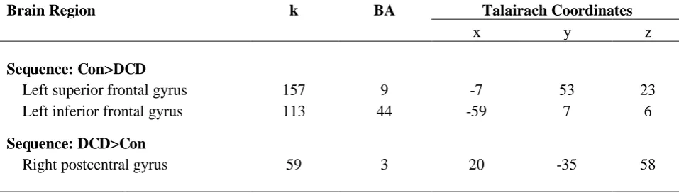
On the finger sequencing task, the control group displayed greater activation in the left superior frontal gyrus
(BA9, Fig. 4a) and the left inferior frontal gyrus (BA44, Fig. 4b), while the DCD group displayed increased
activation in the right postcentral gyrus (BA3, Fig. 4c). Coordinates and volumes of significant clusters on the
finger sequencing task are presented in Table 1. There were no significant activation differences between groups
in the hand clenching condition.
INSERT TABLE 1 ABOUT HERE
INSERT FIGURE 4 ABOUT HERE
Discussion
contribute to poor execution of movement tasks and the increased prevalence of motor overflow. The tasks
included in this study were both complex, one requiring individualized synchronization of each digit and the
other precise movements of all four digits. Even though there were visual prompts on screen to assist with the
timing of tasks, participants were unable to see their hand during scanning and had to rely on sensory input from
sources such as proprioception and touch. Of the two tasks, finger sequencing initiated the most contralateral
motor overflow and it was also the task where three distinct areas of activation difference were seen between the
two groups.
A large cluster of deactivation was found in the DCD group in the left superior frontal gyrus (SFG),
encompassing the lateral and superior portion of BA9. Lesion studies have revealed that the left SFG plays an
integral role in working memory, is active during the highest level of executive processing, and is involved in
spatially oriented processing (Du Boisgueheneuc et al. 2006), all key elements necessary for the execution of the
finger sequencing task used in this study. The finding of decreased activation in the left SFG in the DCD group
is consistent with Zwicker et al. (2010), indicating that children with DCD potentially have a deficit in this
region. The findings also provide evidence to support behavioural studies reporting poorer performance on
working memory tasks in this population (Piek et al. 2007).
The SFG forms part of an extensive cortical-subcortical network, the cerebellar-thalamic-prefrontal
network, predominantly involved in higher order functioning. Even though studying activation of subcortical
regions was beyond the scope of our study, it is impossible to rule out that the hypoactivity seen in the SFG was
not related to deficits extending to and from other regions. Deficits in the structure and function of the
cerebellar-thalamic-prefrontal network have been reported in other clinical populations presenting with
abundant motor overflow, in particular in schizophrenia. Studies have revealed that patients with schizophrenia
have reduced volumes in the superior frontal gyrus and cerebellum, and these reduced volumes have been
moderately correlated with abundant neurological soft signs (Mouchet-Mages et al. 2011; Venkatasubramanian
et al. 2008)which typically includes the measurement of motor overflow. According to the cognitive dysmetria
model of schizophrenia, disrupted connectivity is thought to occur between cortical regions and the cerebellum
which has negative consequences for linking perception, retention, retrieval and response (Andreasen et al.
1996). This ultimately influences cognitive systems including memory, attention and execution and their related
sub-processes such as inhibition (Venkatasubramanian et al. 2008). Even though schizophrenia and DCD are
two distinctly different conditions, there are similarities in certain motor symptoms and measures of cognitive
network level. Cerebellar deficits in DCD have long being suspected (Zwicker et al. 2009), and there is some
preliminary evidence to suggest slight morphological differences (Mariën et al. 2010) and underactivation of the
cerebellum (Zwicker et al. 2011), however further research is needed to examine whether this region and its
associated networks play a role in this disorder.
The second area of activation difference seen between the two groups in the present study was in the
posterior part of the left inferior frontal gyrus (IFG), a finding once again consistent with Zwicker et al. (2010).
Some may consider activation in this area to be quite an unusual finding, certainly when this area is normally
associated with speech production. Interestingly, this area is also involved in the imagination, imitation and
observation of complex hand movements, forming part of the mirror neuron system (MNS, Binkofski et al.
1999a, 199b). The MNS is thought to play a key role in our ability to model the behavior and action of others,
the most widely used form of learning. The participants involved in this study were certainly imitating the hand
actions observed during scanning, with the images presented individually within each condition; thus, it is
highly likely that the MNS was active during the performance of the tasks. Even though the participants with
DCD were able to perform the required actions during scanning, the decreased activity seen in the IFG may
reflect a potential deficit in the development and functioning of the MNS. The MNS was recently hypothesized
to be an underlying cause of motor impairments in children with DCD (Werner et al. 2012) with supportive
evidence coming from studies reporting that children with DCD have difficulty learning and performing
complex imitative gestures (Ozbič andFilipčič 2010). These findings however are not consistent with other
studies reporting no such difficulties in gestural and imitative performance (Dewey et al. 2007). Therefore, there
is a need for further research to examine whether children with DCD truly have difficulties learning complex
imitative tasks and, at the cortical level, if there is a deficit in the functioning of parietal-premotor networks
where the MNS is thought to exist.
The final area of activation difference in the present study was in the right postcentral gyrus, with the
DCD group displaying increased activation in this area. This area plays an important role in the ongoing
processing of somatic sensation. Because the children had to rely heavily on sensory information to execute the
sequencing task, it is possible that the increased activation seen in the DCD group may have reflected increased
reliance on this information to execute the task correctly. It may also reflect sensory integration deficits
frequently seen in this population (Elbason et al. 2012). Interestingly, this finding is not consistent with Zwicker
et al. (2010), who found decreased activation in the left postcentral gyrus in children with DCD when
present study, Mostofsky et al.(2006) reported decreased activation of the right postcentral gyrus in children
with attention deficit hyperactivity disorder (ADHD), a population known to display abundant motor overflow
and some overlap with other motor symptoms. The differences found between studies may reflect task exposure,
with children in the present study introduced to the behavioral tasks during their familiarization session two
weeks prior to scanning and having the opportunity to practice at home. This may have facilitated the increased
utilization of this sensory area and enabled children to execute the tasks with greater ease.Other potential
reasons for the conflicting results could simply relate to task differences and the higher demand for sensory
processing in the present study.
While this study has revealed some interesting findings concerning potential cortical deficits in
children with DCD, there were no obvious activation deficits to explain the abundant motor overflow seen. As
mentioned, functioning of the SFG may be implicated based on moderate correlations between this area and
neurological soft signs in previous research, but when comparing it to other studies examining motor overflow,
the findings differ. Mostofsky et al. (2006) reported a smaller extent of activation in the contralateral primary
motor cortex of children with ADHD, which they attributed to insufficient neuronal activity necessary for the
mobilisation of interhemispheric inhibition, a proposed causal mechanism for motor overflow. Interestingly, if
this insufficiency existed, one may expect to see differing activation in the ipsilateral cortex also, but this was
not seen. In the present study, no differences were found in the cortical activation patterns of the contralateral
motor cortex between the two groups. Ipsilateral activation was seen in the premotor and motor areas; however
this did not differ between the two groups. This ipsilateral activation could potentially be related to transcallosal
inhibitory activation, but may also reflect bilateral planning and execution needed for the execution of complex
unimanual tasks.
Even though fMRI certainly has the potential to explore motor symptoms linked to deficits like
inhibition, other techniques may reveal more distinct differences in activation profiles. For instance, transcranial
magnetic stimulation, capable of separating excitatory and inhibitory activation, may provide more detailed
information concerning suspected inhibitory deficits in this population. This, along with further neuroimaging
studies, are certainly an area worth pursuing in the future to help better understand the nature of this complex
movement condition. Limitations of our work include the cortical area studied, with the scan volume not
extending into sub-cortical areas. Further research examining these areas may provide greater insight into
deficits in cognitive systems and their associated neural functions at a network level. In addition, despite
displayed considerable amounts of motor overflow during the hand clenching task. This may have impacted our
ability to clearly identify areas contributing to abundant motor overflow across the two tasks studied. Finally,
selection of children with DCD was based on one of the four diagnostic criteria from the DSM-IV criteria
(American Psychiatric Association, 2000), with poor motor performance (<5th percentile) established using the MABC-2.
The present study has provided valuable confirmatory evidence to previous neuroimaging studies,
adding to the growing body of literature demonstrating differences in neurological functioning in children with
DCD. Specifically, it has provided evidence to support dysfunction in cortical regions associated with working
memory and executive functioning, along with preliminary evidence to support suspected deficits in the MNS.
This study is also one of the first to quantify a neurological symptom concurrently during scanning.
Acknowledgements
The authors would like to thank the radiology staff from Sir Charles Gairdner Hospital and Princess Margaret
hospital involved in this project, and the children and parents for their time and participation. We would also
like to thank Mag Design and Engineering who created the motion sensor glove.
Compliance with Ethical Standards
Funding: This project was funded by a Research Development Award from the University of Western Australia.
Conflict of interest: The authors declare they have no conflict of interest.
Ethical approval: All procedures performed in studies involving human participants were in accordance with
the ethical standards of the Human Research Ethics Committees at the University of Western Australia
(RA/4/1/2572) and Princess Margaret Hospital for Children (1804) and with the 1964 Helsinki declaration and
its later amendments or comparable ethical standards.
Informed consent: Informed consent was obtained from all parents/guardians and verbal assent from individual
References
1. American Psychiatric Association (2000) Diagnostic and Statistical Manual of Mental Disorders: 4th edition, text revision. Washington DC: American Psychiatric Association.
2. American Psychiatric Association (2013) Diagnostic and Statistical Manual of Mental Disorders: 5th edition. Washington DC: American Psychiatric Association.
3. Andreasen NC, O’Leary DS, Cizadlo T, Arndt S, Rezai K, Boles Ponto LL, et al. (1996) Schizophrenia and
cognitive dysmetria: A positron-emission tomography study of dysfunctional prefrontal-thalamic-cerebellar
circuity. Proc Nat Acad Sci 3: 9985-990.
4. Binkofski F, Buccino G, Posse S, Seitz RJ, Rizzolatti G, et al. (1999a) A fronto-parietal circuit for object
manipulation in man: evidence from an fMRI-study. Eur J Neurosci 11: 3276–3286.doi:
10.1046/j.1460-9568.1999.00753.x
5. Binkofski F, Buccino G, Stephan KM, Rizzolatti G, Seitz RJ, et al. (1999b) A parieto-premotor network for
object manipulation: evidence from neuroimaging. Exp Brain Res 128: 210–213.doi:10.1007/s002210050838
6. Blank R, Smits-Engelsman B, Polatajko HJ, Wilson PH (2012) Recommendations on the definition, diagnosis
and intervention of developmental coordination disorder. Dev Med Child Neurol 54:54-93. doi:
10.1111/j.1469-8749.2011.04171.x
7. Cairney J, Veldhuizen S, Szatmari P. (2010). Motor coordination and emotional-behavioral problems in
children. Curr Opin Psychiatr 23: 324-29.doi: 10.1097/YCO.0b013e32833aa0aa.
8. Dewey D, Cantell M, Crawford SG (2007) Motor and gestural performance in children with autism spectrum
disorders, developmental coordination disorder, and/or attention deficit hyperactivity disorder. J Int
Neuropsychol Soc 13: 246-56. doi: 10.10170S1355617707070270
9. Du Boisgueheneuc F, Levy R, Volle E, Seassau M, Duffau H, Kinkingnehun S, et al. (2006) Functions of the
left superior frontal gyrus in humans: A lesion study. Brain 129: 3315-328.DOI:10.1093/brain/awl244
10. Elbasan B, Kayihan H, Dusgun I (2012) Sensory integration and activities of daily living in children with
developmental coordination disorder. Ital J Pediatr 38: 14. doi: 10.1186/1824-7288-38-14.
11. Henderson SE, Sugden DA, Barnett AL (2007) Movement assessment battery for children-2 second edition.
London, UK: The Psychological Corporation.
12. Kuhtz-Buschbeck J, Krumlinde Sundholm L, Eliasson A, Forssberg H (2000) Quantitative assessment of
mirror movements in children and adolescents with hemiplegic cerebral palsy. Dev Med Child Neurol 42:
13. Largo RH, Caflisch JA, Hug F, Muggli K, Molnar AA, Molinari L (2001) Neuromotor development from 5
to 18 years. Part 2: Associated movements. Dev Med Child Neurol 43: 444-53.doi:
10.1111/j.1469-8749.2001.tb00740.x
14. Largo RH, Fischer JE, Rousson V (2003) Neuromotor development from kindergarten age to
adolescence:developmental course and variability. Swiss Med Wkly 133: 193-99.
15. Kashiwagi M, Iwaki S, Narumi Y, Tamai H, Suzuki S (2009) Parietal dysfunction in developmental
coordination dsiroder. NeuroReport 20: 1319-324.
doi: 10.1097/WNR.0b013e32832f4d87
16. Licari M, Larkin D, Miyahara (2006) The influence of developmental coordination disorder and attention
deficits on associated movements in children. Hum Mov Sci 25: 90-99.doi: 10.1016/j.humov.2005.10.012
17. Licari M, Larkin, D. (2008). Increased associated movement: Influence of attention deficits and movement
difficulties. Hum Mov Sci 27: 310-24.doi:10.1016/j.humov.2008.02.013
18. MacNeil LK, Xavier P, Garvey MA, Gilbert DL, Ranta ME, Denckla MB, et al. (2011) Quantifying
excessive mirror overflow in children with attention-deficit/hyperactivity disorder. Neurol 76: 622-28. doi:
10.1212/WNL.0b013e31820c3052
19.Mariën P, Wackenier P, De Surgeloose D, De Deyn PP, Verhoeven J (2010) Developmental coordination
disorder: disruption of the cerebello-cerebral network evidenced by SPECT. Cerebellum 9: 405-10.doi:
10.1007/s12311-010-0177-6
20. Mostofsky SH, Rimrodt SL, Schafer JGB, Boyce A, Goldberg MC, Pekar JJ, et al. (2006) Atypical motor
and sensory cortex activation in attention-deficit/hyperactivity disorder: A functional magnetic resonance
imaging study of simple sequential finger tapping. Biol Psychiat 59: 48-56.doi: 0.1016/j.biopsych.2005.06.011
21. Mouchet-Mages S, Rodrigo S, Cachia A, Mouaffak F, Oliel JP, Meder JF, et al. (2011) Correlations of
cerebello-thalamo cortical-prefrontal structure and neurological soft signs in patients with first-episode
psychosis. Acta Psychiat Scand 123: 451-58. doi: 10.1111/j.1600-0447.2010.01667.x.
22. Oldfield RC (1971) The assessment and analysis of handedness: the Edinburgh inventory. Neuropsychologia
9: 97-113. doi:10.1016/0028-3932(71)90067-4
23. Ozbič M, Filipčič T (2010) Complex imitation of gestures in school-aged children with learning difficulties.
Kinesiology 4: 44-55.
24. Piek JP, Dyck MJ, Francis M, Conwell A. (2007) Working memory, processing speed, and set-shifting in
children with developmental coordination disorder and attention-deficit-hyperactivity disorder. Dev Med Child
25. Querne L, Berquin P, Vernier-Hauvette M-P, Fall S, Deltour L, Meyer M-E, et al. (2008) Dysfunction of the
attentional brain network in children with developmental coordination disorder: A fMRI study. Brain Res 1244:
89-102. doi: 10.1016/j.brainres.2008.07.066.
26. Venkatasubramanian G, Jayakumar PN, Gangadhar BN (2008) Neuroanatomical correlates of neurological
soft signs in antipsychotic-naive schizophrenia. Psychiat Res-Neuroim 164: 215-22.doi:
10.1016/j.pscychresns.2007.12.021.
27. Werner JM, Cermak SA, Aziz Zadeh L (2012) Neural Correlates of Developmental Coordination Disorder:
The Mirror Neuron System Hypothesis in children and adolescents. Behav Brain Sci 2: 258-68.doi:
10.4236/jbbs.2012.22029
28. Wolraich ML. (2013) Vanderbilt ADHD Parent Rating Scale. American Academy of Pediatrics and the
National Initiative for Children’s Healthcare Quality. Cambridge: MA.
29. World Health Organisation (2012) ICD-10 Classifications of Mental and Behavioural Disorder: Clinical
Descriptions and Diagnostic Guidelines. Geneva: World Health Organisation.
30. Zwicker JG, Missiuna C, Boyd LA. (2009) Neural correlates of developmental coordination disorder: a
review of hypotheses. J Child Neurol 24:1273–1281.doi: 10.1177/0883073809333537
31. Zwicker J, Missiuna C, Harris R, Boyd LA (2010) Brain activation of children with developmental
coordination disorder is different than peers. Pediatr 126:e678-86.
doi: 10.1542/peds.2010-0059
32. Zwicker J, Missiuna C, Harris R, Boyd LA (2011) Brain activation associated with motor skill practice in
children with developmental coordination disorder: an fMRI study. Int J Dev Neurosci 29:145-52. doi:
Fig. 1 Images displayed for the finger sequencing (a.) and hand clenching (b.) tasks
Fig. 3 Total mean displacement (motor overflow) on the finger sequencing and hand clenching tasks in the DCD
and Control groups
* p<0.05, ** p<0.001
Fig. 4 Increased activation in the Con>DCD in the left superior frontal gyrus (a.) and left inferior frontal gyrus
(b.), and increased activation in the DCD>Con in the right postcentral gyrus (c.) on the finger sequencing task.
[image:17.595.90.520.552.740.2]