0022-538X/05/$08.00⫹0 doi:10.1128/JVI.79.1.419–427.2005
Mutations in the RNase H Primer Grip Domain of Murine Leukemia
Virus Reverse Transcriptase Decrease Efficiency and
Accuracy of Plus-Strand DNA Transfer
Jean L. Mbisa, Galina N. Nikolenko, and Vinay K. Pathak*
HIV Drug Resistance Program, National Cancer Institute at Frederick, Frederick, MarylandReceived 20 May 2004/Accepted 19 August 2004
The RNase H primer grip of human immunodeficiency virus type 1 (HIV-1) reverse transcriptase (RT) contacts the DNA primer strand and positions the template strand near the RNase H active site, influencing RNase H cleavage efficiency and specificity. Sequence alignments show that 6 of the 11 residues that constitute the RNase H primer grip have functional equivalents in murine leukemia virus (MLV) RT. We previously showed that a Y586F substitution in the MLV RNase H primer grip resulted in a 17-fold increase in substitutions within 18 nucleotides of adenine-thymine tracts, which are associated with a bent DNA confor-mation. To further determine the effects of the MLV RNase H primer grip on replication fidelity and viral
replication, we performed additional mutational analysis. Using either-galactosidase (lacZ) or green
fluo-rescent protein (GFP) reporter genes, we found that S557A, A558V, and Q559L substitutions resulted in statistically significant increases in viral mutation rates, ranging from 2.1- to 3.8-fold. DNA sequencing
analysis of nonfluorescent GFPclones indicated that the mutations in RNase H primer grip significantly
increased the frequency of deletions between the primer-binding site (PBS) and sequences downstream of the PBS. In addition, quantitative real-time PCR analysis of reverse transcription products revealed that the mutant RTs were substantially inefficient in plus-strand DNA transfer relative to the wild-type control. These results indicate that the MLV RNase H primer grip is an important determinant of in vivo fidelity of DNA synthesis and suggest that the mutant RT was unable to copy through the DNA-RNA junction of the minus-strand DNA and the tRNA because of its bent conformation resulting in error-prone plus-minus-strand DNA transfer.
Genetic diversity is a hallmark of retroviral populations re-sulting from a high rate of mutations during viral replication (4, 37, 39). This genetic variation is of clinical significance because it is the basis for antiviral drug resistance and escape from host immune responses readily exhibited by retroviruses such as human immunodeficiency virus type 1 (HIV-1) (12, 21, 25, 27, 35, 36). The rapid evolution of retroviruses is also an impediment to the design of broadly effective vaccines against HIV-1 (11, 40). Although host cell DNA polymerases and RNA polymerase II are involved in the replication of retrovi-rus genomes, error-prone replication by the virally encoded reverse transcriptase (RT) is most likely a major contributor to the high mutation rate of retroviruses (20).
The structure of RT and inherent nature of the reverse transcription process likely play an important role in the low fidelity of RT. Unlike most high-fidelity cellular DNA poly-merases, RT lacks a classical 3⬘-5⬘ exonuclease proofreading activity. Other structural features also known to affect RT fidelity include positioning of the template-primer complex at the polymerase active site that is dictated by contacts with RT residues and the local geometry of the polymerase active site (3, 15, 44). In addition, reverse transcription of the retroviral RNA genome requires two template-switching events, namely, minus-strand DNA transfer and plus-strand DNA transfer. It is hypothesized that in order to accommodate these two essential
events, RT has evolved to possess low template affinity and processivity (39), which can inadvertently result in template-switching mutations (30). Taken together, these factors con-tribute to the error-prone nature of DNA synthesis by RT and to the high mutation rate of retroviruses.
In spite of the potential role of RT structure in the accuracy of DNA synthesis, only a few studies have characterized the structural determinants of RT fidelity in vivo (13, 14, 22–24, 37, 44). These studies have identified the polymerase active site YXDD motif and other domains, such as the deoxynucleoside triphosphate (dNTP)-binding site, to be important in replica-tion fidelity. Mutareplica-tions in the YXDD motif of murine leuke-mia virus (MLV) and human T-cell leukeleuke-mia virus type 1 were associated with changes in fidelity of DNA synthesis (13, 22). Mutational analysis of the dNTP-binding site and flanking res-idues of both MLV and HIV-1 has revealed the importance of the domain in replication fidelity. Mutating the residue that binds the base and ribose moiety of the incoming dNTP F155 to W in MLV and its HIV-1 homolog Y115 to A resulted in significant increases in the in vivo mutation rate, as did muta-tion of MLV dNTP-binding site flanking residue L151 to F (14, 24). In contrast, mutation of HIV-1 dNTP-binding site residue Q151 to N increased the accuracy of DNA synthesis (24). Intriguingly, mutations in the finger and primer grip subdo-mains of HIV-1, such as K65R, D76V, R78A, and W229A, affected the in vivo replication fidelity by increasing the accu-racy of DNA synthesis (24). In addition, HIV-1 RTs resistant to 3⬘-azido-3⬘-deoxythymidine were shown to increase the in vivo mutation rate (23).
Structural determinants of RT fidelity are not restricted to
* Corresponding author. Mailing address: HIV Drug Resistance Program, NCI-Frederick, P.O. Box B, Bldg. 535, Rm. 334, Frederick, MD 21702-1201. Phone: (301) 846-1710. Fax: (301) 846-6013. E-mail: [email protected].
419
on November 8, 2019 by guest
http://jvi.asm.org/
residues close to the polymerase active site. We recently report-ed that the Y586F substitution in the MLV RNase H domain resulted in an approximately fivefold increase in the MLV mutation rate in vivo, which is the highest reported to date (44). Residue Y586 of MLV is equivalent to HIV-1 residue Y501, a constituent of the recently described RNase H primer grip domain, which contacts the DNA primer strand and po-sitions the template strand near the RNase H active site, in-fluencing RNase H cleavage efficiency and specificity (31, 34). Mansky et al. also recently reported that the HIV-1 mutant Y501W results in a 2.7-fold increase in the in vivo mutation rate (24). Sequence alignments indicate that six out of 11 HIV-1 RNase H primer grip residues have functional equivalents in the MLV RNase H domain (34). In the present study, muta-tional analysis of several residues in the MLV RNase H primer grip was carried out to further determine the role of this domain in MLV replication fidelity in vivo. The results show that mutation of certain residues in this domain result in an increased frequency of deletions between the primer-binding site (PBS), and sequences downstream, indicating an error-prone plus-strand DNA transfer.
MATERIALS AND METHODS
Plasmids, retrovirus vectors, and mutagenesis.Plasmids pLGPS and pRMBNB express MLVgagandpolgenes from a truncated long terminal repeat (⌬LTR) promoter (14, 26). Plasmid pSV␣3.6 encodes the ␣ subunit of the murine Na⫹,K⫹-ATPase gene and confers resistance to ouabain (19). Plasmid pGN-MLV-GFFP-IHy is a derivative of pES-GFFP (38) in which the NotI-NgoMIV fragment containing the GFFP-IRES-neocassette was replaced by the PpuMI-BglII fragment from pKD-HIV-GFFP-IHy (29) containing the cytomegalovirus (CMV)-GFFP-IRES-hygrocassette. Plasmid pHCMV-G expresses the G enve-lope protein of vesicular stomatitis virus from a CMV promoter (43). The S557A, A558V, Q559L, Y586A, and T590A substitution mutations in the MLV RNase H primer grip domain of pRMBNB were generated by using a QuikChange site-directed mutagenesis kit (Stratagene). By using an alanine-scanning mutagenesis strategy, we substituted most of the residues with alanines, with the exception of A558 and Q559. The A558 residue, which was already an alanine was substituted with a valine, and Q559 was substituted with a leucine because of ease of primer design. The presence of the desired mutations and the absence of other mutations were verified by restriction enzyme digestions, followed by DNA sequencing.
Cells, transfections, and infections.D17 dog osteosarcoma cells and 293T cells were obtained from the American Type Culture Collection. The D17-based cell lines ANGIE P or A3GFP11, which contain a single GA-1 or MP-1 provirus, respectively, also express an amphotropic MLV envelope (13, 44). The GN-MLV-GFFP cell line contains a single MLV-based provirus derived from the vector pGN-MLV-GFFP-IHy. To construct the cell line, 293T cells were cotrans-fected with vector pGN-MLV-GFFP-IHy, helper construct pLGPS, and enve-lope construct pHCMV-G. The resulting pseudotyped virus was used to infect 293T cells. After hygromycin selection, several nonfluorescent cell clones (veri-fied by flow cytometry) were isolated, expanded, and characterized by Southern blot analysis and infection assays. A cell clone containing one full-length provirus and producing the best virus titer was named GN-MLV-GFFP and used as a virus producer cell line. Cells were grown in Dulbecco modified Eagle medium (HyClone Laboratories, Inc.) supplemented with 6% calf serum (D17 cells) or 10% fetal calf serum (293T cells), 50 U of penicillin (Gibco)/ml, and 50g of streptomycin (Gibco)/ml. Transfection of ANGIE P and A3GFP11 cells was performed by using the dimethyl sulfoxide-Polybrene method (18). Transfection of 293T cells was carried out by using calcium-phosphate precipitation (33) (CalPhos Transfection Kit; Clontech). Infections were performed in the presence of Polybrene (50g/ml).
In vivo single-replication-cycle fidelity assay. ANGIE P or A3GFP11 cells were plated at a density of 2⫻105
cells per 60-mm-diameter dish and 24 h later were cotransfected with either wild-type or mutant pRMBNB and pSV␣3.6. The transfected cells were selected for resistance to ouabain (10⫺7
M), and the resistant colonies were pooled and expanded. The culture medium from pooled cells containing either GA-1 or MP-1 virus was used to infect D17 target cells plated at a density of 2⫻105cells per 60-mm-diameter dish as previously
described (13). After selection for resistance to G418, GA-1-infected D17 cells were stained with X-Gal (5-bromo-4-chloro-3-indolyl--D-galactopyranoside), andlacZinactivation was determined by counting blue and white colonies, whereas MP-1-infected cells were observed under a fluorescence microscope to determine the frequency ofGFPinactivation (8, 13, 44).
Isolation of single nonfluorescent cell clones by FACS.G418-resistant colonies from MP-1 virus-infected cells were pooled, passaged, and subjected to fluores-cence-activated cell sorting (FACS) (CloneCyt Plus System, FACSVantage SE; BD Biosciences) to isolate individual nonfluorescent clones that did not express functionalGFP. The clones were sorted into 96-well plates, and the nonfluores-cent phenotype was subsequently verified by fluorescence microscopy.
Isolation of genomic DNA, PCR, and sequence analysis.After FACS, single nonfluorescent cell clones grown in 96-well plates were expanded into 24-well plates and then into 60-mm-diameter dishes. The cell clones were harvested and lysed to isolate genomic DNA by using an Bio-Rad AquaPure Genomic DNA Isolation Kit (Bio-Rad). Provirus-specific DNA fragments were amplified by PCR by using Takara Hot-StartTaqDNA polymerase (Takara Mirus Bio., Inc.) and the following sets of forward and reverse primers encompassing the region between the 5⬘LTR and internal ribosome entry site (IRES) of MP-1 provirus: MP-1623F (5⬘-T CACTCCTTCTCTAGGCGCCGGAATTGG-3⬘) and MP-2390R (5⬘-GGAATTG GCCGCTCACTTGTACAGCTCG-3⬘), U3-5538F (5⬘-CCAATCAGTTCGCTTC TCGCT-3⬘) and MP-2390R, or U3-5538F and MP-2938R (5⬘-GTTCAATCATGC GAAACGATCC-3⬘).
Quantitative real-time PCR analysis.For real-time PCR analysis, we used virus produced by transfection of the GN-MLV-GFFP cell line because it pro-duced high-titer virus. GN-MLV-GFFP cells were cotransfected with either wild-type or mutant pRMBNB and pHCMV-G. Medium was changed 24 h posttrans-fection, and virus was collected 24 h later. The virus was used to infect 293T target cells for 1, 3, and 6 h after which cells were washed once with PBS. For harvesting cells at 3 and 6 h postinfection, fresh medium was added to the cells at 1 h postinfection. Total cellular DNA was extracted from infected cells by using the QIAmp DNA Blood Minikit (Qiagen). DNA from⬃105cells was used for each real-time PCR assay with an ABI Prism 7700 sequence detector (Applied Biosys-tems). The primer and probe sets for the RU5,hygro, and U5-⌿regions and the PCR conditions used were as previously described (10, 42). We used a primer and probe set designed to detect the human porphobilinogen deaminase gene to normalize for the amount of DNA analyzed in the real-time PCR experiments (41). Threefold dilutions of the MLV-based vector pMMQD3 (6) were used to generate a standard curve ranging from 17 to 1,000,000 copies of DNA per PCR assay. The same dilutions were used to generate a standard curve for each primer and probe set, which allowed accurate measurement of relative amounts of DNA products detected by different sets. The amount of each PCR product was determined from a standard curve generated with that particular primer and probe set. The correlation coefficient for all standard curves was⬎0.99.
To normalize for the efficiency of initiation of viral DNA synthesis by the different viruses, we quantified the amount of viral RNA from the viral prepa-rations that were used for infection by RT-PCR, as previously described (10).
RESULTS
Effect of mutating MLV RNase H primer grip residues on
RT replication fidelity.We previously showed that mutating
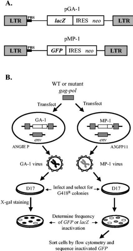
the MLV RNase H primer grip domain residue Y586 to F significantly decreased replication fidelity (44). To further de-termine the role of this domain on replication fidelity, we generated the following mutations: S557A, A558V, Q559L, Y586A, and T590A, encompassing five of the six residues that constitute the domain. The effect of these mutations on the in vivo MLV mutation rate was individually assessed and com-pared to that of wild-type RT by using a previously described single-round replication assay in which thelacZgene served as a mutation reporter gene (Fig. 1). Briefly, GA-1 is an MLV-based vector that expresseslacZandneofrom the viral LTR promoter; theneo open reading frame is translated from an IRES. ANGIE P cells express the amphotropic MLV envelope and contain a single GA-1 provirus. Plasmid pSV␣3.6 confers resistance to ouabain. The ANGIE P cells were cotransfected with either wild-type or mutantgag-polconstructs and pSV␣3.6.
on November 8, 2019 by guest
http://jvi.asm.org/
After selection with ouabain, virus-producing cells were pooled and the virus produced from the pooled cells was used to infect target D17 cells. The infected cells were selected for resistance to G418, a neomycin analog, and the resulting colonies were stained with X-Gal. The frequency oflacZinactivation was then deter-mined by counting white and blue colonies.
Viruses containing wild-type RT inactivated lacZat a fre-quency of 5.5% ⫾ 0.5% (Table 1), which is comparable to
results obtained previously (13, 14, 44). In contrast, three of the five mutants (S557A, A558V, and Q559L) significantly increased the mutation rate by 2.1- to 3.8-fold (P ⬍ 0.003; Student’sttest) relative to wild-type RT. The two mutants with the largest effect on fidelity, S557A and A558V, also dramat-ically reduced virus titers by ca. 100- and 500-fold, respectively, compared to wild-type RT (6.5⫻ 103 CFU/ml). In contrast,
Q559L only reduced virus titers by twofold. Of the remaining two mutants, T590A did not significantly change the frequency oflacZinactivation or the virus titer (P⬎0.1; Studentttest), whereas the effect on replication fidelity by Y586A, a noncon-servative mutation compared to the previously described Y586F, could not be determined because it reduced virus titers by⬎10,000-fold compared to wild-type RT. Thus, the majority of residues within the MLV RNase H primer grip reduced replication fidelity. Using a previously described assay (13), we also determined the RT activities of the S557A, Q559L, and T590A mutant RTs compared to wild-type RT. We found that the S557A mutation had the largest effect on RT activity (21% of wild-type), whereas Q559L and T590A had RT activities of 69 and 56%, respectively, compared to wild-type RT. The RT activities were normalized to the amount of capsid protein quantified by Western blotting (data not shown). That the RT activities were within fivefold of the wild type and that wild-type levels of processed capsid proteins were present on Western blots indicated that the mutations did not significantly influence proteolytic processing. Thus, virus titers of mutants were reduced to a greater extent than RT activities, suggesting that other steps in reverse transcription were also affected by these mutations.
Analysis of mutations generated by the S557A mutant with
GFPas a reporter gene.To analyze the nature of replication
[image:3.585.54.267.69.471.2]errors made by mutant RTs, we characterized the mutations generated by S557A because it had one of the highest mutation
FIG. 1. Single-replication-cycle in vivo fidelity assay. (A) Structures of MLV-based vectors pGA-1 and pMP-1. The schematic shows the relative positions of the LTRs, PBS, and reporter geneslacZandGFP. The neomycin resistance gene (neo) is expressed from an IRES. (B) Experimental protocols. Wild-type (WT) or mutant MLVgag-pol
constructs were cotransfected separately with pSV␣3.6 into ANGIE P or A3GFP11 cell lines. Both cell lines stably express an amphotropic MLV envelope (env) and an integrated MLV-based provirus express-ing eitherlacZ(GA-1 provirus) orGFP(MP-1 provirus), respectively. The GA-1 or MP-1 viruses produced were used to infect D17 target cells. G418-resistant (G418R) infected cell clones were quantified to determine the frequency of inactivation of thelacZorGFPgenes. In addition,GFPvirus-infected cell clones were pooled, individual non-fluorescent cells were isolated by flow cytometry, and the nature of the
[image:3.585.302.541.481.588.2]GFP-inactivating mutations was characterized by PCR amplification, followed by DNA sequencing.
TABLE 1. Effect of mutating MLV RNase H primer grip residues on RT replication fidelity
MLV RT genotype
Virus titer (CFU/ml)a
No. of white colonies/
total no. of coloniesb
Frequency (%) oflacZ
inactivation⫾
SEMc
Relative change in inactivation
oflacZd
Wild type (6.5⫾3.8)⫻103 59/1,109 5.5⫾0.5 1.0
S557A (6.4⫾2.6)⫻101 635/3,643 18.4⫾2.4e 3.3
A558V (1.4⫾0.1)⫻101 76/337 21.1⫾2.0e 3.8
Q559L (3.5⫾1.1)⫻103 281/2,394 11.7⫾0.2e 2.1
Y586A ⬍1 NCf NAg NA
Y586Fh (3.7⫾0.6)⫻102 582/2,203 26.4⫾3 5.4
T590A (9.0⫾5.9)⫻103 50/719 6.7⫾0.5 1.2
aThe average virus titers⫾the standard errors of the mean were determined by serial dilutions and infections, followed by counting the G418Rcolonies.
bThat is, the number of mutant colonies that displayed a white-colony phe-notype and the total number of colonies counted in two to three independent experiments.
cThe frequency oflacZinactivation was calculated as follows: (number of white colonies in each experiment/total number of colonies)⫻100.
dThe relative change in inactivation oflacZgene was calculated as follows: the frequency oflacZinactivation observed with mutant MLV RT/the frequency of
lacZinactivation observed with wild-type MLV RT.
eThe frequency oflacZinactivation was significantly higher than for the wild type (P⬍0.003; Studentttest).
fNo colonies were present after G418 selection, signifying a⬎104-fold reduc-tion in titer.
gNA, not applicable.
hData were from reference 44 [the wild-type virus titer was (9.9⫾1.6)⫻103, and the mutant frequency was 4.9%⫾0.2%].
on November 8, 2019 by guest
http://jvi.asm.org/
rates and a sufficiently high virus titer to permit the experiment to be performed. We usedGFPas a reporter gene because it is smaller than thelacZgene (and therefore amenable to PCR amplification) and because it affords easier isolation of cells expressing inactivatedGFPgenes by FACS. MP-1 is an MLV-based vector that expressesGFPandneofrom the LTR pro-moter; theneoopen reading frame is translated from an IRES. A3GFP11 is a cell line that expresses the amphotropic MLV envelope and contains a single MP-1 provirus (44). The wild-type or S557Agag-pol-expressing construct was cotransfected with pSV␣3.6 into the A3GFP11 cell line (Fig. 1). The virus produced was then used to infect D17 target cells, and G418-resistant colonies were selected. The frequency ofGFP inactiva-tion was determined by examining the G418-resistant colonies by fluorescence microscopy. The frequency of GFPinactivation by wild-type RT was 0.87%⫾0.31%, which is comparable to the results obtained previously (44). In contrast, S557A mutant RT exhibited a 3.4-fold higher frequency ofGFPinactivation, which was not statistically different from that obtained by using
lacZas a reporter gene (3.3-fold higher; Table 1).
To determine the nature of the mutations introduced into
GFPby the wild-type or S557A mutant RT, individual non-fluorescent clones were isolated by FACS, the mutatedGFP
sequences were amplified by PCR, and their DNA sequences were determined as previously described (44). However, in the present study we amplified not only theGFPgene but also the region between the 5⬘LTR andneogene of the MP-1 provirus, which includes theGFPopen reading frame, with different sets of primers. This strategy enabled us to amplify provirus-spe-cific DNA from the majority of nonfluorescent clones because the minimal neomycin-resistant integrated provirus would re-quire both the promoter in the 5⬘LTR andneoexpression. To rule out disproportionate clonal expansion, clones from the same infection possessing the same inactivating mutations were counted only once even though this strategy could have underestimated potential mutational hotspots. Taken together, this approach provided an accurate representation of the
spec-trum ofGFP-inactivating mutations introduced by wild-type and mutant RTs.
S557A mutant is associated with an increase in deletion
mutations between PBS andGFPor IRES.DNA sequencing
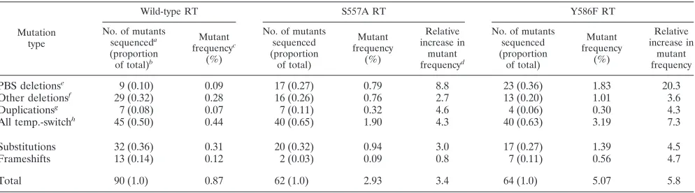
of amplified proviral DNA from nonfluorescent clones showed that both S557A and wild-type RTs introduced a wide range of mutations including deletions, duplications, substitutions, and frameshifts (Table 2). The relative changes in mutant frequen-cies for frameshift and substitution mutations introduced by the S557A RT compared to wild-type RT were modest (0.8-and 3.0-fold, respectively). In contrast, mutations associated with RT template-switching events (deletions and duplica-tions) accounted for the largest increase in mutant frequency by the S557A mutant RT relative to wild-type RT (1.90% versus 0.44%, representing a 4.3-fold increase). Of the tem-plate-switching mutations introduced by the S557A mutant, the highest increase in relative mutant frequency was for de-letions, which ranged from 963 to 2,222 bp in length, between the PBS and sequences inGFPor IRES (an 8.8-fold increase relative to wild-type RT), which was a much higher increase than for the other classes of mutations that had only 0.8- to 4.6-fold changes in the relative mutant frequency.
Sequencing data showed that, although the 5⬘-end junctions of the PBS deletions contained various sequences, they com-prised sequences within a 24-nt region ranging from a few nucleotides upstream of the start of the PBS up to and includ-ing the last nucleotide of the 18-nucleotide (nt) PBS (Fig. 2). This result indicated that the PBS region is a hotspot for template-switching mutations associated with the S557A mu-tant RT and that incomplete copying of the PBS followed by an error-prone plus-strand transfer could be involved in generat-ing the mutations. In contrast, even though the 3⬘-end junc-tions of the delejunc-tions also varied, they contained sequences from a region more than 1,300 bp in length, ranging from the start of theGFPopen reading frame to the end of the IRES, which suggested that no specific acceptor sequences were re-quired for the template switch. Interestingly, 35% of the
dele-TABLE 2. Spectrum ofGFP-inactivating mutations associated with wild-type, S557A, and Y586F RTs
Mutation type
Wild-type RT S557A RT Y586F RT
No. of mutants sequenceda (proportion of total)b
Mutant frequencyc
(%)
No. of mutants sequenced (proportion
of total)
Mutant frequency
(%)
Relative increase in
mutant frequencyd
No. of mutants sequenced (proportion
of total)
Mutant frequency
(%)
Relative increase in
mutant frequency
PBS deletionse
9 (0.10) 0.09 17 (0.27) 0.79 8.8 23 (0.36) 1.83 20.3
Other deletionsf 29 (0.32) 0.28 16 (0.26) 0.76 2.7 13 (0.20) 1.01 3.6
Duplicationsg 7 (0.08) 0.07 7 (0.11) 0.32 4.6 4 (0.06) 0.30 4.3
All temp.-switchh 45 (0.50) 0.44 40 (0.65) 1.90 4.3 40 (0.63) 3.19 7.3
Substitutions 32 (0.36) 0.31 20 (0.32) 0.94 3.0 17 (0.27) 1.39 4.5
Frameshifts 13 (0.14) 0.12 2 (0.03) 0.09 0.8 7 (0.11) 0.56 4.7
Total 90 (1.0) 0.87 62 (1.0) 2.93 3.4 64 (1.0) 5.07 5.8
aThat is, the number of mutants containingGFP-inactivating mutations identified by DNA sequencing.
bThat is, the proportion of mutants identified by DNA sequencing containing a specific type ofGFP-inactivating mutation (e.g., the proportion of mutants with PBS deletions for wild-type RT is 9⫼90⫽0.10).
cMutant frequencies were determined by multiplying the proportion of mutants sequenced by the overall mutant frequency (e.g., the mutant frequency for PBS deletions for wild-type RT is 0.10⫻0.87⫽0.09).
dThat is, the fold increase in mutant frequency for mutant RTs relative to the wild-type RT (e.g., the relative increase in the frequency of PBS deletions for S557A is 0.82⫼0.09⫽9.1).
eNonfluorescent clones containing deletion mutations between the PBS andGFPor IRES. fNonfluorescent clones containing deletion mutations other than PBS deletions.
gNonfluorescent clones containing duplicated sequences.
hTotal of all nonfluorescent clones containing template-switching mutations, CPBS deletions, other deletions, and duplications.
on November 8, 2019 by guest
http://jvi.asm.org/
[image:4.585.45.542.80.218.2]tion junctions contained no homology, whereas the rest had a homology of⬍4 nt. Taken together, the sequencing data indi-cated that the increase in the mutation rate associated with the S557A mutant RT was largely due to a unique class of
tem-plate-switching mutations involving deletions between the PBS and sequences downstream that were a result of error-prone plus-strand DNA transfer.
The Y586F mutant is also associated with deletion
muta-tions between PBS andGFPorIRES.Our previous analysis of
GFP-inactivating mutations associated with the Y586F mutant concentrated on mutations introduced within theGFPgene by using a single set of primers at the 5⬘and 3⬘ends of the gene for PCR amplification. This approach could have missed the
GFP-inactivating mutations involving the PBS. To determine whether deletion mutations involving the PBS or other types of mutations are associated with the Y586F mutant, we used the PCR primers in the 5⬘LTR andneoto amplify provirus-specific DNA from Y586F mutant-infected cells. Similar to S557A mutant analysis, this approach resulted in amplification of pro-virus-specific DNA from a majority of Y586F-infected non-fluorescent clones. DNA sequencing of the PCR products also showed that, similar to the S557A mutant, mutations associ-ated with the RT template-switching events accounted for the largest increase in relative mutant frequency relative to wild-type RT (3.19% versus 0.44%, representing a 7.3-fold in-crease). Furthermore, deletions between the PBS andGFPor IRES accounted for the largest increase in mutant frequency among all of the template-switch mutations, representing a 20.3-fold increase in the relative mutant frequency. In contrast, the other classes of mutations had modest increases in relative mutant frequency of 3.6- to 4.7-fold. Examination of the dele-tion juncdele-tions showed that they were similar to those associ-ated with the S557A mutant, suggesting that they were derived by a similar mechanism (Fig. 2).
Both Y586F and S557A increase the frequency of
substitu-tion mutasubstitu-tions near adenine-thymine tracts (A tracts). We
previously showed that the Y586F mutation increases the fre-quency of substitution mutations in regions associated with nucleotide sequences AAAA, TTTT, or AATT, known as A tracts (44). A tracts are known to induce bends in DNA (5). Analysis of substitution mutations introduced by the Y586F mutant in the present study showed that 22 of 32 substitution mutations (69%) were within 18 nt of A tracts (the total in-cludes 17 mutants from Table 2 and 15 mutants from a second separate experiment [data not shown]). The distance between the polymerase active site and the RNase H cleavage site in an HIV-1 crystal structure in complex with a template-primer and dNTP substrate is 18 nt (16). If the substitution mutations were distributed randomly, we would expect 39% to be located within 18 nt of an A tract inGFP(278 of 717 nt); thus, the 69% frequency is significantly higher than that expected by random distribution (P⬍ 0.0003; 2 test) and is similar to the 81%
observed previously (44). Analysis of substitution mutations induced by the S557A mutant showed that 60% of the muta-tions were within 18 nt of an A tract (12 of 20 mutamuta-tions), which is also significantly higher than that expected by random distribution (P⫽0.05;2test). In contrast, the proportion of
substitution mutations associated with A tracts with wild-type RT was 50% (16 of 32 mutations), which is not statistically different from that expected by random distribution (P⫽0.2;
2test). Therefore, these results indicate that the Y586F and
S557A mutants increased the frequency of substitution muta-tions near A tracts.
FIG. 2. Sequence analysis of deletion mutations between the PBS and GFP or IRES. The complete wild-type (WT) PBS sequence (shaded nucleotides) with flanking sequences is shown compared to the deletion junctions of the nine wild-type, 17 S557A, and 23 Y586F nonfluorescent clones with deletions between the PBS andGFP or IRES. The number of deleted nucleotides for each clone is indicated in the last column. Short direct repeats at the deletion junctions are shown in boldface. Nucleotide numbers above the sequences refer to the number beginning at the start of the 5⬘LTR. Seven clones with an insertion from a different part of the provirus between the junctions are shown with an asterisk after the clone number. Clone numbers were designated by the number of the infection plate followed by the number of the clone.
on November 8, 2019 by guest
http://jvi.asm.org/
Quantitative real-time PCR analysis of viral DNA synthesis
by S557A and Y586F mutant viruses.We hypothesized that the
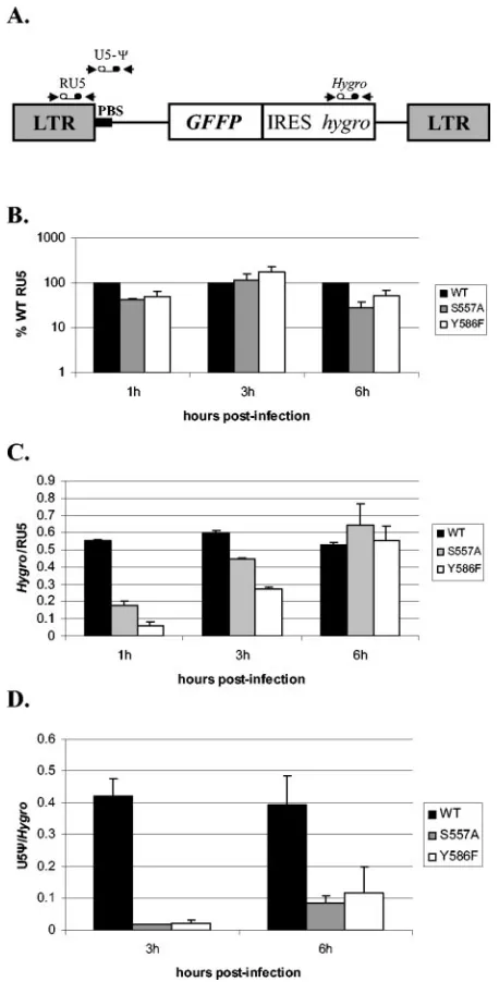
incomplete copying of the PBS by both S557A and Y586F mutant RTs was due to an inability of the mutant RTs to copy through the DNA-RNA junction of the minus-strand DNA and the tRNA because of its bent conformation (32). We postulated that the incomplete copying was followed by an error-prone plus-strand DNA transfer, resulting in deletions between the PBS andGFPor IRES. To test this hypothesis, we analyzed the effects of RNase H primer grip mutations on initiation of viral DNA synthesis. This step involves the RNase H primer grip passing through a RNA-DNA junction. In con-trast to the junction encountered during plus-strand strong-stop DNA synthesis, this junction is part of the primer strand and is formed during the initiation process by the extension of the tRNA primer. However, the RNase H primer grip has to pass through it because it lags behind the polymerase active site. In addition, we also investigated the effects of RNase H primer grip mutations on plus- and minus-strand DNA transfer steps. Wild-type or mutantgag-pol-expressing constructs were cotransfected with the envelope plasmid pHCMV-G into GN-MLV-GFFP cells, which contain an integrated copy of an MLV-based provirus. Virus was collected 48 h after transfec-tion and used to infect target 293T cells. We monitored initi-ation of viral DNA synthesis and DNA strand-transfer events by analyzing products of reverse transcription at 1, 3, and 6 h postinfection by quantitative real-time PCR assay. Initiation of DNA synthesis was observed by analyzing early reverse tran-scription products with an RU5 primer and probe set (Fig. 3A). RNA was isolated from a fraction of the viral preparation used for infection, quantified by RT-PCR, and used to nor-malize the amount of RU5 product. The amount of RU5 product from cells infected with mutant virus was then com-pared to the amount from cells infected with wild-type virus (set at 100%) (Fig. 3B). This analysis showed that initiation of DNA synthesis was not significantly affected by the S557A and Y586F mutations.
Primer and probe sets in thehygroand the U5-⌿region were used to detect products after minus- and plus-strand DNA transfer, respectively (Fig. 3A). To measure minus-strand DNA transfer efficiency, the amount ofhygroDNA synthesized was compared to the amount of RU5 DNA (Fig. 3C). To measure plus-strand DNA transfer efficiency, the amount of U5-⌿ DNA synthesized was compared to the amount ofhygroDNA (Fig. 3D). We measured strand transfer efficiency up to 6 h postinfection because our previous results have indicated that the majority of reverse transcription events occur within the first 6 h (42). The analysis showed that minus-strand DNA transfer was less efficient at 1 and 3 h postinfection for both S557A and Y586F mutant viruses by as much as three- and ninefold, respectively, relative to wild-type virus, but was sim-ilar for all three viruses at 6 h postinfection. This result indi-cates that minus-strand DNA transfer occurred slowly by the mutant viruses but was as efficient as that by wild-type virus later during infection. In contrast, plus-strand DNA transfer was significantly less efficient for both S557A and Y586F by 22-and 18-fold at 3 h postinfection 22-and by five- 22-and threefold at 6 h postinfection, respectively. The significant deficiency in plus-strand DNA transfer by the mutant viruses by using the quantitative real-time PCR assay was consistent with the
mu-FIG. 3. Effect of S557A and Y586F mutations on viral DNA syn-thesis. (A) Locations of real-time PCR primer-probe sets used are shown above the structure of the MLV-GFFP vector. (B) Initiation of viral DNA synthesis by mutant viruses was determined by analyzing the amount of RU5 product at 1, 3, and 6 h postinfection relative to wild-type virus (set at 100%). 293T target cells were infected by virus produced by transfection of the GN-MLV-GFFP helper cell line. The amount of RU5 product was normalized to the amount of viral RNA in each viral preparation used for infection, which was determined by RT-PCR. Two independent experiments were analyzed, and each DNA sample was analyzed by real-time PCR twice. (C) Efficiency of minus-strand DNA transfer by mutant viruses was determined by an-alyzing the amount ofhygrogene product relative to RU5 product at 1, 3, and 6 h postinfection for each virus relative to wild-type virus. The experiments were carried out as described in panel B. (D) The effi-ciency of plus-strand DNA transfer by mutant viruses was determined by analyzing the amount of U5-⌿product relative tohygrogene prod-uct at 3 and 6 h postinfection for each virus relative to wild-type virus. The experiments were carried out as described in panel B. The error bars in all of the graphs represent standard error of the mean.
on November 8, 2019 by guest
http://jvi.asm.org/
[image:6.585.307.536.79.530.2]tation analysis data showing a defect in plus-strand DNA trans-fer by both S557A and Y586F mutants.
DISCUSSION
The data presented in this study confirm and extend our previous finding that the MLV RNase H primer grip domain plays a significant role in viral replication fidelity (44). Using
lacZorGFPas the reporter gene, we found that mutation of three of the six residues that constitute the domain resulted in 2.1- to 3.8-fold increases in the in vivo mutation rates. Al-though most of the mutations in the RNase H primer grip resulting in an increase in the mutation rate also showed sig-nificant decreases in virus titers, we do not believe that the former is a consequence of the latter. We have previously reported mutations in RT that significantly reduced virus titers but did not affect mutation rates (14). DNA sequencing anal-ysis ofGFP-inactivating mutations introduced by the mutant RTs showed that the increases in the mutation rate were pri-marily due to an increase in the frequency of deletions between the PBS andGFPor IRES. In the present study, we amplified provirus-specific DNA from nonfluorescent clones by using primers between the 5⬘LTR andneo. Because expression of genes from the provirus would require the 5⬘LTR promoter and expression ofneoto overcome G418 selection, this strategy enabled DNA amplification from the smallest possible neomy-cin-resistant integrated provirus and resulted in amplification of DNA from⬎90% of nonfluorescent clones. Thus, this ap-proach provided an accurate representation of the spectrum of
GFP-inactivating mutations introduced by wild-type and mu-tant RTs.
We observed that the increase in the frequency of deletions between the PBS andGFPor IRES in mutant RT viruses was the result of an error-prone plus-strand DNA transfer that is often preceded by an incomplete copying of the tRNA se-quences that anneal to the PBS (see proposed model, Fig. 4). The incomplete copying of the tRNA primer sequences could be due to the inability of the RT to copy through the DNA-RNA junction of the minus-strand DNA and the tDNA-RNA be-cause of its bent conformation (32). The premature termina-tion may occur after copying through the DNA-RNA junctermina-tion because the RNase H primer grip lags behind the polymerase active site and may be unable to progress through the bend in the duplex at the DNA-RNA junction. The global structure of the duplex formed by the chimeric DNA-tRNA strand and the cDNA strand of both MLV and HIV-1 has been shown to be significantly distorted (9, 32). The duplex assumes an H-form structure at the DNA:RNA hybrid portion and a B-form struc-ture at the DNA:DNA end, resulting in structural discontinuity at the junction. In the MLV duplex, this discontinuity causes a change in the direction of the helix with a bend of 18⫾3°, a large negative buckle at the junction base-pair step T5䡠 A14-T6䡠a13, and a gradual increase in the minor groove width from the DNA section toward the hybrid section (32). Plus-strand DNA transfer is then error-prone, possibly due to a decrease in the length of homology between the plus-strand strong-stop DNA and the complementary minus-strand DNA region or due to weaker interactions between the mutant RT and the template-primer. Although our proposed model de-picts minus-strand DNA transfer to occur to the copackaged
RNA that has not initiated DNA synthesis, it is possible that minus-strand DNA transfer could occur intramolecularly or to a copackaged RNA that has initiated DNA synthesis. In this case, minus-strand DNA synthesis could continue not by pair-ing RU5 sequences but by pairpair-ing the complete or incomplete PBS sequence at the deletion junction with the PBS sequence in the minus-strand DNA. Alternatively, the deletions could form as a result of incomplete minus-strand DNA synthesis (for ex-ample, due to slow processivity of mutant RTs), forcing the plus-strand strong-stop DNA to anneal to the 3⬘ end of the prematurely terminated minus-strand DNA resulting in dele-tion of the region between the PBS and the terminadele-tion point of minus-strand DNA synthesis (Fig. 4). Both mechanisms would lead to errors occurring during plus-strand DNA transfer.
Our previous finding showed an increased frequency of sub-stitution mutations by the Y586F mutant near A tracts (44). Similar to DNA-RNA junctions, A tracts are associated with a bent DNA conformation at their junction with a G/C base pair, which results in a narrowed minor groove (5). Analysis of sub-stitution mutations associated with the Y586F and S557A mu-tants in the present study also showed an increased frequency of substitution mutations within 18 nt of A tracts relative to wild-type RT. This result indicates that MLV RNase H primer grip mutants are unable to induce a proper conformation of the template-primer duplex at the polymerase active site when a bent conformation is present, resulting in error-prone repli-cation.
Our hypothesis that error-prone plus-strand DNA transfer is responsible for the increase in deletion mutations between the PBS andGFP or IRES was tested further by comparing the efficiency of plus-strand DNA transfer by the mutant and wild-type RTs, by using a quantitative real-time PCR assay. This analysis showed that the efficiency of accurate plus-strand DNA transfer was significantly decreased in mutant RT viruses relative to wild-type virus, which is consistent with the observed increase in deletions. In contrast, the efficiency of minus-strand DNA transfer, although diminished in mutant viruses at early time points, was similar to that of wild-type virus by 6 h post-infection. It is possible that the longer region of homology in minus-strand stop DNA compared to plus-strand strong-stop DNA makes minus-strand DNA transfer more efficient than plus-strand DNA transfer in mutant viruses despite the weaker RT and template-primer interactions.
RT encounters another RNA-DNA junction during initia-tion of viral DNA synthesis, which is formed by the tRNA primer and nascent DNA strand. In this case, the hybrid duplex consists of the chimeric tRNA-DNA strand and the template RNA strand. In contrast to the significant effect of RNase H primer grip mutations on plus-strand DNA transfer, their ef-fect on initiation of DNA synthesis was insignificant relative to wild-type virus. One explanation for this difference could be that the RNA-DNA junction is formed during initiation of minus-strand DNA synthesis and becomes part of the primer strand, whereas the DNA-RNA junction during plus-strand strong-stop DNA synthesis is part of the template strand. Al-ternatively, the MLV RNase H primer grip mutants could be better at copying through the DNA-RNA junction when it is in a duplex with an RNA strand rather than a DNA strand. The crystal structure of the HIV-1 hybrid duplex formed by a chi-meric DNA-tRNA strand and template RNA strand has been
on November 8, 2019 by guest
http://jvi.asm.org/
reported to have an A-form structure with minor structural perturbations at the r(cpa)䡠d(TpG) base pair step (28). In-triguingly, HIV-1 RNase H primer grip mutants have been shown to significantly affect initiation of viral DNA synthesis but not plus-strand DNA transfer (17). The fact that HIV-1 RT is a heterodimer, whereas MLV RT is a monomer and that the residues constituting the HIV-1 RNase H primer grip re-side in both the p66 and p51 subunits (7, 34), could influence the nature of RT and template-primer interactions by the two viruses. The MLV RT structure has recently been reported (7), but un-fortunately the RNase H domain is not sufficiently resolved to directly examine whether the contacts between the RNase H primer grip residues and the DNA primer strand are similar to
those observed in HIV-1. The mutations in the RNase H primer grip could also indirectly affect viral DNA synthesis by affecting RNase H activity. However, in vitro studies have shown that mutations at A558 and Q559 of MLV RT do not affect its RNase H activity (1, 2). In contrast, the RNase H activity of the Y586F mutant is reduced by 20-fold. The effect on RNase H activity of mutations at S557 has not been determined.
[image:8.585.58.530.67.444.2]In summary, our findings indicate that the MLV RNase H primer grip domain plays a significant role in dealing with structural constraints in the template-primer complex intro-duced by A tracts or DNA-RNA junctions. This role not only ensures proper positioning of the template-primer at the poly-merase active site, resulting in incorporation of the correct
FIG. 4. Proposed mechanism for the formation of deletions between the PBS andGFPor IRES. Initiation of DNA synthesis is proposed to start from only one of the two copackaged RNA genomes labeled a. Minus-strand DNA transfer then occurs onto the copackaged RNA labeled b, which did not initiate reverse transcription, resulting in minus-strand DNA that has R, U5, and PBS at its 3⬘end. Plus-strand DNA synthesis initiates from the PPT as expected but fails to copy through the DNA-RNA junction of the minus-strand DNA and the tRNA because of its bent conformation, resulting in incomplete copying of the PBS (stripped arrowhead) and error-prone plus-strand DNA transfer into theGFPgene or IRES because of decreased homology and weaker RT-template interactions. The R and U5 regions from the minus-strand DNA then hybridize with the complementary R and U5 regions from the plus-strand strong-stop DNA, forming a loop from the PBS intoGFPor IRES that is excised and repaired by the host DNA repair machinery. The completion of plus-strand DNA synthesis downstream ofGFPor IRES and minus-strand DNA synthesis upstream of the R region results in the formation of a double-stranded DNA with a deletion between the PBS andGFPor IRES that is subsequently integrated into the host genome to form a deleted provirus. Alternatively, error-prone plus-strand DNA transfer could occur because of incomplete minus-strand DNA synthesis, which would also result in deletions between the PBS and the sequences downstream of the premature minus-strand DNA synthesis termination point. RNA molecules are shown in gray, whereas DNA molecules are shown in black.
on November 8, 2019 by guest
http://jvi.asm.org/
nucleotides, but also helps in preventing premature dissocia-tion of the RT from the template-primer duplex, thereby fa-cilitating efficient and accurate DNA synthesis.
ACKNOWLEDGMENTS
We especially thank Wei-Shau Hu for intellectual input throughout the project and Anne Arthur for expert editorial help. We also thank Patricia Henry, Sook-Kyung Lee, and Evguenia Svarovskaia for critical reading of the manuscript.
This study was supported by the HIV Drug Resistance Program, National Cancer Institute.
REFERENCES
1.Blain, S. W., and S. P. Goff.1995. Effects on DNA-synthesis and transloca-tion caused by mutatransloca-tions in the RNase-H domain of Moloney murine leu-kemia virus reverse transcriptase. J. Virol.69:4440–4452.
2.Blain, S. W., and S. P. Goff.1993. Nuclease activities of Moloney murine leukemia virus reverse transcriptase: mutants with altered substrate speci-ficities. J. Biol. Chem.268:23585–23592.
3.Chowdhury, K., N. Kaushik, V. N. Pandey, and M. J. Modak.1996. Eluci-dation of the role of Arg 110 of murine leukemia virus reverse transcriptase in the catalytic mechanism: biochemical characterization of its mutant en-zymes. Biochemistry35:16610–16620.
4.Coffin, J. M.1995. HIV population dynamics in vivo: implications for genetic variation, pathogenesis, and therapy. Science267:483–489.
5.Crothers, D. M., T. E. Haran, and J. G. Nadeau.1990. Intrinsically bent DNA. J. Biol. Chem.265:7093–7096.
6.Dang, Q., and W. S. Hu.2001. Effect of homology length in the repeat region on minus-strand DNA transfer and retroviral replication. J. Virol.75:809–820. 7.Das, D., and M. M. Georgiadis.2004. The crystal structure of the monomeric
reverse transcriptase from Moloney murine leukemia virus. Structure12: 819–829.
8.Delviks, K. A., W. S. Hu, and V. K. Pathak.1997. Psi(⫺) vectors: murine leukemia virus-based self-inactivating and self-activating retroviral vectors. J. Virol.71:6218–6224.
9.Fedoroff, O. Y., M. Salazar, and B. R. Reid.1996. Structural variation among retroviral primer-DNA junctions: solution structure of the HIV-1 negative-strand Okazaki fragment r(gcca)d(CTGC)䡠d(GCAGTGGC). Biochemistry 35:11070–11080.
10.Fu, W., and W. S. Hu.2003. Functional replacement of nucleocapsid flanking regions by heterologous counterparts with divergent primary sequences: effects of chimeric nucleocapsid on the retroviral replication cycle. J. Virol. 77:754–761.
11.Gaschen, B., J. Taylor, K. Yusim, B. Foley, F. Gao, D. Lang, V. Novitsky, B. Haynes, B. H. Hahn, T. Bhattacharya, and B. Korber.2002. AIDS: diversity considerations in HIV-1 vaccine selection. Science296:2354–2360. 12.Goulder, P. J. R., C. Brander, Y. H. Tang, C. Tremblay, R. A. Colbert, M. M.
Addo, E. S. Rosenberg, T. Nguyen, R. Allen, A. Trocha, M. Altfeld, S. Q. He, M. Bunce, R. Funkhouser, S. I. Pelton, S. K. Burchett, K. McIntosh, B. T. M. Korber, and B. D. Walker.2001. Evolution and transmission of stable CTL escape mutations in HIV infection. Nature412:334–338.
13.Halvas, E. K., E. S. Svarovskaia, and V. K. Pathak.2000. Development of an in vivo assay to identify structural determinants in murine leukemia virus reverse transcriptase important for fidelity. J. Virol.74:312–319.
14.Halvas, E. K., E. S. Svarovskaia, and V. K. Pathak.2000. Role of murine leukemia virus reverse transcriptase deoxyribonucleoside triphosphate-bind-ing site in retroviral replication and in vivo fidelity. J. Virol.74:10349–10358. 15.Harris, D., P. N. S. Yadav, and V. N. Pandey.1998. Loss of polymerase activity due to Tyr to Phe substitution in the YMDD motif of human im-munodeficiency virus type-1 reverse transcriptase is compensated by Met to Val substitution within the same motif. Biochemistry37:9630–9640. 16.Huang, H. F., R. Chopra, G. L. Verdine, and S. C. Harrison.1998. Structure
of a covalently trapped catalytic complex of HIV-1 reverse transcriptase: implications for drug resistance. Science282:1669–1675.
17.Julias, J. G., M. J. McWilliams, S. G. Sarafianos, E. Arnold, and S. H. Hughes.2002. Mutations in the RNase H domain of HIV-1 reverse tran-scriptase affect the initiation of DNA synthesis and the specificity of RNase H cleavage in vivo. Proc. Natl. Acad. Sci. USA99:9515–9520.
18.Kawai, S., and M. Nishizawa.1984. New procedure for DNA transfection with polycation and dimethyl sulfoxide. Mol. Cell. Biol.4:1172–1174. 19.Kent, R. B., J. R. Emanuel, Y. Benneriah, R. Levenson, and D. E. Housman.
1987. Ouabain resistance conferred by expression of the cDNA for a murine Na⫹,K⫹-ATPase alpha-subunit. Science237:901–903.
20.Kim, T., R. A. Mudry, Jr., C. A. Rexrode II, and V. K. Pathak.1996. Ret-roviral mutation rates and A-to-G hypermutations during different stages of retroviral replication. J. Virol.70:7594–7602.
21.Malim, M. H., and M. Emerman.2001. HIV-1 sequence variation: drift, shift, and attenuation. Cell104:469–472.
22.Mansky, L. M.2000. In vivo analysis of human T-cell leukemia virus type 1 reverse transcription accuracy. J. Virol.74:9525–9531.
23.Mansky, L. M., and L. C. Bernard.2000. 3⬘-Azido-3⬘-deoxythymidine (AZT) and AZT-resistant reverse transcriptase can increase the in vivo mutation rate of human immunodeficiency virus type 1. J. Virol.74:9532–9539. 24.Mansky, L. M., E. Le Rouzic, S. Benichou, and L. C. Gajary.2003. Influence
of reverse transcriptase variants, drugs, and Vpr on human immunodefi-ciency virus type 1 mutant frequencies. J. Virol.77:2071–2080.
25.Mellors, J. W., H. Z. Bazmi, R. F. Schinazi, B. M. Roy, Y. Hsiou, E. Arnold, J. Weir, and D. L. Mayers.1995. Novel mutations in reverse transcriptase of human immunodeficiency virus type 1 reduce susceptibility to foscarnet in lab-oratory and clinical isolates. Antimicrob. Agents Chemother.39:1087–1092. 26.Miller, A. D., and C. Buttimore.1986. Redesign of retrovirus packaging
cell-lines to avoid recombination leading to helper virus production. Mol. Cell. Biol.6:2895–2902.
27.Moore, C. B., M. John, I. R. James, F. T. Christiansen, C. S. Witt, and S. A. Mallal.2002. Evidence of HIV-1 adaptation to HLA-restricted immune responses at a population level. Science296:1439–1443.
28.Mueller, U., G. Maier, A. M. Onori, L. Cellai, H. Heumann, and U. Heine-mann.1998. Crystal structure of an eight-base pair duplex containing the 3⬘-DNA-RNA-5⬘junction formed during initiation of minus-strand synthesis of HIV replication. Biochemistry37:12005–12011.
29.Nikolenko, G. N., E. S. Svarovskaia, K. A. Delviks, and V. K. Pathak.2004. Antiretroviral drug resistance mutations in human immunodeficiency virus type 1 reverse transcriptase increase template-switching frequency. J. Virol. 78:8761–8770.
30.Pathak, V. K., and H. M. Temin.1990. Broad-spectrum of in vivo forward mutations, hypermutations, and mutational hotspots in a retroviral shuttle vector after a single replication cycle: deletions and deletions with insertions. Proc. Natl. Acad. Sci. USA87:6024–6028.
31.Rausch, J. W., D. Lener, J. T. Miller, J. G. Julias, S. H. Hughes, and S. F. J. Le Grice.2002. Altering the RNase H primer grip of human immunodefi-ciency virus reverse transcriptase modifies cleavage specificity. Biochemistry 41:4856–4865.
32.Salazar, M., O. Y. Fedoroff, and B. R. Reid.1996. Structure of chimeric duplex junctions: solution conformation of the retroviral Okazaki-like frag-ment r(ccca)d(AATGA)䡠d(TCATTTGGG) from Moloney murine leuke-mia virus. Biochemistry35:8126–8135.
33.Sambrook, J., and D. W. Russell.2001. Molecular cloning: a laboratory manual, 3rd ed. vol. 3. Cold Spring Harbor Laboratory Press, Cold Spring Harbor, N.Y.
34.Sarafianos, S. G., K. Das, C. Tantillo, A. D. Clark, J. Ding, J. M. Whitcomb, P. L. Boyer, S. H. Hughes, and E. Arnold.2001. Crystal structure of HIV-1 reverse transcriptase in complex with a polypurine tract RNA:DNA. EMBO J.20:1449–1461.
35.Shankar, P., M. Russo, B. Harnisch, M. Patterson, P. Skolnik, and J. Lieber-man.2000. Impaired function of circulating HIV-specific CD8⫹T cells in chronic human immunodeficiency virus infection. Blood96:3094–3101. 36.Stebbing, J., S. Patterson, and F. Gotch.2003. New insights into the
immu-nology and evolution of HIV. Cell Res.13:1–7.
37.Svarovskaia, E. S., S. R. Cheslock, W. H. Zhang, W. S. Hu, and V. K. Pathak. 2003. Retroviral mutation rates and reverse transcriptase fidelity. Front. Biosci.8:D117–D134.
38.Svarovskaia, E. S., K. A. Delviks, C. K. Hwang, and V. K. Pathak.2000. Structural determinants of murine leukemia virus reverse transcriptase that affect the frequency of template switching. J. Virol.74:7171–7178. 39.Temin, H. M.1993. Retrovirus variation and reverse transcription: abnormal
strand transfers result in retrovirus genetic variation. Proc. Natl. Acad. Sci. USA90:6900–6903.
40.van der Groen, G., P. N. Nyambi, E. Beirnaert, D. Davis, K. Fransen, L. Heyndrickx, P. Ondoa, G. Van der Auwera, and W. Janssens.1998. Genetic variation of HIV type 1: relevance of interclade variation to vaccine devel-opment. AIDS Res. Hum. Retrovir.14(Suppl. 3):S211–S221.
41.Voronin, Y. A., and V. K. Pathak.2004. Frequent dual initiation in human immunodeficiency virus-based vectors containing two primer-binding sites: a quantitative in vivo assay for function of initiation complexes. J. Virol.78: 5402–5413.
42.Voronin, Y. A., and V. K. Pathak.2003. Frequent dual initiation of reverse transcription in murine leukemia virus-based vectors containing two primer-binding sites. Virology312:281–294.
43.Yee, J. K., A. Miyanohara, P. Laporte, K. Bouic, J. C. Burns, and T. Fried-mann.1994. A general method for the generation of high-titer, pantropic retroviral vectors: highly efficient infection of primary hepatocytes. Proc. Natl. Acad. Sci. USA91:9564–9568.
44.Zhang, W. H., E. S. Svarovskaia, R. Barr, and V. K. Pathak.2002. Y586F mutation in murine leukemia virus reverse transcriptase decreases fidelity of DNA synthesis in regions associated with adenine-thymine tracts. Proc. Natl. Acad. Sci. USA99:10090–10095.