RESEARCH
Bead-based assays to simultaneously
detect multiple human inherited blood
disorders associated with malaria
Lynn Grignard
1*, Catherine Mair
1, Jonathan Curry
2, Laleta Mahey
2, Guide J. H. Bastiaens
3, Alfred B. Tiono
4,
Joseph Okebe
5, Sam A. Coulibaly
4, Bronner P. Gonçalves
1, Muna Affara
5, Alphonse Ouédraogo
4,
Edith C. Bougouma
4, Guillaume S. Sanou
4, Issa Nébié
4, Kjerstin H. W. Lanke
3, Sodiomon B. Sirima
4,
Umberto d’Alessandro
5,6, Taane G. Clark
7,8, Susana Campino
7, Teun Bousema
1,3and Chris Drakeley
1Abstract
Background: Glucose-6-phosphate dehydrogenase deficiency (G6PDd), haemoglobin C (HbC) and S (HbS) are
inherited blood disorders (IBD) common in populations in malaria endemic areas. All are associated to some degree with protection against clinical malaria whilst additionally G6PDd is associated with haemolysis following treatment with 8-aminoquinolines. Measuring the prevalence of these inherited blood disorders in affected populations can improve understanding of disease epidemiology. Current methodologies in epidemiological studies commonly rely on individual target amplification and visualization; here a method is presented to simultaneously detect the poly-morphisms and that can be expanded to include other single nucleotide polypoly-morphisms (SNPs) of interest.
Methods: Human DNA from whole blood samples was amplified in a novel, multiplex PCR reaction and extended
with SNP-specific probes in an allele specific primer extension (ASPE) to simultaneously detect four epidemiologically
important human markers including G6PD SNPs (G202A and A376G) and common haemoglobin mutations (HbS
and HbC). The products were hybridized to magnetic beads and the median fluorescence intensity (MFI) was read on MAGPIX® (Luminex corp.). Genotyping data was compared to phenotypical data generated by flow cytometry and to established genotyping methods.
Results: Seventy-five samples from Burkina Faso (n = 75/78, 96.2%) and 58 samples from The Gambia (n = 58/61, 95.1%) had a G6PD and a HBB genotype successfully assigned by the bead-based assay. Flow cytometry data available for n = 61 samples further supported the concordance between % G6PD normal/deficient cells and genotype.
Conclusions: The bead based assay compares well to alternative measures of genotyping and phenotyping for
G6PD. The screening is high throughput, adaptable to inclusion of multiple targets of interest and easily standardized.
Keywords: Malaria, Glucose-6-phosphate dehydrogenase deficiency, Haemoglobin S, Haemoglobin C, Multiplex
detection
© The Author(s) 2019. This article is distributed under the terms of the Creative Commons Attribution 4.0 International License (http://creat iveco mmons .org/licen ses/by/4.0/), which permits unrestricted use, distribution, and reproduction in any medium, provided you give appropriate credit to the original author(s) and the source, provide a link to the Creative Commons license, and indicate if changes were made. The Creative Commons Public Domain Dedication waiver (http://creat iveco mmons .org/ publi cdoma in/zero/1.0/) applies to the data made available in this article, unless otherwise stated.
Open Access
*Correspondence: [email protected]
1 Department of Immunology and Infection, London School of Hygiene &
Tropical Medicine, London, UK
Background
Plasmodium parasites have co-evolved with human hosts and exert a considerable evolutionary pressure on muta-tions that confer a degree of protection against malaria
[1–5]. Examples of such mutations include
polymor-phisms in the X-linked glucose-6-phosphate dehydroge-nase (G6PD) gene and in the HBB gene. G6PD is essential for the protection of cells against oxidative damage [6] and G6PD deficiency is common in malaria endemic areas [7] due to its association with protection of some manifestations of severe malaria [8]. The repertoire of G6PD deficiency molecular markers is extensive and displays considerable spatial heterogeneity [7, 9]. One of
the most common G6PD variants in sub-Saharan Africa,
the A− variant, requires two single nucleotide changes
[10]. There are three common A− variants, an A376G
substitution (present in all variants) may occur in com-bination with G202A, T968C or A542T substitutions. In The Gambia for example, the combinations of A376G/ G202A, A376G/T968C and A376G/A542T are present in 1.8%, 2.1% and 1% of the population, respectively, while the A376G/G202A variant and A376G without additional
G6PD mutations are commonly found in Burkina Faso
[11]. G6PD A− homo-/hemizygosity is associated with a
pronounced 88% loss in enzyme activity whilst G6PD A+
is associated with only a mild 15% loss in enzyme activity [12]. The level of enzyme activity has consequences for the tolerability of some anti-malarial drugs; 8-aminoqui-nolines can cause haemolytic anaemia in G6PD deficient individuals [13, 14].
Polymorphisms in the HBB gene, haemoglobin S
(HbS, sickle cell trait) and haemoglobin C (HbC) pro-tect against severe malaria [15]. The SNPs are located on chromosome 11 and result in the substitution of a gluta-mate residue to a valine or lysine residue in the ß-globin chain, respectively. HbAS heterozygote sickle cell trait carriers have a selective advantage in malaria endemic regions as they are protected against severe malaria
syn-dromes [16]. Homozygous HbSS individuals, however,
suffer from a serious blood disorder, sickle cell anaemia. Heterozygous HbAC individuals show some protection against severe malaria whereas homozygotes (HbCC) are strongly protected against malaria and, as opposed to HbSS homozygotes, only show symptoms of mild anae-mia [17, 18]. Polymorphisms in the HBB gene may also increase the likelihood that malaria-infected individuals infect mosquitoes [18, 19].
Considering the implications conferred by human inherited blood disorders (IBD) on malaria disease risk and transmission potential, operationally attrac-tive genotyping tools are of value. Current approaches typically assess a single IBD and whilst basic size elec-trophoresis for HbC/HbS is practical, G6PD can require
multiple PCR’s and visualizations on agarose gel. The assay described here simultaneously assesses four
epi-demiologically important human markers: G6PD G202A
and A376G, HbS and HbC. Outcomes of the
MagPlex-TAG™ microsphere assay on samples from
malaria-exposed populations in Burkina Faso and in The Gambia are compared with established genotyping assays.
Methods
DNA from field samples
Study design and whole blood collection were performed as previously described [20]. Briefly, whole blood was col-lected from males aged 18–45 years in Banfora (Burkina Faso) from August 2014 to November 2015 and from males aged over 10 years in Basse Santa Su, Upper River Region, The Gambia from December 2015 to April 2016. All participants were screened for glucose-6-phosphate
enzyme activity using CareStart™ G6PD rapid
diagnos-tic test (G6PD RDT; Access Bio, Inc. Somerset, USA) and the Fluorescent Spot Test (FST) [21]. Both studies were conducted to investigate the safety of primaquine hence only males who had the G6PD enzyme activity measured by point-of-care tests were recruited. A small subset of samples (n = 6) from female participants was made avail-able from another study in Burkina Faso [22] to test the assay on females.
DNA was extracted from 50 µl whole blood from Bur-kina Faso using the Total NA High Performance kit on a MagNA Pure LC instrument (Roche, Switzerland) and eluted in 50 µl. DNA from The Gambia was extracted from 100 µl cryopreserved blood samples using the DNA Blood Mini kit (Qiagen, Germany) and eluted in 100 µl.
Assay optimizations
The assay required optimization at the PCR level, the allele specific primer extension (ASPE) level and the median fluorescence intensity (MFI) acquisition level. At the PCR level, the primer concentration (100–300 nM each), the annealing temperature (52.6–65.0 °C) and the template input volume (1–5 µl) were adjusted. At the ASPE level, annealing temperature (51.0 – 57.5 °C) and template volume (1–7 µl) were adjusted in addition to the Biotin-14-dCTP concentration (5–15 µM). At the MFI acquisition level the volume of ASPE product hybridized to beads (1–5 µl) and the concentration of SAPE conju-gate (2.5–10 µg/ml) were adjusted. Bovine serum albu-min (0.1%, BSA) was added before reading the plate on the MagPix to decrease buffer background MFI levels (Additional file 1: Table S1).
Genomic PCR amplification
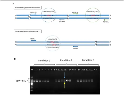
The gene contains multiple exons and the SNPs tested (G202A and A376G) are located on exons 5 and 6, respectively. The HBB gene is located on the short arm of chromosome 11 and the SNPs of interest (HbS and HbC) are located one base pair apart on exon 1 (Fig. 1a). Three fragments of 619 bp, 769 bp and 802 bp were amplified in the multiplex PCR reaction (Fig. 1b).
Five microliter of extracted DNA was amplified in a 50 µl HotStarTaq Mastermix (Qiagen, Germany) reaction
containing 100 nM (each) of G6PD primers and 300 nM
(each) of HBB primers (Table 1). The G6PD primers were
designed using Primer3 software. For the HBB
amplifi-cation, published primers were used [23]. After ampli-fication, primers and nucleotides from the genomic PCR were removed by adding 3 µl ExoSAP-IT reagent (Applied Biosystems, USA) to 7.5 µl of PCR product.
Allele specific primer extension (ASPE)
Five microliter of cleaned PCR product was added to 20 µl ASPE reaction mix containing 250 nM (each) of
a
b
M 1 2 3 4 5 6 N N 9 10 11 12 13 14 N N 17 18 19 20 21 22 N N
Condion 1 Condion 2 Condion 3
550 – 850
Fig. 1 Genomic location and amplification of SNP targets. a Top: Representation of the multi-exon G6PD gene on the X chromosome. The G202A and the A376G markers are amplified by the primer pairs G202A Fw (forward)/G202A Rv (reverse) and A376G Fw (forward) and A376G Rv (reverse). The G202A allele specific primer extension (ASPE) probes (G202 and A202) anneal to the sense strand and either have a G or an A at their 3′ end (see red highlighted letters in brackets) and the A376G probes (A376 and G376) anneal to the sense strand and either have an A or a G at their 3′
[image:3.595.57.539.241.610.2]ASPE probes and 200 µM Biotin-14-dCTP (Invitrogen, UK). The ASPE probes were designed using Primer3 soft-ware and included the allele specific sequence at their 3′
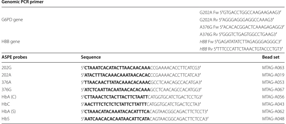
end. The tag sequences and magnetic beads were cho-sen according to the manufacturer’s recommendations (Table 1).
Hybridization and MagPix microsphere multiplex assay Five µl ASPE reaction was hybridized to 2500 beads of
each set, washed twice in 1 × Tm buffer (0.2 M NaCl,
0.1 M Tris, 0.8% TritonX-100, pH 8.0) and incubated for 15 min at 37 °C in 1 × Tm containing 0.1% bovine serum albumin (BSA) and 2.5 µg/ml Streptavidin, R-Phyco-erythrin Conjugate (SAPE, Invitrogen, UK). Fifty µl of the reaction was read at 37 °C on a MagPix (Luminex corp., USA). The G6PD and HBB genotypes were called accord-ing to net MFI of the wild type allele compared to the mutated allele (Additional file 1: Table S3A and B). All field samples were repeated at least once. The assay was optimized on positive control DNA to maximize the fluo-rescent signal and minimize background.
Flow cytometry for percentage (%) G6PD positive cells In The Gambia, red blood cells (RBC) were cryopre-served using established protocols [24, 25] and stored at − 80 °C (n = 61 samples, G6PD status confirmed by CareStart™ G6PD RDT, Additional file 2: Table S7). The samples were thawed and re-suspended in equal volume additive (2.5% glucose, 0.9% sodium chloride, 0.027%
adenine, 0.75% mannitol) before measuring the % G6PD positive cells by a variation of the methaemoglobin test described in detail in [24, 26]. Samples were examined on a FACScailbur cytometer (Benson & Dickenson) (no gating, 10,000 events, in the FL1 channel 533 +− 30 nm). Analysis was conducted on FlowJo 10.1, and the SED (Super-Enhanced Dmax Subtraction Algorithm, FlowJo 10.1, USA) was calculated. RBCs from anonymous blood donors from the London School of Hygiene and Tropical Medicine were used as controls.
Results
Multiplex HBB and G6PD genotyping on field samples
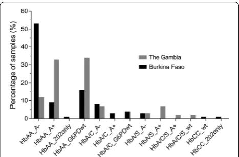
Seventy-five samples from Burkina Faso (n = 75/78,
96.2%) and fifty-eight samples from The Gambia (n = 58/61, 95.1%) had a G6PD and a HBB genotype suc-cessfully assigned by the bead-based assay (Fig. 2). Three samples from Burkina Faso and two samples from The Gambia were excluded due to net MFI values below the arbitrary cut-off of mean MFI plus two standard devia-tions (mean MFI + 2SD) (Additional file 1: Table S2). This cut-off was chosen to unambiguously separate positive fluorescence signal from background signal. One sam-ple from The Gambia failed to amplify in the initial PCR reaction. The G6PD and HBB genotypes of the remaining samples were called according to net MFI of the wild type allele compared to the mutated allele (Additional file 1: Table S3A and B). In Burkina Faso, the majority of
sam-ples (53.3%, 40/75) were HBB wild type and contained
Table 1 Primers and Probes
PCR primer sequences and allele specific primer extension (ASPE) probe details. The top of the table contains the primers for genomic PCR amplification and the bottom part of the table contains the ASPE probes. The probe sequence that is complimentary to the anti-tag sequence coupled to the magnetic beads is highlighted in bold. The bead sets were chosen according to the Tag-It® Oligo Design Software v.3.00 (courtesy of Luminex corp., USA)
Genomic PCR primer
G202A Fw 5′GTG ACC TGG CCA AGA AGA AG3′
G6PD gene G202A Rv 5′AGG GAG GGA GGC CAAAG3′
A376G Fw 5′ACA CAC GGA CTC AAA GAG AGG3′
A376G Rv 5′GGG TCT GAG TGG CCT GAA G3′
HBB gene HBB Fw 5′GAG ATA TAT CTT AGA GGG AGGGC3′
HBB Rv 5′TTT CCC ATT CTA AAC TGT ACC CTG T3′
ASPE probes Sequence Bead set
[image:4.595.54.541.106.317.2]both the 202A and 376G SNPs (A−). Two samples con-tained the 202A (mutation) SNP in the absence of the 376G (mutation) SNP. HbAS was detected in 2.7% (2/75) and HbAC in 14.7% (11/75) of samples. Two individuals were homozygous HbCC.
In The Gambia, 32.8% (19/58) were HBB wild type and
202G (wild type) and 376G (mutation, A+); HbAS was
detected in 13.8% (8/58) and HbAC in 10.3% (6/58) of samples. Two individuals were heterozygous carriers for both the HbC and HbS alleles. Homozygous HbSS was not detected in either population.
To determine assay performance in G6PD normal or
heterozygous female individuals, a small subset of sam-ples (n = 6) from a separate study conducted in Burkina Faso [22] were tested. G6PD SNPs were successfully gen-otyped in all samples irrespective of gender (Additional file 1: Table S4).
G6PD genotyping method comparison
The genotyping data generated here was compared to
TaqMan OpenArray genotyping data [13] and KASP
chemistry data [27] available for all samples (Burkina
Faso, n = 78 and The Gambia, n = 61). Eight samples
failed in the KASP assay and three samples failed in the TaqMan assay. The allele calls for all available samples agreed by all assays, only a single KASP genotyping call disagreed (data available for 139 sample pairs, Additional file 2: Table S6).
G6PD genotype and phenotype comparison
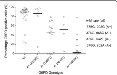
Sixty-one cryopreserved red blood samples from The Gambia (males only) were analysed by flow cytometry for percentage (%) G6PD positive cells, used here as a proxy of G6PD enzyme activity (Additional file 2: Table S7). There were no indications of haemolysis in samples upon visual inspection. For four samples (4/61) the % of G6PD positive cells was below that of the negative controls and was set to zero. Flow cytometry results were stratified by G6PD genotype (Additional file 1: Figure S2). G6PD wild type samples (202G and 376A; n = 23) had the greatest % G6PD positive cells (median 89.5%, IQR, 77.1–93.7); defi-cient A− samples (202A and 376G; n = 13) displayed the lowest % G6PD positive cells (median 2.37%, IQR 0.9– 16.7). The A+ samples (202A and 376G; n = 25, median G6PD positive cells 55.3%, IQR 4.81–79.4) contained two populations of cells: one similar to wild type sam-ples (n = 17, median 72.3%, IQR 55.3–86.3) and the other similar to A− samples (n = 8, median 1.51%, IQR 0.005– 3.84). Interestingly, outliers were detected in the wild type populations and in the A− populations (Additional file 1: Figure S2). Three out of four wild type samples (0%, 22.5% and 40.3% G6PD positive cells respectively) with lower than expected % G6PD positive cells tested deficient by CareStart G6PD RDT. The fourth wild type outlier contained 26.0% G6PD positive cells and tested
normal by CareStart G6PD RDT. The four A− samples
with higher than expected % G6PD positive cells con-tained 16.7%, 29.3%, 40.5% and 77.1% G6PD positive cells respectively and all four samples tested deficient by Car-eStart G6PD RDT (Additional file 2: Table S7).
Genotyping data generated by PCR-based KASP chem-istry was available for all samples with cryopreserved RBC data (n = 61, Sepulveda and Grignard, pers. comm., and [27]) and contained the two additional alleles com-monly found in The Gambia; the Betica-Selma vari-ant (T968C) and the Svari-antamaria varivari-ant (A542T). The two alleles were only present in samples that also had the 376G mutation and the samples were hence classed
as A− genotypes. The 376G/968C (n = 12) and the
376G/542T (n = 4) mutations had a median % G6PD
pos-itive cells of 46.3% (IQR 1.5–64.0) and 52.3% (IQR 23.8–
70.4), respectively. Only nine samples were A+ (376G
only) and these had a median % G6PD positive cells of 86.3 (IQR 14.5–95.6). Now, only three A+ genotypes had low % G6PD positive cells, suggesting that some of the outliers in this group were explained by alternative SNPs (968C and 542T) affecting G6PD activity (Fig. 3).
Sample analysis and cost
Using the microsphere assay for the four markers, data for 96 samples was available within 7 h at an estimated Fig. 2 Summary of positive genotyping results of samples from
[image:5.595.57.291.87.241.2]cost per sample of $ 4.30 (calculated December 2017, Additional file 1: Table S5). Standard protocols for G6PD
and HBB genotyping involve a PCR reaction followed
either by a restriction digestion (PCR–RFLP) or Sanger sequencing [23, 28]. The cost per sample for the four markers using standard protocols was estimated at $ 5.84 per sample with 40 working hours processing time.
Discussion
This study describes a method for simultaneous
assess-ment of G6PD-deficiency markers and HBB
polymor-phisms. These were successfully genotyped from small volumes (5 µl) of DNA extracted from human blood. Gene segments were amplified in a single, multiplex PCR reaction and genotyped by magnetic bead-based allele specific multiplex assay. The assay is fast, relatively inex-pensive and performed as well as established genotyping methods.
Assessing G6PD deficiency status is relevant to predict the susceptibility of individuals for the disadvantageous haemolytic consequences of treatment with
8-amino-quinolines like primaquine and tafenoquine [29–33].
Although G6PD phenotyping assays [21, 34, 35] may be most informative in this respect and allow meaningful assessments of varying levels of G6PD due to random X-inactivation or lyonization [24–26, 36, 37], genotyping approaches are frequently used in population research to plan public health interventions [11, 38]. Here, a multi-plex assay that concurrently detects common SNPs for
the A− G6PD variant and HBB mutations was
success-fully developed using a mix of previously published and
novel oligonucleotides [23] and optimized to maximize
the SNP specific fluorescence signal. Using this micro-sphere multiplex assay, samples from G6PD deficient and G6PD normal individuals were assessed for muta-tions with good agreement with conventional genotyping methods. To assess the correlation in between G6PD gen-otype and G6PD enzyme activity, the % G6PD-positive cells was assessed for different G6PD genotypes. In line
with published data, the A− genotype (376G and 202A)
had the lowest percentage G6PD positive cells compared to wild type and A+ individuals [39]. A small proportion of genetically deficient individuals did not have a lower percentage G6PD positive cells and, conversely, a
frac-tion of genetically G6PD normal individuals displayed
unexpected low percentage G6PD positive cells. Includ-ing additional SNPs that have been associated with lower G6PD activity in The Gambia [8], improved the correla-tion between percentage G6PD positive cells and geno-types. This suggests that genotyping approaches that focus only on 376G and 202A may misclassify individuals with low G6PD enzyme activity. The inability to explain all variation in G6PD activity by the examined SNPs sug-gests that other (currently unidentified) genetic variants that affect G6PD status may exist.
The current genotyping results were based on popula-tions enriched for G6PD deficient individuals and find-ings thus do not represent G6PD deficiency prevalence in the two countries. Whilst the current selective sample set does not allow unbiased population prevalence esti-mates, the HbS gene was more common in The Gambia than Burkina Faso (13.8% vs. 2.7%) and the HbC trait was more common in Burkina Faso (14.7% vs. 10.3%) [16, 40].
The assay presented here has an advantage that it can be adapted to multiple SNPs of interest and is increas-ingly used for detecting SNPs associated with drug
resistance in parasites [41, 42]. The assay does not
detect large deletions such as either of the thalassaemias (α-thalassaemia and β-thalassaemia), which is one of its limitations. At present the assay contains two common
G6PD SNPs and two HBB gene polymorphisms, but
the flexible multiplex nature of the assay allows users to include additional markers. The assay is less time con-suming and labour intensive than traditional end-point PCR/agarose gel genotyping methods and requires less operator time. Up to 50 markers can be tested in a single reaction and markers can be mixed and matched accord-ing to population of interest and parasite epidemiology.
Conclusions
The microsphere multiplex assay presented in the current study may play a role in addressing the increasing need to test for human and parasite genetic changes because of their impact on disease progression and malaria Fig. 3 Percentage G6PD positive cells by flow cytometry by G6PD
[image:6.595.58.291.88.238.2]epidemiology. The multiplex nature of the assay in addi-tion to the fast turn-around time and the relatively low cost, make it attractive for assessment of multiple genetic markers in large-scale epidemiological studies.
Additional files
Additional file 1: Table S1. Optimisation flow chart: A. Genomic PCR, B. ASPE (allele specific primer extension) and C. Hybridisation of ASPE reactions to microspheres. Optimal conditions in black. Table S2. Cut-off MFI values for each marker. The mean plus two standard deviations (Mean
+ 2SD) of the background MFI signal of each marker was chosen as a cut-off value. An MFI signal greater than the cut-off scored a positive result. Table S3. Examples of how the net median fluorescence intensity (MFI) is recorded for each allele and the percentage wild type MFI (WT %) is calculated. A. For G6PD (G202A and A376G) in males (males only have a single copy of the G6PD gene), above 75% wild type MFI signifies that the sample is wild type and below 25% wild type MFI signifies that the gene carries a mutation. In males, if the percentage wild type MFI is between 25%-75%, the genotyping needs to be repeated. In HBB (HbC and HbS), above 75% wild type MFI signifies homozygous wild type and below 25% wild type MFI signifies a mutation on both alleles. A percentage wild type MFI and a percentage mutated MFI in between 25% and 75% signi-fies that the sample is heterozygous and contains both a wild type and mutated allele. The G6PD genotypes are as follows: A- samples carry both the 202A and 376G mutations and the A+ samples carry the 202G wild type allele and the 376G mutation. B. The HBB genotypes can be either homozygous HbAA, HbCC or HbSS. Heterozygous genotypes contain a combination of genotyping calls, for example, HbA/C samples are heterozygous samples with a wild type allele (HbA) and a mutated HbC allele. This sample does not contain the HbS mutation on either of its two alleles. An HbA/C/S genotype is heterozygous for the both HbC and HbS alleles. Samples with net MFI values below the mean MFI plus two stand-ard deviations (Mean + 2SD) for that marker are highlighted with a * (see Table S2). These samples were excluded from further analysis. Table S4.
G6PD and HBB genotyping on samples from female participants. Six samples from females were analysed to show that the assay is working for homozygous and heterozygous women. Table S5. Pricing and time.
Figure S1. Example of comparison of G6PD activity in normal (left) and deficient (right) individuals. The grey-shaded peaks correspond to samples and the blue-outlined peaks correspond to untreated negative controls. The log-10 fluorescence in the FL1 channel (533 ± 30 nm) is measured (x-axis). The SE Dmax % positive and the Overton % positive detect the % of positive cells against the control. The SE Dmax additionally accounts for outliers. Figure S2. Flow cytometry results by G6PD genotype. The per-centage of G6PD positive cells was plotted by G6PD genotype generated by magnetic bead-based multiplex assay; wild type (wt, 202G and 376A), A+ (202G and 276G) and A- (202A and 376G). The % G6PD positive cells were calculated using FlowJo version 10 Super-Enhanced Dmax Subtrac-tion (SED) algorithm.
Additional file 2: Table S6. G6PD G202A & A376G genotyping compari-son. The MagPix genotyping calls were compared to data generated by TaqMan OpenArray and KASP PCR-based genotyping. Wild type calls are 202G and 376A, A- calls are 202A and 376G and A+ calls are 202G and 376G. A dot (.) denotes samples for which genotyping was unsuccessful and samples marked with a * had low net MFI values (net MFI < net MFI +
2SD). Table S7. G6PD percentage positive cells (G6PD % positive), geno-typing amd CareStart G6PD RDT results for all samples from The Gambia. Samples were initially stratified by RDT result. The KASP genotypes include additional SNPs, T968C and A542T, that have been associated with decreased G6PD enzyme activity in The Gambia. The samples containing the mutant alleles are highlighted in green and the % G6PD positive cells are marked in green. Samples labelled with a * did not have a MagPix genotype assigned and hence had their KASP G6PD (G202A and A376G) used in this comparison.
Abbreviations
G6PDd: glucose-6-phosphate dehydrogenase deficiency; HbC: haemoglobin C; HbS: haemoglobin S; IBD: inherited blood disorders; SNPs: single nucleotide polymorphisms; ASPE: allele specific primer extension; MFI: median fluo-rescence intensity; G6PD: glucose-6-phosphate dehydrogenase; RDT: rapid diagnostic test; FST: Fluorescent Spot Test; RBC: red blood cell; SED: Super-Enhanced Dmax Subtraction.
Authors’ contributions
LG, TB and CD designed the study. LG, CM and KL extracted the DNA. LG developed the molecular bead-based assays. LG and CM performed the molecular assays. CM performed the flow cytometry. LG, CM, TB and CD ana-lysed the data. TGC, SC and JC selected the targets for the KASP genotyping. JC and LM designed and performed the KASP genotyping and analysed the data. GJHB, ABT, SAC, BPG, MA, AO, ECB, GSS, IN, SB, CD, TB and UdA designed the field studies and collected the samples. LG, TB and CD wrote the first draft of the manuscript. All authors read and approved the final manuscript.
Author details
1 Department of Immunology and Infection, London School of Hygiene &
Tropical Medicine, London, UK. 2 LGC Genomics, Hoddesdon, Hertfordshire,
UK. 3 Department of Medical Microbiology, Radboud University Medical
Cen-tre, Nijmegen, The Netherlands. 4 Department of Biomedical Sciences, Centre
National de Recherche et de Formation sur le Paludisme, Ouagadougou, Burkina Faso. 5 Disease Control & Elimination Theme, Medical Research Council
Unit at London School of Hygiene and Tropical Medicine, Fajara, The Gambia.
6 Department of Disease Control, London School of Hygiene and Tropical
Medicine, London, UK. 7 Department of Pathogen Molecular Biology, London
School of Hygiene and Tropical Medicine, London, UK. 8 Department of
Infec-tious Disease Epidemiology, London School of Hygiene and Tropical Medicine, London, UK.
Acknowledgements
We thank the study participants, members of the field teams, and staff of the Banfora Hospital in Burkina Faso and the Basse Health Centre in The Gambia. We thank Wouter Graumans and Marion Ybema-Antoine for their help with sample processing. We thank Nuno Sepúlveda for his help and input with the selection of G6PD SNPs.
Competing interests
The authors declare that they have no competing interests.
Availability of data and materials
The datasets used and/or analysed during the current study are available from the corresponding author on reasonable request.
Ethics approval and consent to participate
The studies received approval of the Interventions Research Ethics Committee of the London School of Hygiene and Tropical Medicine (#6523 for Burkina Faso; #8487 for The Gambia), Comité d’Ethique pour la Recherche en Santé, Ministère de la Santé du Burkina Faso (#2014-02-09) and Comité Technique d’Examen des Demandes d’Autorisation d’Essais Cliniques, Ministère de la Santé du Burkina Faso (#50002520146EC00000) and The Gambia Government/ MRC Joint Ethics Committee (SCC 1391, 17 August 2015). Both studies were registered at ClinicalTrials.gov (NCT02174900 and NCT02654730). Written informed consent was obtained from all participants or from their parents or guardians prior to screening and enrolment.
Funding
Publisher’s Note
Springer Nature remains neutral with regard to jurisdictional claims in pub-lished maps and institutional affiliations.
Received: 5 November 2018 Accepted: 12 January 2019
References
1. Feng Z, Smith DL, McKenzie FE, Levin SA. Coupling ecology and evolution: malaria and the S-gene across time scales. Math Biosci. 2004;189:1–19.
2. Flint J, Hill AV, Bowden DK, Oppenheimer SJ, Sill PR, Serjeantson SW, et al. High frequencies of alpha-thalassaemia are the result of natural selection by malaria. Nature. 1986;321:744–50.
3. Hamblin MT, Thompson EE, Di Rienzo A. Complex signatures of natural selection at the Duffy blood group locus. Am J Human Genet. 2002;70:369–83.
4. Modiano D, Luoni G, Sirima BS, Simpore J, Verra F, Konate A, et al. Haemo-globin C protects against clinical Plasmodium falciparum malaria. Nature. 2001;414:305–8.
5. Piel FB, Patil AP, Howes RE, Nyangiri OA, Gething PW, Williams TN, et al. Global distribution of the sickle cell gene and geographical confirmation of the malaria hypothesis. Nat Commun. 2010;1:104.
6. Minucci A, Moradkhani K, Hwang MJ, Zuppi C, Giardina B, Capoluongo E. Glucose-6-phosphate dehydrogenase (G6PD) mutations database: review of the “old” and update of the new mutations. Blood Cells Mol Dis. 2012;48:154–65.
7. Howes RE, Battle KE, Satyagraha AW, Baird JK, Hay SI. G6PD deficiency: global distribution, genetic variants and primaquine therapy. Adv Parasi-tol. 2013;81:133–201.
8. Clarke GM, Rockett K, Kivinen K, Hubbart C, Jeffreys AE, Rowlands K, et al. Characterisation of the opposing effects of G6PD deficiency on cerebral malaria and severe malarial anaemia. Elife. 2017;6:e15085.
9. Manjurano A, Sepulveda N, Nadjm B, Mtove G, Wangai H, Maxwell C, et al. African glucose-6-phosphate dehydrogenase alleles associated with protection from severe malaria in heterozygous females in Tanzania. PLoS Genet. 2015;11:e1004960.
10. Yoshida A, Beutler E, Motulsky AG. Human glucose-6-phosphate dehy-drogenase variants. Bull World Health Organ. 1971;45:243–53. 11. Howes RE, Dewi M, Piel FB, Monteiro WM, Battle KE, Messina JP, et al.
Spatial distribution of G6PD deficiency variants across malaria-endemic regions. Malar J. 2013;12:418.
12. Clark TG, Fry AE, Auburn S, Campino S, Diakite M, Green A, et al. Allelic heterogeneity of G6PD deficiency in West Africa and severe malaria susceptibility. Eur J Hum Genet. 2009;17:1080–5.
13. Dicko A, Brown JM, Diawara H, Baber I, Mahamar A, Soumare HM, et al. Primaquine to reduce transmission of Plasmodium falciparum malaria in Mali: a single-blind, dose-ranging, adaptive randomised phase 2 trial. Lancet Infect Dis. 2016;16:674–84.
14. Eziefula AC, Bousema T, Yeung S, Kamya M, Owaraganise A, Gabagaya G, et al. Single dose primaquine for clearance of Plasmodium falciparum gametocytes in children with uncomplicated malaria in Uganda: a ran-domised, controlled, double-blind, dose-ranging trial. Lancet Infect Dis. 2014;14:130–9.
15. Goncalves BP, Gupta S, Penman BS. Sickle haemoglobin, haemoglobin C and malaria mortality feedbacks. Malar J. 2016;15:26.
16. Goncalves BP, Sagara I, Coulibaly M, Wu Y, Assadou MH, Guindo A, et al. Hemoglobin variants shape the distribution of malaria para-sites in human populations and their transmission potential. Sci Rep. 2017;7:14267.
17. Ringelhann B, Hathorn MK, Jilly P, Grant F, Parniczky G. A new look at the protection of hemoglobin AS and AC genotypes against Plasmo-dium falciparum infection: a census tract approach. Am J Hum Genet. 1976;28:270–9.
18. Mangano VD, Kabore Y, Bougouma EC, Verra F, Sepulveda N, Bisseye C, et al. Novel insights into the protective role of hemoglobin S and C against Plasmodium falciparum parasitemia. J Infect Dis. 2015;212:626–34.
19. Robert V, Tchuinkam T, Mulder B, Bodo JM, Verhave JP, Carnevale P, et al. Effect of the sickle cell trait status of gametocyte carriers of Plasmo-dium falciparum on infectivity to anophelines. Am J Trop Med Hyg. 1996;54:111–3.
20. Bastiaens GJH, Tiono AB, Okebe J, Pett HE, Coulibaly SA, Goncalves BP, et al. Safety of single low-dose primaquine in glucose-6-phosphate dehydrogenase deficient falciparum-infected African males: two open-label, randomized, safety trials. PLoS ONE. 2018;13:e0190272. 21. Beutler E, Blume KG, Kaplan JC, Lohr GW, Ramot B, Valentine WN.
International Committee for Standardization in Haematology: recommended methods for red-cell enzyme analysis. Br J Haematol. 1977;35:331–40.
22. Mooney JP, Barry A, Goncalves BP, Tiono AB, Awandu SS, Grignard L, et al. Haemolysis and haem oxygenase-1 induction during persis-tent “asymptomatic” malaria infection in Burkinabe children. Malar J. 2018;17:253.
23. Gouagna LC, Bancone G, Yao F, Yameogo B, Dabire KR, Costantini C, et al. Genetic variation in human HBB is associated with Plasmodium falcipa-rum transmission. Nat Genet. 2010;42:328–31.
24. Kahn M, LaRue N, Bansil P, Kalnoky M, McGray S, Domingo GJ. Cryopreser-vation of glucose-6-phosphate dehydrogenase activity inside red blood cells: developing a specimen repository in support of development and evaluation of glucose-6-phosphate dehydrogenase deficiency tests. Malar J. 2013;12:286.
25. Kahn M, Ward WH, LaRue N, Kalnoky M, Pal S, Domingo GJ. Maintaining specimen integrity for G6PD screening by cytofluorometric assays. J Histochem Cytochem. 2015;63:454–8.
26. Shah SS, Diakite SA, Traore K, Diakite M, Kwiatkowski DP, Rockett KA, et al. A novel cytofluorometric assay for the detection and quantification of glucose-6-phosphate dehydrogenase deficiency. Sci Rep. 2012;2:299. 27. Dombrowski JG, Souza RM, Curry J, Hinton L, Silva NRM, Grignard L, et al.
G6PD deficiency alleles in a malaria-endemic region in the Western Brazil-ian Amazon. Malar J. 2017;16:253.
28. Zuniga MA, Mejia RE, Sanchez AL, Sosa-Ochoa WH, Fontecha GA. Glucose-6-phosphate dehydrogenase deficiency among malaria patients of Honduras: a descriptive study of archival blood samples. Malar J. 2015;14:308.
29. Baird JK, Battle KE, Howes RE. Primaquine ineligibility in anti-relapse therapy of Plasmodium vivax malaria: the problem of G6PD deficiency and cytochrome P-450 2D6 polymorphisms. Malar J. 2018;17:42. 30. Recht J, Ashley EA, White NJ. Use of primaquine and
glucose-6-phos-phate dehydrogenase deficiency testing: divergent policies and practices in malaria endemic countries. PLoS Negl Trop Dis. 2018;12:e0006230. 31. Rueangweerayut R, Bancone G, Harrell EJ, Beelen AP, Kongpatanakul S,
Mohrle JJ, et al. Hemolytic potential of tafenoquine in female volunteers heterozygous for glucose-6-phosphate dehydrogenase (G6PD) defi-ciency (G6PD Mahidol variant) versus G6PD-normal volunteers. Am J Trop Med Hyg. 2017;97:702–11.
32. Taylor WR, Naw HK, Maitland K, Williams TN, Kapulu M, D’Alessandro U, et al. Single low-dose primaquine for blocking transmission of Plasmo-dium falciparum malaria—a proposed model-derived age-based regimen for sub-Saharan Africa. BMC Med. 2018;16:11.
33. Watson J, Taylor WRJ, Bancone G, Chu CS, Jittamala P, White NJ. Implica-tions of current therapeutic restricImplica-tions for primaquine and tafenoquine in the radical cure of vivax malaria. PLoS Negl Trop Dis. 2018;12:e0006440. 34. Bancone G, Kalnoky M, Chu CS, Chowwiwat N, Kahn M, Malleret B, et al.
The G6PD flow-cytometric assay is a reliable tool for diagnosis of G6PD deficiency in women and anaemic subjects. Sci Rep. 2017;7:9822. 35. Brewer GJ, Tarlov AR, Alving AS. Methaemoglobin reduction test: a new,
simple, in vitro test for identifying primaquine-sensitivity. Bull World Health Organ. 1960;22:633–40.
36. Ley B, Alam MS, O’Donnell JJ, Hossain MS, Kibria MG, Jahan N, et al. A comparison of three quantitative methods to estimate G6PD activity in the Chittagong Hill Tracts, Bangladesh. PLoS ONE. 2017;12:e0169930. 37. Taylor JD, Briley D, Nguyen Q, Long K, Iannone MA, Li MS, et al. Flow
cyto-metric platform for high-throughput single nucleotide polymorphism analysis. Biotechniques. 2001;30(661–6):8–9.
•fast, convenient online submission
•
thorough peer review by experienced researchers in your field
• rapid publication on acceptance
• support for research data, including large and complex data types
•
gold Open Access which fosters wider collaboration and increased citations maximum visibility for your research: over 100M website views per year
•
At BMC, research is always in progress.
Learn more biomedcentral.com/submissions
Ready to submit your research? Choose BMC and benefit from:
39. Okebe J, Amambua-Ngwa A, Parr J, Nishimura S, Daswani M, Takem EN, et al. The prevalence of glucose-6-phosphate dehydrogenase deficiency in Gambian school children. Malar J. 2014;13:148.
40. Piel FB, Howes RE, Patil AP, Nyangiri OA, Gething PW, Bhatt S, et al. The distribution of haemoglobin C and its prevalence in newborns in Africa. Sci Rep. 2013;3:1671.
41. Carnevale EP, Kouri D, DaRe JT, McNamara DT, Mueller I, Zimmerman PA. A multiplex ligase detection reaction-fluorescent microsphere assay for
simultaneous detection of single nucleotide polymorphisms associ-ated with Plasmodium falciparum drug resistance. J Clin Microbiol. 2007;45:752–61.