Copyright © 2003, American Society for Microbiology. All Rights Reserved.
JC Virus-Induced Changes in Cellular Gene Expression
in Primary Human Astrocytes
Sujatha Radhakrishnan, Jessica Otte, Sahnila Enam, Luis Del Valle,
Kamel Khalili,* and Jennifer Gordon
Center for Neurovirology and Cancer Biology, College of Science and Technology, Temple University, Philadelphia, Pennsylvania 19122
Received 3 December 2002/Accepted 9 May 2003
Cell-type-specific transcription of the JC virus (JCV) promoter in glial cells initiates a series of events leading to viral replication in the brain and the development of the fatal demyelinating disease progressive multifocal leukoencephalopathy (PML) in patients with neurologic complications due to infection with human immunodeficiency virus type 1. Here we employed an in vitro infection of primary cultures of human astrocytes to compare the transcriptional profile of cellular genes after JCV infection by using an oligonucleotide-based microarray of 12,600 genes. Transcription of nearly 355 genes was enhanced and expression of 130 genes was decreased to various degrees. Many transcripts that were increased upon JCV infection were found to encode proteins with properties that suggest their involvement in cell proliferation, including cyclin A and cyclin B1;
signaling pathways, such as transforming growth factorreceptor 1, platelet-derived growth factor receptor
and fibroblast growth factor family receptor; and other regulatory events, such as inflammatory responses, including cyclo-oxygenase-2 (Cox-2). Microarray-based data for several cell cycle-regulatory genes were further examined by using Western blot analysis of in vitro infected astrocytes harvested early and late during the infection. Results demonstrate that protein levels of all upregulated genes were found to increase at some point during the infection time course. In parallel, immunohistochemical assessment of cell cycle proteins, including cyclins A, B1, E, and Cdk2, showed positive staining of astrocytes within PML lesions of brain tissue from patients with neuro-AIDS. Microarray analysis was found to be a useful predictor of gene expression in infected cells; however, it may not directly correlate with protein levels during infection with JCV.
The human polyomavirus, JC (JCV), is the etiologic agent for the fatal demyelinating disease of the central nervous sys-tem, progressive multifocal leukoencephalopathy (PML) (for review see references 5 and 11). Once a rare disease seen primarily in patients with impaired immune systems due to myeloproliferative and lymphoproliferative disorders, the inci-dence of PML has been significantly increased due to the AIDS epidemic (27). Clinical studies show that⬎4% of AIDS patients with neurologic problems are diagnosed with PML (1, 2, 6, 7, 10, 18, 19, 20, 26, 31, 32). JCV infects ⬎65% of the human population by early childhood with no major clinical symptoms (16, 23, 28, 30). Reactivation of the viral genome that preferentially occurs in oligodendrocytes and astrocytes leads to cytolytic destruction of the myelin-producing oligo-dendrocytes and degeneration of the myelin sheath in white matter. The chief pathological features of PML are the pres-ence of altered oligodendrocytes whose nuclei are two to three times the size of normal cells and are filled with virions and viral antigens (34–36); giant, bizarre astrocytes with pleomor-phic, hyperchromatic nuclei; and occasional mitotic figures. Early studies revealed that expression of the viral genome is controlled by transcription factors that are enriched and/or active in oligodendrocytes and astrocytes, permitting produc-tion of the viral early protein, i.e., large T antigen and its
various isoforms (15). Once T antigen is expressed, the virus enters into a lytic cycle that consists of active viral DNA rep-lication and late gene transcription coding for the capsid pro-teins, VP1, VP2, and VP3, and the auxiliary Agnoprotein in oligodendrocytes. Astrocytic cells are semipermissive for JCV infection in that they allow for expression of T antigen and late gene expression to a limited extent but are not the major source of virion production in vivo (5, 13). However, several studies have demonstrated the ability of JCV to infect many cell types in the central nervous system (CNS) in addition to oligodendrocytes, including neurons, astrocytes, and macro-phages (8, 26).
Studies that use human cell lines and animal models have provided important information on the various functions asso-ciated with the viral early protein, T antigen. For example, T antigen has the ability to associate with several cellular pro-teins that are implicated in the control of the cell cycle and proliferation. The association of T antigen with p53 and pRb is believed to be involved, at least in part, in the transforming ability of JCV in cell culture and its tumorigenicity in experi-mental animals (9, 16, 17, 24, 29). Accordingly, JCV T antigen has been reported in several human brain tumors, particularly medulloblastoma, leading to speculation about JCV involve-ment in CNS neoplasias. Furthermore, the JCV Agnoprotein may deregulate cell growth by altering the expression of several cell cycle-associated proteins (17). To gain more comprehen-sive information of the cellular events that are affected during the course of JCV infection, we employed microarray technol-ogy and compared the level of expression of cellular genes in JCV-infected and uninfected human primary astrocytes.
Sev-* Corresponding author. Mailing address: Center for Neurovirology and Cancer Biology, College of Science and Technology, Temple Uni-versity, 1900 North 12th St., 015-96, Room 203, Philadelphia, PA 19122. Phone: (215) 204-0678. Fax: (215) 204-0679. E-mail: kamel [email protected].
10638
on November 8, 2019 by guest
http://jvi.asm.org/
eral of the genes whose expression was affected by JCV infec-tion are involved in cell cycle progression and receptors for several growth factors and are classified as transcription factors and/or cofactors.
A distinction should be made between the function of JC viral proteins in transformed cells and tumor tissues versus during the course of viral infection. Of note, T antigen has been shown to interact with tumor suppressor proteins and cell cycle regulators may be dysregulated in tumor cells in vitro and in vivo. However, little information has been reported on the effect of JCV on cellular proteins during infection. While it has been previously demonstrated that p53 protein accumulates in JCV-infected cells within PML-afflicted brains and that these same cells may overexpress PCNA and Ki67, as well as cyclins A and B1 (3, 4). However, the significance of these findings has not been fully explored. In addition to T antigen, the viral Agnoprotein seems to have a regulatory role in the viral lytic cycle and may impact host function. Earlier studies have re-vealed that the 8-kDa Agnoprotein has a perinuclear cytoplas-mic localization and may shuttle between the nucleus and cytoplasm (22). In more recent studies, it has been demon-strated that JCV Agnoprotein has the ability to associate with T antigen and controls viral gene expression and replication in human glial cells (12). As anticipated, the structural capsid protein, VP1 may also be detected in oligodendrocytes and, to a lesser extent, bizarre astrocytes (13). In order to determine the effect of JCV infection on the astrocytic cells, we per-formed gene expression analysis with gene array technology.
Toward this end, primary human fetal astrocytes were in-fected with the Mad1/SVEdelta strain of JCV, which has been described previously (29). This hybrid JCV contains the se-quences for all JCV coding regions and a modified noncoding region in which the distal portion of the second 98-bp repeat sequence has been replaced with an analogous portion of a 72-bp repeat sequence of the simian virus 40 noncoding region. Infections were performed at a multiplicity of infection of 1, and production of the viral protein at 0, 5, 10, and 15 days after infection was monitored by Western blotting and immunocy-tochemistry. As shown in Fig. 1, T antigen was first detected at 5 days after infection and its level increased during the course of infection (Fig. 1C, compare lane 3 to lanes 4 and 5). The late gene products, including VP1 and Agnoprotein, were also de-tected at days 5, 10, and 15 after infection (Fig. 1F and I, respectively). Results from immunocytochemistry showed nu-clear accumulation of T antigen and VP1 in the infected cells 15 days after infection (Fig. 1B and E). In accordance with earlier observations, Agnoprotein showed a strong presence in the cytoplasm around the nuclei of the infected cells (Fig. 1H). Total cellular RNAs were isolated from uninfected cells as well as from cells on day 15 postinfection to determine the profile of gene expression by using the Affymetrix U95A Hu-man GeneChip. All protocols were conducted as described in the Affymetrix GeneChip Expression Analysis Technical Man-ual. Total RNA was prepared by the RNeasy Total RNA Isolation Kit according to the manufacturer’s instructions (Qiagen). Briefly, 20g of total RNA was converted to first-strand cDNA by using Superscript II reverse transcriptase primed by a poly(T) oligomer that incorporated the T7 pro-moter (Superscript Choice Kit; Invitrogen). Second-strand cDNA synthesis was followed by in vitro transcription for
lin-ear amplification of each transcript and incorporation of bio-tinylated CTP and UTP. Biobio-tinylated cRNA was synthesized by using the High Yield RNA Transcript labeling kit (Enzo Diagnostics) by incorporating biotinylated CTP and UTP and was then purified with RNeasy affinity columns (Qiagen).
The subsequent steps that include fragmentation and hy-bridization of cRNA to probe arrays (Affymetrix U95A) con-taining approximately 12,600 human gene sequences were per-formed by Research Genetics (Huntsville, Ala.) according to Affymetrix protocols. cRNA was fragmented and was tested for quality control by using Affymetrix Test 2 arrays. The cRNA products were fragmented to 200 nucleotides or less, heated at 99°C for 5 min, and hybridized for 16 h at 45°C to human U95A microarrays. The microarrays were then washed at low (6⫻SSPE [1⫻SSPE is 0.18 M NaCl, 10 mM NaH2PO4,
and 1 mM EDTA {ph 7.7}]) and high (100 mM morpholine-ethanesulfonic acid and 0.1 M NaCl) stringencies and were stained with streptavidin-phycoerythrin. Fluorescence was am-plified by adding biotinylated antistreptavidin and an addi-tional aliquot of streptavidin-phycoerythrin stain. A confocal scanner was used to collect a fluorescence signal at a 3-m resolution after excitation at 570 nm. The average signal from two sequential scans was calculated for each microarray fea-ture.
Affymetrix Microarray Suite 5.0 was used to quantitate ex-pression levels for targeted genes, and default values provided by Affymetrix were applied to all analysis parameters. Border pixels were removed, and the average intensity of pixels within the 75th percentile was computed for each probe. The average of the lowest 2% of probe intensities occurring in each of 16 microarray sectors was set as background and was subtracted from all features in that sector. Probe pairs were scored posi-tive or negaposi-tive for detection of the targeted sequence by comparing signals from the perfect match and mismatch probe features. The number of probe pairs meeting the default dis-crimination threshold ( ⫽0.015) was used to assign a call of absent, present, or marginal for each assayed gene, andPwas calculated to reflect confidence in the detection call. A weighted mean of probe fluorescence (corrected for nonspe-cific signal by subtracting the mismatch probe value) was cal-culated by using the One-Step Tukey’s Biweight Estimate. This signal value, a relative measure of the expression level, was computed for each assayed gene. Global scaling was applied to allow comparison of gene signals across multiple microarrays: after exclusion of the highest and lowest 2%, the average total chip signal was calculated and was used to determine what scaling factor was required to adjust the chip average to an arbitrary target of 150. All signal values from one microarray were then multiplied by the appropriate scaling factor. Detect-ing a change of⬎or⬍2.0-fold on data normalized with Af-fymetrix Microarray Suite v5.0 was considered significant, in accordance with Affymetrix product specifications. Chip repli-cates are considered unnecessary, as repeated hybridizations with the same target have been shown to be highly reproduc-ible (21, 33). Normalized data were then analyzed by using GeneSpring v4.2 software. Since each set of experimental con-ditions was created in duplicate with use of different prepara-tions of primary cells and viral stocks, all genes shown to be classified as up- or downregulated under each set of virus-infected conditions were compared with genes under
on November 8, 2019 by guest
http://jvi.asm.org/
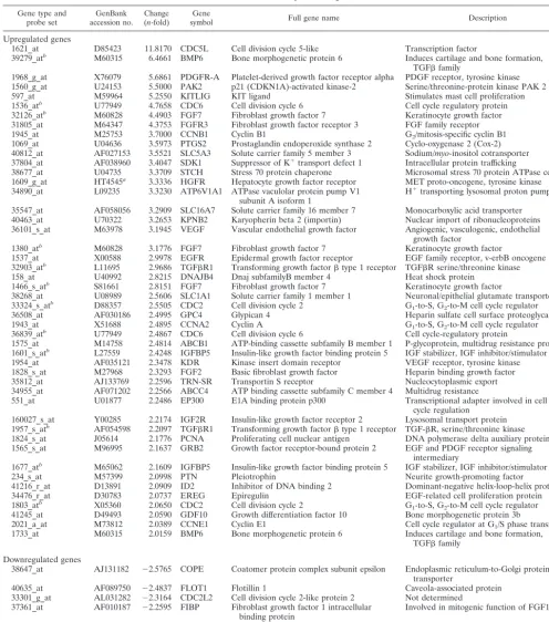
infected conditions. Calculated mean changes (n-fold) of the duplicates are presented. Among the tested DNA sequences, we found that transcription of 355 genes was enhanced to various extents, while expression of nearly 130 genes was di-minished due to viral infection. Among the genes whose ex-pression was affected by viral infection, several belonged to cell cycle regulators, transcriptional activators, cytokines and im-munomodulators, and genes involved in signal transduction. Table 1 represents a subset of genes annotated as cell cycle-regulatory proteins in databases supporting GeneSpring gene lists. The complete list of 355 upregulated and 130 down-regulated genes can be found at http://www.temple.edu/cnvcb /supplemental.htm. Many of the genes represented in the list
have unknown functions, represent cDNA clones or expressed sequence tags, or represent genes whose expression has not previously been linked to infection with JCV.
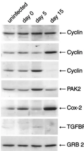
[image:3.603.47.539.70.423.2]In order to validate the gene expression data, we selected a small subset of several affected genes that were involved in cell cycle or cellular proliferation such as cyclin A, cyclin B1, cyclin E, PAK2, Cox-2, and transforming growth factorR1 (TGFR1) to assess the level of their proteins during the course of JCV infection of primary astrocytes by Western blot analysis. As shown in Fig. 2, levels of cyclin A increased slightly at 5 days but returned to normal levels at 15 days after infection, while RNA levels were found to be increased 2.5-fold at 15 days postinfection. A significant increase in the levels of cyclin B1
FIG. 1. Western blot and immunocytological analysis of viral proteins in JCV-infected primary human fetal astrocytic cells. Primary human fetal astrocytes (kindly provided by Avindra Nath) plated into tissue culture flasks were infected with the Mad1/SVEdelta strain of JCV at a multiplicity of infection of 1 in the absence of serum for 3 h at 37°C. The Mad1/SVEdelta JCV is a modification of the Mad-1 strain of JCV, which has been described previously (29). After infection, cells were washed and were refed with Dulbecco modified Eagle medium–F12 supplemented with 15% fetal calf serum. Cells were harvested on days 0, 5, 10, and 15 postinfection. Representative phase-contrast images of the cells on day 15 are shown (A, D, and G). Primary human fetal astrocytes at 15 days postinfection seeded in polylysine-coated chamber slides were fixed with ice-cold acetone, and expression of the viral proteins was analyzed by immunocytochemistry by using pAb416 antibody (1:1,000 dilution; Oncogene Research Products) for the detection of T antigen (B), rabbit polyclonal antibody for detection of VP1 (1:1,000 dilution, Lee Biomolecular) (E), and rabbit polyclonal antibody for detection of Agnoprotein (1:3,000 dilution) (14) (H). Proteins were detected with fluorescein-conjugated secondary antibodies. Western blot analysis was performed in parallel on extracts prepared from uninfected astrocytes or infected cells harvested 0, 5, 10, and 15 days postinfection by using the antibodies described above to detect T antigen (1:1,000) (C), VP1 (1:1,000 dilution) (B), and Agnoprotein (1:3,000 dilution) (C). Proteins were separated by sodium dodecyl sulfate-polyacrylamide gel electrophoresis followed by transfer to a polyvinyl difluoride membrane, and bands were visualized by autoradiography by using alkaline-phosphatase conjugated secondary antibodies and CDP-Star substrate according to the manufacturer’s instructions (NEN-Dupont).
on November 8, 2019 by guest
http://jvi.asm.org/
and Cox-2 was observed at 15 days postinfection, which is consistent with the observed 3.7- and 3.6-fold increases in RNA levels, respectively. Cyclin E and PAK2 showed a subtle increase at the early phase of viral infection followed by a
[image:4.603.42.539.79.642.2]decrease at the late phase of infection. This is in contrast with the results from RNA analysis by microarray, where 2- and 5.5-fold increases in cyclin E and PAK2 RNA, respectively, at 15 days postinfection were observed. A similar pattern was also
TABLE 1. Gene and probe designations
Gene type and
probe set accession no.GenBank Change(n-fold) symbolGene Full gene name Description
Upregulated genes
1621_at D85423 11.8170 CDC5L Cell division cycle 5-like Transcription factor
39279_atb M60315 6.4661 BMP6 Bone morphogenetic protein 6 Induces cartilage and bone formation, TGFfamily
1968_g_at X76079 5.6861 PDGFR-A Platelet-derived growth factor receptor alpha PDGF receptor, tyrosine kinase 1560_g_at U24153 5.5000 PAK2 p21 (CDKN1A)-activated kinase-2 Serine/threonine-protein kinase PAK 2 597_at M59964 5.2550 KITLIG KIT ligand Stimulates mast cell proliferation 1536_atb U77949 4.7658 CDC6 Cell division cycle 6 Cell cycle regulatory protein 32126_atb M60828 4.4903 FGF7 Fibroblast growth factor 7 Keratinocyte growth factor 31805_at M64347 4.3753 FGFR3 Fibroblast growth factor receptor 3 FGF family receptor 1945_at M25753 3.7000 CCNB1 Cyclin B1 G2/mitosis-specific cyclin B1 1069_at U04636 3.5973 PTGS2 Prostaglandin endoperoxide synthase 2 Cyclo-oxygenase 2 (Cox-2) 40812_at AF027153 3.5521 SLC5A3 Solute carrier family 5 member 3 Sodium/myo-inositol cotransporter 37804_at AF038960 3.4047 SDK1 Suppressor of K⫹transport defect 1 Intracellular protein trafficking
38677_at U04735 3.3709 STCH Stress 70 protein chaperone Microsomal stress 70 protein ATPase core 1609_g_at HT4545a 3.3336 HGFR Hepatocyte growth factor receptor MET proto-oncogene, tyrosine kinase 34890_at L09235 3.3230 ATP6V1A1 ATPase vaculolar protein pump V1
subunit A isoform 1 H
⫹transporting lysosomal proton pump
35547_at AF058056 3.2909 SLC16A7 Solute carrier family 16 member 7 Monocarboxylic acid transporter 40463_at U70322 3.2653 KPNB2 Karyopherin beta 2 (importin) Nuclear import of ribonucleoproteins 36101_s_at M63978 3.1945 VEGF Vascular endothelial growth factor Angiogenic, vasculogenic, endothelial
growth factor
1380_atb M60828 3.1776 FGF7 Fibroblast growth factor 7 Keratinocyte growth factor
1537_at X00588 2.9978 EGFR Epidermal growth factor receptor EGF family receptor, v-erbB oncogene 32903_atb L11695 2.9686 TGFR1 Transforming growth factortype 1 receptor TGFR serine/threonine kinase 158_at U40992 2.8215 DNAJB4 Dnaj subfamilyB member 4 Heat shock protein
1466_s_atb S81661 2.8151 FGF7 Fibroblast growth factor 7 Keratinocyte growth factor
38268_at U08989 2.5606 SLC1A1 Solute carrier family 1 member 1 Neuronal/epithelial glutamate transporter 33324_s_atb D88357 2.5505 CDC2 Cell division cycle 2 G
1-to-S, G2-to-M cell cycle regulator 36508_at AF030186 2.4995 GPC4 Glypican 4 Heparin sulfate cell surface proteoglycan 1943_at X51688 2.4895 CCNA2 Cyclin A G1-to-S, G2-to-M cell cycle regulator 36839_atb U77949 2.4867 CDC6 Cell division cycle 6 Cell cycle-regulatory protein
1575_at M14758 2.4814 ABCB1 ATP-binding cassette subfamily B member 1 P-glycoprotein, multidrug resistance protein 1 1601_s_atb L27559 2.4248 IGFBP5 Insulin-like growth factor binding protein 5 IGF stabilizer, IGF inhibitor/stimulator 1954_at AF035121 2.3478 KDR Kinase insert domain receptor VEGF receptor, tyrosine kinase 1828_s_at M27968 2.3293 FGF2 Basic fibroblast growth factor Heparin binding growth factor 35812_at AJ133769 2.2596 TRN-SR Transportin S receptor Nucleocytoplasmic export 34955_at AF071202 2.2566 ABCC4 ATP binding cassette subfamily C member 4 Multidrug resistance
551_at U01877 2.2486 EP300 E1A binding protein p300 Transcriptional adapter involved in cell cycle regulation
160027_s_at Y00285 2.2174 IGF2R Insulin-like growth factor receptor 2 Lysosomal transport protein 1957_s_atb AF054598 2.2097 TGFR1 Transforming growth factortype 1 receptor TGF-R, serine/threonine kinase 1824_s_at J05614 2.1776 PCNA Proliferating cell nuclear antigen DNA polymerase delta auxiliary protein 1565_s_at M96995 2.1637 GRB2 Growth factor receptor-bound protein 2 EGF and PDGF receptor signaling
intermediary
1677_atb M65062 2.1609 IGFBP5 Insulin-like growth factor binding protein 5 IGF stabilizer, IGF inhibitor/stimulator 234_s_at M57399 2.0998 PTN Pleiotrophin Neurite growth-promoting factor 41216_r_at D13891 2.0909 ID2 Inhibitor of DNA binding 2 Dominant-negative helix-loop-helix protein 34476_r_at D30783 2.0737 EREG Epiregulin EGF-related cell proliferation protein 1803_atb X05360 2.0650 CDC2 Cell division cycle 2 G
1-to-S, G2-to-M cell cycle regulator 41245_at D49493 2.0590 GDF10 Growth differentiation factor 10 Bone morphogenetic protein 3b
2021_a_at M73812 2.0389 CCNE1 Cyclin E1 Cell cycle regulator at G1/S phase transition 1733_at M60315 2.0159 BMP6 Bone morphogenetic protein 6 Induces cartilage and bone formation,
TGFfamily Downregulated genes
38647_at AJ131182 ⫺2.5765 COPE Coatomer protein complex subunit epsilon Endoplasmic reticulum-to-Golgi protein transporter
40635_at AF089750 ⫺2.4837 FLOT1 Flotillin 1 Caveola-associated protein 33301_g_at AL031282 ⫺2.3164 CDC2L2 Cell division cycle 2-like protein 2 Not determined
37361_at AF010187 ⫺2.2595 FIBP Fibroblast growth factor 1 intracellular
binding protein Involved in mitogenic function of FGF1 160027_s_at Y00285 ⫺2.2174 IGF2R Insulin-like growth factor 2 receptor Lysosomal transport protein
aGene number from the Institute for Genomic Research.
bAppears on list more than once. Multiple probe sets for this gene are present on the DNA array.
on November 8, 2019 by guest
http://jvi.asm.org/
observed when TGFR1 protein levels were examined during the course of JCV infection. As seen in Fig. 2, the levels of the protein at 5 days were increased while a decrease was observed at day 15 as the infection cycle progressed; RNA levels for TGFR1 were increased 2.2-fold. Surprisingly, the level of a nonspecific protein such as Grb-2 remained fairly constant during the course of infection, even though an increase in RNA levels of approximately 2.2-fold was observed at 15 days postinfection. This may be due to stability of the mRNA or to posttranscriptional regulation of Grb-2. In summary, with the exception of Grb-2, all proteins examined showed an increase either at 5 or 15 days postinfection, though the increase was not sustained during the course of the infection. Several of the proteins, including cyclin A, cyclin E, PAK2, and TGFR1, showed an increase on day 5 and a decrease on day 15 postin-fection, with increases of ⬎2-fold in RNA levels at day 15, suggesting that either these RNAs are not efficiently translated at the later stages of viral infection and/or that the proteins are rapidly degraded at the later times of the infection cycle. Cyclin B1 and Cox-2, however, showed increases at both the RNA and protein levels 15 days postinfection.
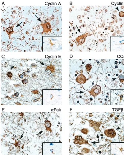
To further determine the ability of microarray in a cell culture infection model to predict up- or downregulation of genes in vivo, immunohistochemistry was performed on
paraf-fin-embedded, formalin-fixed tissue sections of brain tissue from a PML patient. Examination of cyclins A, B1, E, and Cox-2 showed enhanced cytoplasmic staining of these proteins in the bizarre astrocytes of PML lesions compared to results from non-PML normal brain, though nuclear staining in PML astrocytes was notably absent (Fig. 3A to D). Similarly, in-creased expression of PAK2 and TGFR1 was evident in as-trocytes of PML compared to that in normal asas-trocytes of non-PML samples (Fig. 3E and F). Interestingly, punctate im-munoreactivity to TGFR1 was also detected in the nuclei of some astrocytes (Fig. 3F). Oligodendrocyte inclusion bodies also showed staining in the perinuclear region or cytoplasm for most proteins analyzed, while nuclear staining of inclusion bodies was also seen with antibodies to cyclin A, cyclin E, PAK2, and TGFR1. These staining patterns in astrocytes corroborate the results from in vitro infection of primary as-trocytes by JCV and indicate that infection of the asas-trocytes by JCV both in cell culture and in the brains of PML patients leads to dysregulation of cell cycle regulators.
The upregulation of a host of cell cycle regulatory proteins has been previously demonstrated in JCV-transformed human tumor cells in vitro and in vivo. However, a distinction should be made between the study of transformed cells, as is the case for most cell lines and certainly for tumor tissues and primary cultures of nontransformed cells. In this regard, limited infor-mation on cellular gene expression in JCV-infected nontrans-formed cells or within PML brain lesions is known. In one report, cyclins A and B1 were shown to be overexpressed in PML astrocytes and oligodendrocytes, as well as positive Ki67 staining (4). While the results presented here may suggest increased cycling of the cells, one must cautiously consider that myelin-producing oligodendrocytes are thought to be termi-nally differentiated and that no present evidence suggests that inclusion-bearing oligodendrocytes are undergoing cell divi-sion. However, the bizarre astrocytes of PML do contain mul-tilobulated nuclei or may even appear multinucleated, suggest-ing that the cells may be passsuggest-ing through some steps of the cell cycle. In any case, the advantage for JCV in controlling the cell cycle is clear. It may be necessary for a double-stranded DNA virus to control the cellular replication machinery normally present during S phase. In this regard, JCV T antigen can orchestrate viral DNA replication and complete a productive infection cycle.
In summary, microarray technology was employed to inves-tigate the expression of cellular genes during the course of infection with JCV. We chose to carry out our study in highly purified human primary astrocytes instead of mixed primary cultures of human fetal brain, which are commonly used for JCV infection in vitro, due to the inherent variations in the cell-type context of each preparation that can significantly af-fect the efficacy of inaf-fection in each experiment and thus the reproducibility of the outcome. Also, it is extremely difficult to obtain highly purified cultures of oligodendrocytes in the large quantities necessary to yield a sufficient amount of RNA for analysis. Thus, our results may not provide a complete picture of events related to cellular gene expression in JCV-infected brain cells, particularly oligodendrocytes. Nevertheless, our re-sults illuminate some of the changes that occur in astrocytes upon JCV infection. The role of astrocytes in PML has not been elucidated, though it is widely accepted that they
repre-FIG. 2. Western blot analysis of extracts from JCV-infected astro-cytes for gene products of selected upregulated genes. We carried out Western blot analysis of protein extracts (50 g) from uninfected primary astrocytes or cells infected with JCV 0, 5, and 15 days after infection with the following mouse monoclonal antibodies: anti-cyclin A (1:1,000, BF683), anti-cyclin B1 (1:1,000, GNS1), and anti-Grb-2 (1:1,000, no. 81). Alternatively we used the following polyclonal bodies: cyclin E (1:1,000, M20), PAK2 (1:1,000, V-19), anti-Cox-2 (1:1,000, C-20), and anti-TGFR1 (1:500, R-20). All antibodies were purchased from Santa Cruz Biotechnology (Santa Cruz, Calif.), with the exception of Grb-2, which was purchased from BD Transduc-tion Laboratories. Western blot analysis was performed as described in the Fig. 1 legend.
on November 8, 2019 by guest
http://jvi.asm.org/
[image:5.603.94.235.70.322.2]sent an abortive infection. Many questions remain to be an-swered, such as whether astrocytes provide any protection to infected oligodendrocytes or may even contribute toward a favorable environment for viral replication. The present study has suggested a number of proteins of known and unknown
[image:6.603.89.489.71.570.2]function whose gene expression has been altered upon infec-tion with JCV. Further study toward understanding different mechanisms that may affect astrocyte function during the course of PML may lead us to understand astrocyte responses to viral infections in the CNS.
FIG. 3. Immunohistochemical analysis of cellular proteins in demyelinated plaques of PML and normal brain tissue. Paraffin-embedded sections of brain tissue lesions from a patient with PML or from normal brain were analyzed for the expression of cyclin A (A), cyclin B1 (B), cyclin E (C), Cox-2 (D), PAK2 (E), and TGFR1 (F). Insets show staining of normal brain tissue depicting a representative astrocyte with various antibodies as indicated in each panel. Bizarre astrocytes exhibit cytoplasmic immunoreactivity for cyclin A, cyclin B1, cyclin E, Cox-2, and PAK2 (arrows). Bizarre astrocytes exhibit cytoplasmic and punctate nuclear immunoreactivity when tested with an antibody for TGFR1 (arrows). Oligodendrocyte inclusion bodies show cytoplasmic or perinuclear immunoreactivity for cyclin E, PAK2, TGFR1, and Cox-2 and nuclear immunoreactivity for cyclin A, cyclin E, and TGFR1 (arrowheads). Immunohistochemistry was performed by using the ABC Vector Elite system and was detected with DAB chromogen as described previously (14). All panels, original magnification⫻1,000.
on November 8, 2019 by guest
http://jvi.asm.org/
We thank past and present members of the Center for Neurovirol-ogy and Cancer BiolNeurovirol-ogy for their insightful discussions and sharing of ideas and reagents. We thank Avindra Nath (Johns Hopkins Univer-sity) for providing purified cultures of human fetal astrocytes, Walter Atwood (Brown University) for providing Mad1/SVEdelta, and Susan Morgello (Mount Sinai School of Medicine) and the Manhattan HIV-1 Brain Bank for providing human brain tissue samples. We also thank C. Schriver for preparation of the manuscript.
This work was made possible by grants awarded by the NIH to L.D.V. and K.K.
REFERENCES
1. Anders, K. H., W. E. Guerra, U. Tomiyasu, M. A. Verity, and H. V. Vinters.
1986. The neuropathology of AIDS: UCLA experience and review. Am. J. Pathol.124:537–558.
2. Antinori, A., A. Ammassari, M. L. Giancola, A. Cingolani, S. Grisetti, R. Murri, L. Alba, B. Ciancio, F. Soldani, D. Larussa, G. Ippolito, and A. De Luca.2001. Epidemiology and prognosis of AIDS-associated progressive multifocal leukoencephalopathy in the HAART era. J. Neurovirol.7:323– 328.
3. Ariza, A., J. L. Mate, A. Fernandez-Vasalo, C. Gomez-Plaza, J. Perez-Piteira, M. Pujol, and J. J. Navas-Palacios.1994. p53 and proliferating cell nuclear antigen expression in JC virus-infected cells of progressive multifocal leu-koencephalopathy. Hum. Pathol.25:1341–1345.
4. Ariza, A., J. L. Mate, M. Isamat, A. Calatrava, A. Fernandez-Vasalo, and J. J. Navas-Palacios.1998. Overexpression of Ki-67 and cyclins A and B1 in JC virus-infected cells of progressive multifocal leukoencephalopathy. J. Neuropathol. Exp. Neurol.57:226–230.
5. Berger, J. R., and M. Concha.1995. Progressive multifocal leukoencepha-lopathy: the evolution of a disease once considered rare. J. Neurovirol.
1:5–18.
6. Berger, J. R., B. Kaszovitz, J. D. Post, and G. Dickinson.1987. Progressive multifocal leukoencephalopathy associated with human immunodeficiency virus infection: a review of the literature with a report of sixteen cases. Ann. Intern. Med.107:78–87.
7. Berger, J. R., A. Chauhan, D. Galey, and A. Nath.2001. Epidemiological evidence and molecular basis of interactions between HIV and JC virus. J. Neurovirol.7:329–338.
8. Boldorini, R., S. Cristina, L. Vago, A. Tosoni, S. Guzzetti, and G. Costanzi.
1993. Ultrastructural studies in the lytic phase of progressive multifocal leukonecephalopathy in AIDS patients. Ultrastruct. Pathol.17:599–609. 9. Chesters, P. M., J. Heritage, and D. J. McCance.1983. Persistence of DNA
sequences of BK virus and JC virus in normal human tissues and in diseased tissues. J. Infect. Dis.147:676–684.
10. Cinque, P., C. Pierotti, M. G. Vigano, A. Bestetti, C. Fausti, D. Bertelli, and A. Lazzarin.2001. The good and evil of HAART in HIV-related progressive multifocal leukoencephalopathy. J. Neurovirol.7:358–363.
11. Clifford, D. B., and E. O. Major.2001. The biology of JC virus and progres-sive multifocal leukoencephalopathy. J. Neurovirol.4:279.
12. Darbinyan, A., N. Darbinian, M. Safak, S. Radhakrishnan, A. Giordano, and K. Khalili.2002. Evidence for dysregulation of cell cycle by human poly-omavirus, JCV, late auxiliary protein. Oncogene21:5574–5581.
13. Del Valle, L., S. Croul, S. Morgello, S. Amini, J. Rappaport, and K. Khalili.
2000. Detection of HIV-1 Tat and JCV capsid protein, VP1, in AIDS brain with progressive multifocal leukoencephalopathy. J. Neurovirol.6:221–228. 14. Del Valle, L., J. Gordon, S. Enam, S. Delbue, S. Croul, S. Abraham, S. Radhakrishnan, M. Assimakoupoulou, C. D. Katsetos, and K. Khalili.2001. Expression of human neurotropic polyomavirus JCV late gene product AGNO protein in human medulloblastoma. J. Natl. Cancer Inst.94:267–273. 15. Frisque, R. J.2001. Structure and function of JC virus T proteins. J.
Neu-rovirol.7:293–297.
16. Gardner, S. D.1977. The new human papovaviruses: their nature and
sig-nificance, p. 93–115.InA. P. Waterson (ed.), Recent advances in clinical virology. Churchill Livingstone, Edinburgh, United Kingdom.
17. Grinnell, B. W., B. L. Padgett, and D. L. Walker.1983. Comparison of infectious JC virus DNAs cloned from human brain. J. Virol.45:299–308. 18. Krupp, L. B., R. B. Lipton, M. L. Swerdlow, N. E. Leeds, and J. Llena.1985.
Progressive multifocal leukoencephalopathy: clinical and radiographic fea-tures. Ann. Neurol.17:344–349.
19. Kure, K., J. L. Llena, W. D. Lyman, R. Soeiro, K. M. Weidenheim, D. Hirano, and W. Dickson.1991. Human immunodeficiency virus-1 infection of the nervous system: an autopsy study of 268 adult, pediatric, and fetal brains. Hum. Pathol.22:700–710.
20. Lang, W., J. Miklossy, J. P. Deruaz, G. P. Pizzolato, A. Probst, T. Schaffner, E. Gessage, and P. Kleihues.1989. Neuropathology of the acquired immune deficiency syndrome (AIDS): a report of 135 consecutive autopsy cases from Switzerland. Acta Neuropathol.77:379–390.
21. Lockhart, D. J., H. Dong, M. C. Byrne, M. T. Follettie, M. V. Gallo, M. S. Chee, M. Mittman, C. Wang, M. Kobayashi, H. Horton, and E. L. Brown.
1996. Expression monitoring by hybridization to high-density oligonucleotide arrays. Nat. Biotechnol.14:1675–1680.
22. Okada, Y., S. Endo, H. Takahashi, H. Sawa, T. Umemura, and K. Na-gashima.2001. Distribution and function of JCV agnoprotein. J. Neurovirol.
7:302–306.
23. Padgett, B. L., and D. L. Walker.1973. Prevalence of antibodies in human sera against JC virus, an isolate from a case of progressive multifocal leu-koencephalopathy. J. Infect. Dis.127:467–470.
24. Padgett, B. L., C. M. Rogers, and D. L. Walker.1977. JC virus, a human polyomavirus associated with progressive multifocal leukoencephalopathy: additional biological characteristics and antigenic relationships. Infect. Im-mun.15:656–662.
25. Safak, M., B. Sadowska, R. Barrucco, and K. Khalili.2002. Functional interaction between JC virus late regulatory Agnoprotein and cellular Y-box binding transcription factor, YB-1. J. Virol.76:3828–3838.
26. Shinohara, T., K. Nagashima, and E. O. Major.1997. Propagation of the human polyomavirus, JCV, in human neuroblastoma cell lines. Virology
228:269–277.
27. Snider, W. D., S. M. Simpson, S. Nielson, J. W. M. Gold, C. E. Metroka, and J. B. Posner.1983. Neurological complications of acquired immune defi-ciency syndrome. Analysis of 50 patients. Ann. Neurol.14:403–418. 28. Taguchi, R., J. Kajioka, and T. Miyamura.1982. Prevalence rate and age of
acquisition of antibodies against JC virus and BK virus in human sera. Microbiol. Immunol.26:1057–1064.
29. Vacante, D. A., R. Traub, and E. O. Major.1989. Extension of JC virus host range to monkey cells by insertion of a simian virus 40 enhancer into the JC virus regulatory region. Virology170:353–361.
30. Walker, D. L., and B. L. Padgett.1983. The epidemiology of human papova-viruses, p. 99–106.InJ. L. Sever and D. L. Madden, (ed.), Polyomaviruses and human neurological disease. Alan R. Liss, New York, N.Y.
31. Wiley, C. A., and J. A. Nelson.1990. Human immunodeficiency virus: infec-tion of the nervous system. Curr. Top. Microbiol. Immunol.160:157–172. 32. Wiley, C. A., M. Grafe, C. Kennedy, and J. A. Nelson.1988. Human
immu-nodeficiency virus (HIV) and JC virus in acquired immune deficiency syn-drome (AIDS) patients with progressive multifocal leukoencephalopathy. Acta Neuropathol.76:338–346.
33. Wodicka, L., H. Dong, M. Mittmann, M. H. Ho, and D. J. Lockhart.1997. Genome-wide expression monitoring in Saccharomyces cerevisiae. Nat. Bio-technol.15:1359–1367.
34. Zu Rhein, G. M.1972. Virions in progressive multifocal leukoencephalopa-thy, p. 2893–2912.InJ. Minkler (ed.), Pathology of the nervous system, vol. 3. McGraw-Hill, New York, N.Y.
35. Zu Rhein, G. M.1969. Association of papova-virions with a human demy-elinating disease (progressive multifocal leukoencephalopathy). Prog. Med. Virol.11:185–247.
36. Zu Rhein, G. M., and S. M. Chou.1968. Papova virus in progressive multi-focal leukoencephalopathy. Res. Publ. Assoc. Nerv. Ment. Dis.44:307–362.