University of Southampton Research Repository
ePrints Soton
Copyright © and Moral Rights for this thesis are retained by the author and/or other
copyright owners. A copy can be downloaded for personal non-commercial
research or study, without prior permission or charge. This thesis cannot be
reproduced or quoted extensively from without first obtaining permission in writing
from the copyright holder/s. The content must not be changed in any way or sold
commercially in any format or medium without the formal permission of the
copyright holders.
When referring to this work, full bibliographic details including the author, title,
awarding institution and date of the thesis must be given e.g.
AUTHOR (year of submission) "Full thesis title", University of Southampton, name
of the University School or Department, PhD Thesis, pagination
UNIVERSITY OF SOUTHAMPTON
FACULTY OF MEDICINE, HEALTH AND LIFE SCIENCES SCHOOL OF MEDICINE
PRENATAL DEVELOPMENT AND LATER
NEUROENDOCRINE CONTROL OF CARDIOVASCULAR
FUNCTION
Alexander Jones BM BSc MRCPCH
Thesis submitted in fulfilment of the requirements for
the degree of Doctor of Philosophy
FEBRUARY 2006
SUPERVISORS
Professor DIW Phillips MA PhD FRCP
Whilst midwife and friends assist a woman labouring, two birth-gazers attempt to predict the long-term outcome of the birth from prevailing environmental characteristics (by charting the position of the stars). Although no validation for astrology, there is now evidence that season
of birth may be predictive of size at birth and later cardiovascular healthY
Jost Amman (Swiss, 1539-91), from KunstbOchlin ('Art booklet'), Sigmund Feyerabund (printer & publisher), 1580, woodcut, Frankfurt.
UNIVERSITY OF SOUTHAMPTON
ABSTRACf
FACULTY OF MEDICINE, HEALTH AND LIFE SCIENCES SCHOOL OF MEDICINE
Doctor of Philosophy
PRENATAL DEVELOPMENT AND LATER NEUROENDOCRINE CONTROL OF CARDIOVASCULAR
FUNCTION
Alexander Jones
Small size at birth, a marker of fetal growth, is associated with hypertension and cardiovascular disease in adulthood. Explanatory mechanisms for this association are not well characterized but may involve alterations of stress response systems such as the hypothalamic-pituitary-adrenal axis (HPAA) and autonomic nervous system (ANS). Animal studies show that adverse prenatal environments lead to sex-specific, lifelong alterations in the activity of these systems both at rest and during stress. The extent to which such prenatal adaptations occur in humans is unknown but may be of clinical importance, given emerging evidence that stress responsivity is a risk factor for cardiovascular disease. The sparse published human data comes from older populations and may be confounded by existing cardiovascular disease. Therefore, I have studied younger populations - young adults from Adelaide, Australia and pre-pubertal children from Southampton, UK. In these studies, healthy individuals, born at term, underwent psychological stress testing whilst measures of ANS, cardiovascular and HPAA
function (salivary cortisol) were recorded. In adults, continuous finger arterial pressure was used to derive indices of autonomic and baroreflex function whilst in children, electrocardiography, impedance cardiography and blood pressure tonometry were used to assess cardiovascular function. Women, but not men, who were small at birth had increased sympathetic activity at rest (r
=
.28, P < .05) and during stress (r = .42, P < .001), reduced parasympathetic activity (r = .22, P < .05) and reduced baroreflex sensitivity (r = .34, P < .01). In boys, birth weight was inversely related to salivary cortisol responses to stress (r = -.56, P < .001) but not to morning cortisol levels, whilst in girls, morning peak cortisol was inversely related to birth weight (r = -.36, P < .05). In boys, lower birth weight was also associated with higher arterial pressure andsystemic vascular resistance, particularly following psychosocial stress (r = -.62, P < .01, and r = -.47, P < .05, respectively). In girls, lower birth weight was associated with greater cardiac sympathetic activation, indicated by shorter pre-ejection period (r = .53, P < .01) and corrected QT interval (r = ,45, P < .01). These associations were
independent of gestational age and potential confounding factors such as obesity, social class and educational achievement. These results suggest that, as in animals,
CONTENTS
Abstract ... iii
Illustrations ... viii
Tables ... x
Declaration of Authorship ... xi
Acknowledgements ... xii
Abbreviations ... xv
Chapter 1. Introduction ... 1
1.1 Developmental Origins of Health and Disease ... 2
1.1.1 The Longstanding Influence of Early Developmental Experience ... 3
1.1.2 Cardiovascular Disease ... 9
1.1.3 Risk Factors Associated with Cardiovascular Disease ... 10
1.1.4 Size at Birth and Risk of Cardiovascular Disease ... 12
1.1.5 Maternal Nutrition and Risk of Cardiovascular Disease ... 20
1.2 Stress ... 22
1.2.1 Stress and Psychosocial Pathways to Cardiovascular Disease ... 25
1.2.1.1 Psychosocial Factors ... 26
1.2.1.2 Chronic Stress ... 29
1.2.1.3 Acute Stress ... 31
1.2.1.4 Stress Responsiveness and Cardiovascular Disease ... 34
1.3 The Hypothalamic Pituitary Adrenal Axis (HPAA) ... 37
1.3.1 Organisation, Function and Control of the HPAA. ... 39
1.3.2 Development of the HPAA ... 45
1.3.3 Developmental Plasticity of the HPAA ... 46
1.4 The Autonomic Nervous System (ANS) ... 49
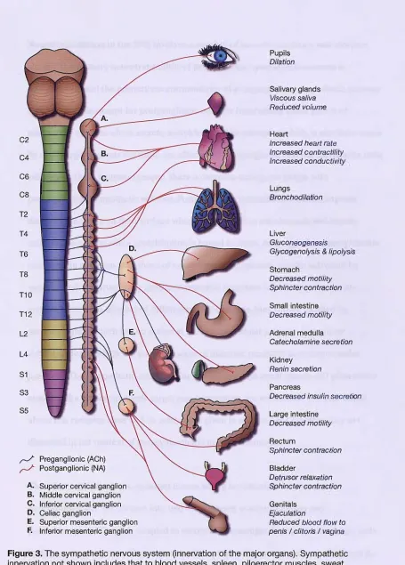
1.4.1 The Sympathetic Nervous System (SNS) ... 51
1.4.1.1 Organisation and Function of the SNS ... 54
1.4.1.2 Development of the SNS ... 60
1.4.1.3 Developmental Plasticity of the SNS ... 63
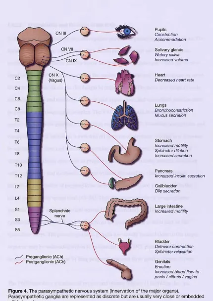
1.4.2 The Parasympathetic Nervous System (PNS) ... 69
1.4.2.1 Organisation and Function of the PNS ... 71
1.4.2.2 Development of the PNS ... 74
1.4.2.3 Developmental Plasticity of the PNS ... 75
1.4.3 The Arterial Baroreflex ... 78
1.4.3.1 Development of the Arterial Baroreflex ... 84
1.4.3.2 Developmental Plasticity of the Arterial Baroreflex ... 85
1.5 Size at Birth and Neuroendocrine Cardiovascular Control in Humans ... 86
1.6 Hypotheses ... 87
Chapter 2. Methodological Issues ... 89
2.1 Stress Induction ... 91
2.1.1 The Trier Social Stress Test for Children (TSST-C) ... 92
2.2 Salivary Cortisol ... 94
2.2.1 Assessment of Baseline Levels ... 95
2.2.1.1 Postal Delivery of Saliva Samples ... 96
2.3 Mobility in Psychological Stress Tests ... 96
2.3.1 Accurate Event Timing ... 99
2.4 Electrocardiography ... 99
2.4.1 Waveform Detection ... 99
2.5 Blood Pressure Measurement ... 103
2.5.1 Finger Arterial Plethysmography ... 104
2.5.2 Radial Arterial Tonometry ... 105
2.5.3 Limitation of Motion Artefact ... 106
2.5.4 Hydrostatic Adjustment for Arm Height... ... 108
2.6 Frequency Analysis of Cardiovascular Signals ... 110
2.6.2 Interpretation of Cardiovascular Spectrum Analysis ... 113
2.6.2.1 Interpretation of Heart Rate Spectra ... 114
2.6.2.2 Interpretation of Blood Pressure Spectra ... 115
2.6.3 The Relationship Between Blood Pressure and Heart Rate ... 116
2.7 Thoracic Impedance Cardiography ... 121
2.7.1 The Parallel Cylinder Model ... 124
2.7.1.1 Limitations of the Model ... 127
2.7.2 Electrode Placement ... 130
2.7.2.1 Accurate Height Measurement on the Surface of the Body ... 132
2.8 The Pilot Study ... 135
2.8.1 Power Calculations ... 136
Chapter 3. Cardiovascular Control in Adults ... 138
3.1 Methods ... 140
3.1.1 Signal Processing ... 140
3.1.1.1 Wavelet Analysis of Heart Period and Systolic Arterial Pressure Variability ... 141
3.1.1.2 Adaptive Autoregressive Modelling of Baroreflex Function ... 142
3.1.2 Statistical Methods ... 143
3.2 Results ... 143
3.3 Discussion ... 149
Chapter 4. Adrenocortical Function in Children ... 155
4.1 Methods ... 156
4.1.1 Statistical Methods ... 159
4.2 Results ... 160
4.3 Discussion ... 165
Chapter 5. Cardiovascular Function in Children ... 169
5.1 Methods ... 171
5.1.1 Signal Processing ... 173
5.1.2 Statistical Methods ... 176
5.2 Results ... 177
5.3 Discussion ... 185
Chapter 6. Conclusions ... 192
References ... 202
Appendix A - Ethical Approval. ... 280
Appendix B - Participant Contact ... 284
Appendix C - Informed Consent ... 293
\ , ~.
, . ' , ' \ . t
ILLUSTRATIONS
Figure 1. Central and peripheral neuroendocrine pathways influencing cardiovascular and
metabolic outcomes during stress ... 38
Figure 2. Sympathetic outflow from the T1 to L2/L3 regions of the spinal cord in humans ... 54
Figure 3. The sympathetic nervous system (innervation of the major organs) ... 56
Figure 4. The parasympathetic nervous system (innervation of the major organs) ... 70
Figure 5. Location and innervation of the principal arterial baroreceptors in the vasculature of the upper thorax and neck ... 82
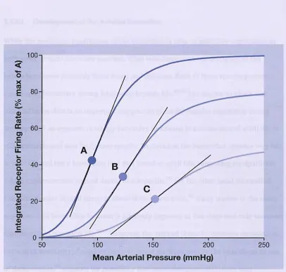
Figure 6. Effect of varying arterial pressure on carotid sinus baroreceptor firing rates in three hypothetical cases with differing baroreceptor sensitivity ... 83
Figure 7. A participant undergoing the Trier Social Stress Test for Children ... 93
Figure 8. A mobile equipment station for the safe continuous recording of cardiovascular parameters ... 97
Figure 9. An event marker for accurate timing of key occurrences during the experimental protocol. ... 98
Figure 10. ECG beat detection and editing using semi-automatic analysis ... 102
Figure 11. The Portapres finger arterial blood pressure plethysmograph ... 104
Figure 12. The sensor assembly of the Vasotrac radial arterial tonometer. ... 106
Figure 13. A heat-moulded plastic wrist splint to minimise the influence of movement on radial arterial pressure measurements ... 107
Figure 14. A cuff and splint system to hold the arm and hand still during ambulatory radial artery tonometry ... 108
Figure 15. A device for hydrostatic correction of radial arterial pressure readings for alterations in the distance between the heart and the wrist. ... 109
Figure 16. Frequency analysis of a physiological time series (heart period) ... 112
Figure 17. Time-frequency analysis of the relationship between systolic arterial pressure and heart period using a bivariate adaptive autoregressive model to determine baroreflex function .
... 120
Figure 18. Electrode positioning and circuitry for thoracic impedance cardiography ... 123
Figure 19. A simplified cylindrical model of the thorax containing uniform blood and tissue compartments for determination of thoracic impedance ... 125
Figure 20. A laser device for determining the height of points on the curved surface of a
standing subject. ... 133
Figure 21. Demonstration of the laser-levelling device in use ... 134
Figure 22. Key cardiovascular parameters in men (N
=
100) and women (N=
68) experiencing stress, grouped by birth weight ... 147Figure 23. Timeline showing median times and durations of the pre- and post-stress periods, and the Trier Social Stress Test for Children ... 158
Figure 24. Geometric mean salivary cortisol profiles during a restful day and a clinic visit for the Trier Social Stress Test for Children in boys and girls. The inset graphs show the relationship
between time-weighted mean cortisol responses (comparing home and clinic visit cortisol
concentrations) and birth weight adjusted for gestational age ... 162
Figure 25. A representative recording of the electrocardiogram (ECG) and first time-derivative
of the impedance cardiogram (dZ/dt) from a single study participant for two heart beats ... 174
TABLES
Table 1. The impact of psychosocial factors on risk of cardiovascular events. ... 27
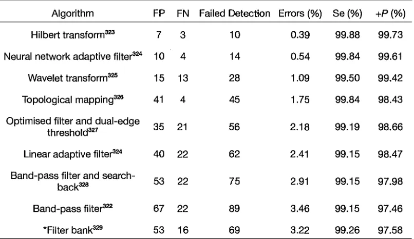
Table 2. Comparison of the performance of the Hilbert transform QRS detection algorithm
with a range of other high performance algorithms ... 100
Table 3. Power calculations at the 90% level for 40, 60 & 80% recruitment rates from a possible
total of 374 children ... 137
Table 4. Geometric mean (geometric SD) of cardiovascular parameters in adults at rest and
during three stress tasks ... 145
Table 5. Normalized regression coefficients relating birth weight to cardiovascular parameters
in adults at rest and during three stress tasks ... 146
Table 6. Normalized regression coefficients relating the size at birth and gestational age of
women (N = 68) to their cardiovascular responses to stressors ... 148
Table 7. Birth and current characteristics of the 8-year-old participants (median and
interquartile range) ... 157
Table 8. Normalised regression coefficients showing associations between birth weight and
salivary cortisol measures during a restful day at home and during a clinic visit for a stress study
in boys and girls ... 160
Table 9. Normalised regression coefficients showing how salivary cortisol measures in children
relate to their postnatal weight gain and their mothers' body composition, smoking history, and
diet in pregnancy ... 164
Table 10. Median (interquartile range) of cardiovascular variables prior to, during and following
stress ... 179
Table 11. Normalised regression coefficients relating birth weight to cardiovascular variables
prior to, during and following stress ... 180
Table U. Normalised regression coefficients showing how cardiovascular function of children
following stress relates to their postnatal weight gain and their mothers' body composition,
smoking history, and diet in pregnancy ... 184
ACKNOWLEDGEMENTS
This thesis would not have been possible without the intellectual, practical and
emotional support of a great many people. To all of these people, I am indebted and
extremely grateful. In the first instance, I thank my supervisors, David Phillips and
John Warner. David showed tremendous faith in me, coming as I did to the Medical
Research Council from my clinical training with little academic experience, and was
unstintingly encouraging and inspiring. He allowed me to broaden the scope of the
original project to explore the cardiovascular physiology that excited me and was
extraordinarily generous both in his intellectual and practical support. John has also
been essential to the successful completion of this work. He encouraged me to follow
my ambition to become an academic and was always available to provide the guidance
and sage advice that I needed to do so successfully.
This thesis would also not have been possible without the contributions of a number of
collaborators. I am extremely grateful to all of my colleagues and the staff at the
Medical Research Council. These include Keith Godfrey, who carried the original study
of the cohort of children that I followed-up and was generous with his advice,
encouragement and intellectual stimulation throughout and Clive Osmond who has
patiently, and with great humour, shepherded me through the tangled paths of
statistical analysis - all of the analyses in this thesis have benefited from his input. I am
also deeply indebted to David Simpson and Alessandro Beda at the Institute of Sound
and Vibration Research. When it became apparent that I would need some heavy duty
signal processing to carry out this work, I was put in touch with David. He
enthusiastically welcomed a physician, with little experience of signal processing, as a
myself transformed from a competent programmer and MATLAB novice to (I hope) a
competent amateur signal processing engineer and MATLAB programmer. For this I
am extremely grateful. In addition, both he and Alessandro have contributed directly
in countless ways to the signal processing that is laid out in this thesis. Although the
bulk of the programming involved was mine, both David and Alessandro contributed
widely to all areas of the software development, signal processing strategies and
debugging. The implementation of the baroreflex algorithm described in section 2.6.3
was entirely Alessandro's work. I look forward to our future collaborations together. I
would also like to thank Alexandra Ward and her collaborators in Adelaide, Australia
(Jeffrey Robinson, Vivienne Moore, Caroline Smith, Meaghan Coyle and Catherine
Gibson) for generously allowing us access to the data from their study. To carry-out the
sort of large-scale study of psychological stress in children that is presented in this
thesis is no small feat and would not have been possible without the practical
contributions of a small army of individuals. In the first instance, I should thank Tracey
Tudball who cheerfully took over the coordination of the study and dealt brilliantly
with the participants and all of the staff involved. I should also thank Peter Goulden,
Jennifer Trewin, Sharon Horne, Lynda Middleton, Jenni Lee, Hannah Ferguson and
Rosemary King who carried out the children's study with me and the numerous staff at
the Wellcome Trust Clinical Research Facility and Medical Research Council who gave
generously of their time to sit on the Trier Social Stress Test panels. Thanks also go to
Pete Wood, Christine Glenn and the staff of the Regional Endocrine Laboratory for
running the cortisol assays with such speed and efficiency, to Pat Taylor and her staff
who carried out all of the DXA scans, to Andy He who spent many hours patiently
writing and debugging the online data collection software for the study protocol, to
Eric Halton (head teacher at Ridgemede School) who welcomed us to his school and
allowed us to recruit children for our pilot study, and to Pete Willcocks and Stephen
Clitheroe in Medical Physics who built the equipment station with me and were
cheerful company as well as expert wielders of the soldering iron. I should also like to
thank all of the participants of the study and particularly the children (from the cohort
and from Ridgemede School). They rose to the challenges of the stress study with
admirable enthusiasm, resilience and courage, often exceeding the expectations of
adults and reminding us why it is essential not to underestimate children's desire,
willingness and ability to participate in clinical research. A number of companies gave
generous contributions to my study. I should like to thank John Kerslake and Cad
bury-Schweppes for donation of large quantities of Trident sugar-free gum and Toys'R'Us,
Tomy, MatteI, and Bandai for their generous donation of toys. In the early stages of
study design, I spent a week at the University of Minnesota and a week at Emory
University where I benefited greatly from helpful discussion with Megan Gunnar,
Bonny Donzella and their colleagues at the former institution and with Carol
Worthman and her colleagues at the latter. I am indebted to them for their generous
hospitality. I am also most grateful for the helpful advice of John Karemaker regarding
non-invasive measures of autonomic nervous system function and of Clemens
Kirschbaum and his wife, Angelika Buske-Kirschbaum who provided much invaluable
advice on the Trier Social Stress Test. David Phillips is the holder of a National
Institute of Child Health and Human Development grant (1 R01 HD41107-01) which
provided much of the financial support for my work.
My final and most heartfelt thanks go to my family. To Dad for all of his belief and
encouragement over the years and to his wife, Linda. Their emotional and financial
support is very much appreciated. Finally, much love and thanks go to my wife,
Stephanie and my son, Elwyn. Their love and support have been both a welcome relief
ABBREVIATIONS
11 J3-HSD
ACTH AGA AngII ANP ANS ApoE AR ARMA BHR BMI BNST BSA cAMP CARDIA CBG CES CNS CO cOT CRH CSS CVP
11 J3-hydroxysteroid dehydrogenase
Baroreflex sensitivity
Adrenocorticotrophic hormone
Appropriate for gestational age
Angiotensin II
Atrial natriuretic peptide
Autonomic nervous system
Apolipoprotein E
Autoregressive
Autoregressive moving-average
Borderline hypertensive rat
Body mass index
Bed nucleus of the stria terminalis
Body surface area
Cyclic adenosine monophosphate
Coronary Artery Risk Development in Young Adults
Corticosteroid binding globulin
Centre for Epidemiologic Studies
Central nervous system
Cardiac output
Corrected QT interval
Corticotrophin releasing hormone
Carotid sinus stimulation
Central venous pressure
DELFIA DNA DXA ECG FFf Ff GABA GR GREs HF HP HPAA HPhf HPlf HPratio HR HRV IGF-l IRSD LDL LF LREC LVET MAP MR
Dissociation-Enhanced Lanthanide Fluorescent Immunoassay
Deoxyribonucleic acid
Dual energy X-ray absorptiometry
Electrocardiogram
Fast Fourier transform
Fourier transform
Gamma-aminobutyric acid
Glucocorticoid receptors
Glucocorticoid Response Elements
High-frequency
Heart period (the interval between adjacent heart beats)
Hypothalamic-pituitary-adrenal axis
High-frequency heart period variability
Low-frequency heart period variability
Ratio of low to high frequency heart period variability
Heart rate
Increment of heart rate from rest to stress
Heart rate variability
Insulin-like growth factor 1
Index of Relative Socioeconomic Disadvantage
Low density lipoprotein
Low-frequency
Local Research Ethics Committee
Left ventricular ejection time
Mean arterial pressure
mRNA MSNA NGF NO PEP PNS POMC PVN RMS RSA RSNA SA SAP SAPlf SGA SNS STR SV SVR SVRI TSST-C VLF VVC WPT WT
Messenger ribonucleic acid
Muscle bed sympathetic nerve activity
Nerve growth factor
Nitric oxide
Pre-ejection period
Parasympathetic nervous system
Pro-opiomelanocortin
Paraventricular nucleus
Root mean square
Respiratory sinus arrhythmia
Renal sympathetic nerve activity
Sinoatrial
Systolic arterial pressure
Increment of systolic arterial pressure from rest to stress
Low-frequency systolic arterial pressure variability
Small for gestational age
Sympathetic nervous system
Systolic time ratio (ratio of PEP to LVET)
Stroke volume
Systemic vascular resistance
Systemic vascular resistance index (SVR normalised by BSA)
Trier Social Stress Test for children
Very low-frequency
Ventral vagal complex
Wavelet packet transform
Wavelet transform
Chapter
1.
INTRODUCTION
The major clinical drive behind the work presented in this thesis is a desire to better
understand cardiovascular disease and its related conditions such as hypertension.
Despite the fact that diseases of the heart and vasculature are amongst the most
prevalent and serious of all human health disorders, and despite the vast body of
research on these conditions, a comprehensive understanding of their development has
still not emerged. This introduction deals with a number of questions which arise from
relatively recent research which departs from the traditional viewpoint that the
significant pathogenesis of cardiovascular disease occurs in mid to late adulthood when
cardiovascular diseases usually appear. Although it is now clear that lifestyle choices,
such as the decision to smoke, lead a sedentary existence or eat a poor diet, are
significant risk factors for cardiovascular disease, is it possible that these apparent
choices are not simply a matter of free will or sociocultural pressures but have potent
underlying biological imperatives too? Do the processes leading to cardiovascular
disease begin in early life, even before birth? If so, are they determined entirely by the
influence of genes or are there mechanisms by which environmental factors may
influence them also? Although stress is known to influence the development of
cardiovascular disease - what is stress? How is it measured and how does a
psychological factor, a process of the mind, connect with cardiovascular disease, a
condition of the body? Finally, is it possible that the structure and function of
physiological pathways that are activated by stress (the wiring between mind and
body) are altered in response to early demands on an individual with a detrimental
Addressing these questions, this introduction proceeds as follows: Initially, I have
summarised the evidence that individual differences in adult physiology and, therefore,
in risk of adult disease, do not simply come from differences in genotype but may be
influenced by a variety of environmental agents during vulnerable periods of
development. As this thesis is focused on cardiovascular disease, I go on to address the
burden of cardiovascular disease, the processes (or risk factors) involved in
cardiovascular disease, and the evidence that cardiovascular disease is related to
prenatal development. Focus is then given to stress and psychosocial processes as
specific cases of factors which influence risk of cardiovascular disease. The issue of the
definition of stress is dealt with and an argument is forwarded that stress, through its
effects on neuroendocrine physiology and behaviour, may affect a whole range of the
risk factors known to determine cardiovascular disease, such as hypertension,
dyslipidaemia, glucose intolerance, and perhaps even lifestyle behaviours. The
physiology of the neuroendocrine pathways involved is then detailed and the
hypotheses of this thesis are stated.
1.1
Developmental Origins of Health and Disease
The search for determinants of health and disease is at the core of modern medical
science. Understanding of non-communicable diseases has come largely from a focus
on lifestyle factors in adult life and from genetics. Recently, however, attention has
shifted to prenatal and childhood development as a third major factor in the origins of
later disease. Large-scale epidemiological observations have led to the hypothesis that
environmental factors acting during vulnerable periods of early development can have
dramatic and lasting effects on the propensity to develop disease in adulthood.9,lo This
is supported by research in developmental biology, evolutionary biology, and animal
and human physiology, which has identified a wide range of potential mechanisms
acting at varied times, from pre-conception to early childhood. Encompassed as the
Developmental Origins of Health and Disease Hypothesis, these findings have led to a
rapidly expanding area of research where better understanding of the processes
involved has potentially significant medical and socioeconomic implications.9 ,l1,12
1.1.1 The Longstanding Influence of Early Developmental Experience
A universal feature of living organisms is their ability to modify their final form and
function during development in response to environmental demands. This is termed
developmental plasticity and is more formally defined as 'the ability of a single
genotype to produce more than one alternative form of structure, physiological state or
behaviour in response to environmental conditions.'13 Without this plasticity, the
survival characteristics of individual organisms would be effectively fixed at
conception, with no capacity for adaptation to a post-conceptual environment that was
'unpredicted' by their genome. Natural selection alone requires a timescale of at least
one generation (usually many more) to adapt to environmental change. Given that the
timescale of dramatic environmental change can often be far shorter than this, it seems
highly unlikely that natural selection would not have favoured the development of
organisms capable of exploiting potential non-genetic mechanisms for adaptation to
environmental change within one generation. In fact, such a process has long been
recognised. Although Darwinism is often equated with a rigid adherence to the idea
that natural selection alone is responsible for adaptation of organisms to their
environment, Darwin showed considerable insight into the concept of developmental
showed much greater variability than wild types, he rejected the contemporary view
that the reproductive system alone produces such variation:
'Some authors believe it to be as much the function of the reproductive
system to produce individual differences, or very slight deviations of
structure, as to make the child like its parents. But the much greater
variability, as well as the greater frequency of monstrosities, under
domestication or cultivation, than under nature, leads me to believe that
deviations of structure are in some way due to the nature of the
conditions of life, to which the parents and their more remote ancestors
have been exposed during several generations.'14
He also identified the reproductive system of the parents as the most likely arena for
environmental conditions to impact on offspring variability:
'1 have remarked ... that the reproductive system is eminently
susceptible to changes in the conditions of life; and to this system being
functionally disturbed in the parents, I chiefly attribute the varying or
plastic condition of the offspring.'14
Darwin felt that the window for such environmental interference in reproduction was
likely to be pre-conceptual and, indeed, evidence for pre-conceptual environmental
influences on offspring development has since emerged with, for example, a study of
mothers who experienced a severe famine in Holland during the Second World War.1S
As expected, this study showed that starvation in pregnancy resulted in a significant
decline in the birth weight of the offspring. Less expected was the finding that birth
weight and perinatal mortality were affected in the subsequent offspring of the
daughters of women who experienced famine during pregnancy as well. Similar
second-generation effects of impaired nutrition during pregnancy have also been
demonstrated in animal studies.l
6-20 One possible explanation for the epidemiological
observations in Holland may be that the famine affected the germ cells of the female
fetuses in utero with subsequent effects on the offspring formed from those germ cells. Studies in animals indicate that inheritance of an epigenetic trait induced by the
environment prior to conception can be produced experimentally and appears to be
mediated by incomplete erasure of deoxyribonucleic acid (DNA) methylation
(imprinting) during meiosis, causing the offspring to express certain genes at a level
similar to their parents.2
1,22 There are other possible explanations for these observations. For example, development of uterine vasculature and structure, and reproductive
endocrine axes may also be altered by an adverse fetal environment.23
Thus, famine
exposure in utero might have detrimental effects on the reproductive physiology of female offspring by these mechanisms, resulting in smaller second-generation offspring.
'Lamarck was the first man whose conclusions on the subject excited
much attention. This justly-celebrated naturalist first published his
views in 1801 ... He first did the eminent service of arousing attention to
the probability of all change in the organic, as well as in the inorganic
world, being the result of law, and not of miraculous interposition ...
With respect to the means of modification, he attributed something to
the direct action of the physical conditions of life, something to the
crossing of already existing forms, and much to use and disuse, that is, to
the effects of habit.'
Although the archetype of Lamarckian evolution was refuted by the work of Darwin
and Gregor Mendel,26 it is now clear that elements of Lamarck's thinking have been
unfairly dismissed. Indeed, there are a number of mechanisms by which acquired traits
may be inherited. Not only are there epigenetic modes of inheritance, as already
discussed, but also entirely non-genomic mechanisms of inheritance where the
development of the offspring is influenced by placental and maternal physiological
conditions with permanent effects on the physiology and behaviour of the offspring
that may continue to have effect over subsequent generations. Furthermore, it is now
clear that the window of sensitivity to these mechanisms varies, is not limited to a
pre-conceptual time period and may include any part of prenatal life or early childhood.
This phenomenon is frequently referred to as developmental programming.
The clearest evidence that non-genomic maternal characteristics and uterine
environment can predetermine adult characteristics of the offspring comes from
animal experiments19
,27-JO where, in contrast to experiments in humans, genetic
homogeneity can be assured, reducing the confounding influence of genetic variability.
These studies show that significant modifications in the offspring's size at birth,
metabolic and cardiovascular function, and behaviour, which have lasting effects on the
offspring into adulthood, can be induced by manipulation of the maternal
environment. Where more than one generation of offspring was studied, it has been
shown that altering maternal nutrition or exercise prenatally, and postnatal nutritional
or handling interventions have significant effects on birth weight,16,31 glucose tolerance20
and function of the hypothalamic-pituitary-adrenal axis (HPAA)32 in subsequent
generations. Thus, the secular trends for increasing birth weight observed in several
populations worldwide33-35 or inter-generational effects of low birth weight or raised
blood pressure, may be partially explained by non-genomic intergenerational
inheritance mediated by these mechanisms.10,36
Not all changes in the offspring induced by the environment may be adaptive, allowing
the offspring to increase their likelihood of survivaL Indeed, changes in the offspring
may simply result from environmental constraint where alternative developmental
pathways are unavailable to the fetus_ However, phenomena such as brain-sparing,37 the
process whereby a fetus diverts supply of nutrients and oxygen away from less
important organs such as the liver, in favour of brain growth during times of impaired
oxygen or nutrient supply from the mother, suggest that fetal development seeks the
best trade-off for survival in response to adverse conditions_ This concept is elaborated
in the womb provokes the developing baby to become thrifty, increasing blood sugars
to promote brain growth at the expense of glycogen storage in the muscles and muscle
growth. This phenotype persists into adulthood, increasing the likelihood of insulin
resistance and type 2 diabetes, particularly in the context of later adiposity. Support for
this theory comes from maternal nutrient restriction studies where altered insulin
homeostasis is seen in the offspring.38The observations that maternal overnutrition also
leads to altered metabolic function in the adult offspring and that the postnatal
nutrient environment is important in determining the extent of prenatal nutrient
influence on the adult offspring, has led to an extension of the Thrifty Phenotype
hypothesis. The Predictive Adaptive Response hypothesis proposes that the
developmental choices made by the fetus in response to environmental influences are
purposeful and designed to better adapt the fetus to the environment they are likely to
encounter in postnatal life. 12 This hypothesis assumes that until our recent evolutionary
past, most pregnant mothers would experience an environment and nutrition that was
likely to continue during their offspring's lifetimes, making a mismatch between fetal
predictive adaptive responses and later reality unlikely. Modern living has created
many more opportunities for such a mismatch to occur and it is proposed that the
consequence of these developmental false predictions is the development of adult
diseases such as cardiovascular disease or type 2 diabetes mellitus and precursors to
these diseases such as dyslipidaemia, impaired glucose tolerance, or vascular
endothelial dysfunction.
1.1.2 Cardiovascular Disease
The World Health Organisation estimates that worldwide, cardiovascular disease
caused the death of 14.7 million people in 1990 and 17 million people in 1999 which
approximates to 30% of all deaths worldwide per annum.39 Cardiovascular disease is the principal cause of death throughout the world except for sub-Saharan Africa where
it is expected to overtake infectious diseases as the leading cause of death in the next
few years. Although traditionally thought of as a disease affecting rich industrialised
nations, 80% of these deaths occur in countries with low or modest gross domestic
product such as India, China, Russia, Argentina and Poland.39
As these countries
become increasingly affluent, the populations are likely to adopt a more Western
lifestyle and therefore a worsening risk profile for cardiovascular disease. Clearly, the
rise in detrimental lifestyle factors such as obesity, smoking and lack of exercise will
worsen the global epidemic. However, the traditional viewpoint that rapid increases in
disease prevalence in developing nations are explained by the combination of these
lifestyle changes with an inherited predisposition to cardiovascular disease is
questionable. Interventional studies targeting these lifestyle factors have failed to
significantly reduce levels of cardiovascular disease.40
This suggests that although these
factors modify risk of developing cardiovascular disease, they do not fully account for
it. Furthermore, rapid changes in disease incidence in a short time period are unlikely
to be explained by changes in gene frequency in these populations. Therefore, until
other factors involved in the pathogenesis of cardiovascular disease are more fully
investigated, it is unlikely that successful interventions aimed at curbing this global
1.1.3 Risk Factors Associated with Cardiovascular Disease
Approximately 75 % of cardiovascular disease can be attributed to conventional risk
factors.41 Of these, the major modifiable risk factors are hypertension, dyslipidaemia, tobacco use, physical inactivity, obesity, diabetes mellitus, and poor diets that are low in
fruit and vegetables and high in saturated fat. Other modifiable risk factors include low
socioeconomic status, mental ill-health, psychosocial stress, excessive alcohol use, and
use of hormonal medications such as the oral contraceptive pill. Although many of
these risk factors are related directly to lifestyle, others, such as hypertension and
diabetes have complex aetiologies and many of these risk factors may interact with one
another to alter overall risk of disease. Furthermore, risk factors that had been
previously thought to be exclusively determined by behaviour may have powerful
biological imperatives underlying these apparent lifestyle choices. For example, obesity
is undoubtedly the consequence of an inappropriately high-energy diet often
accompanied by inactivity, but until recently, these factors were thought to be entirely
the consequence of individual lifestyle choices. Now, there is growing evidence to
support the view that appetite regulation and physical activity are influenced by the
prenatal nutritional environment and may be modulated in early life by postnatal
nutrition.4
2-46 In addition, hypercaloric diets in later life amplify the effect of prenatal nutrition on later sedentary behaviour suggesting that these biological and behavioural
factors are strongly interlinked.43
,47,48 Animal studies have shown that fetal adipose
tissue development is altered by maternal nutrition such that undernutrition during
fetal life leads to adult obesity, hyperinsulinaemia, hyperleptinaemia, reduced
locomotor activity and hyperphagia, particularly where offspring are fed high-energy
diets. These effects can be reversed by administration of leptin, suggesting that fat
signalling is important in controlling appetite and sedentary behaviour in the
offspring.49,so It might be argued that if these processes prove to be significant in human
development, they may provide an explanation for the poor record of large
interventional studies that have attempted to reduce cardiovascular risk status by
altering lifestyle choices.40,sl
Although cardiovascular disease typically occurs in middle-age or later, many of these
modifiable risk factors appear in childhood with a worrying worldwide trend towards
earlier onset of obesity, smoking, poor diet, and lack of exercise. Physical activity
decreases dramatically around age 10, particularly in girls, leading to obesity.52 Obesity
in childhood is increasing rapidly, not only in North America and Europe, but also in
traditionally lean popUlations such as the Chinese and Japanese. Worldwide, it is
estimated that 18 million children under the age of five are overweight and 14% of
13-15 year-old students smoke.41
With this degree of risk, it is unsurprising that features of
cardiovascular disease are now also found in young children. However, there has long
been evidence that even in children without such risk factors, pathological changes
associated with cardiovascular disease appear in early childhood. In post-mortem
studies of children after accidental death, features of coronary atherosclerosis such as
fibrous plaques and fatty streaks are frequently found, particularly in children whose
risk factors include smoking, dyslipidaemia, hypertension, and obesity.53 There is
growing evidence that blood pressure status, too, is established in early life.54
Longitudinal studies following individuals though childhood and into adulthood show
that individual blood pressure rankings, relative to the peer group, remain fairly stable.
This phenomenon is known as tracking. There is a growing awareness that many of the
risk factors for cardiovascular disease have their origins in early life and that they may
be driven by biological as well as sociological factors. Despite this, relatively few
studies have targeted interventions, intended to reduce cardiovascular risk, at children
carried out in adults. Although they are potentially informative, studies in adults may
not provide an adequate picture of the processes involved in generation of
cardiovascular disease due to the confounding influence of adult factors such as the
vascular pathology resulting from those processes, the cumulative influence of varied
lifetime experiences on, for example, stress response systems, and the marked
moderating effects of post-pubertal hormone levels on cardiovascular function and
control.
1.1.4 Size at Birth and Risk of Cardiovascular Disease
It has long been known that small size at birth is associated with an increased risk of
cardiovascular disease such as myocardial infarction or cerebrovascular disease such as
stroke. To better understand these associations, many international studies have
examined the relationship between size at birth and a range of conditions known to
predispose to the development of cardiovascular disease. Specifically, small size at birth
is associated with a cluster of disorders known as the metabolic syndrome which
substantially increase the risk of development of cardiovascular disease. These
disorders include glucose intolerance, type 2 diabetes, dyslipidaemia, insulin resistance
and raised blood pressure.55
Evidence from over 80 epidemiological studies shows that low birth weight is
associated with increased blood pressure and conditions related to raised blood
pressure such as vascular endothelial dysfunction, glucose intolerance, insulin
resistance and cardiovascular disease, in childhood and into adult life.9
.56-58 Such studies have been fundamental to the advancement of the Developmental Origins of Health
and Disease hypothesis but have necessarily relied upon measures of size at birth such
as birth weight to indicate impaired fetal growth. On occasion, this has led to
misunderstanding their results to represent a causal role of small size at birth itself in
the development of adult diseases. However, as studies of twins,S9-6Z siblings,63,64 first
cousins,65 and intergenerational pairs of first births66
suggest that the heritable
component of birth weight is small, it is felt generally to be an imperfect but useful
reflection of adverse intrauterine influences to which fetal growth is highly sensitive.67
Indeed, whilst variations in genotype are unlikely to explain large variations in size at
birth on a population level, it is not entirely clear what the major determinants of size
at birth are or at which stages during fetal life they have their greatest impact.
Therefore, whilst most studies in humans have had to rely, perhaps necessarily, upon
birth weight and other markers of size at birth as predictors of later disease processes
in the offspring, animal studies have been more directed at the factors which possibly
underlie both the alterations in size at birth and the programming of later disease, such
as maternal nutrition.19
,68,69These studies suggest that effects on outcome measures such
as postnatal blood pressure, arterial endothelial function, glucose tolerance and HPAA
responsivity of prenatal manipulations such as maternal nutrient restriction may be
demonstrated in the absence of alterations in offspring birth weight. This has led to
questions about the role of birth weight in the developmental origins of later disease.
Such studies make it clear that birth weight need not be affected for programming to
occur but where birth weight is altered, it is unclear whether this is a coincidental and
unreliable effect of underlying processes which also set about the development of later
disease by growth-independent pathways or whether the degree to which size at birth
Interestingly, a recent study of sheep was carried out to address the hypothesis that a
reduction of size at birth was not on the causal pathway between maternal
malnutrition and adult disease.7o The pregnant ewes in this study were severely
undernourished for 10 or 20 days in late gestation and compared to a group of
normally fed ewes. To the authors' surprise, maternal nutrition in late gestation was not
a significant determinant of blood pressure, glucose tolerance or insulin-like growth
factor 1 (IGF-1) response to growth hormone challenge in the offspring once their
birth weight and current weight were taken into account. However, a reduced birth
weight did predict raised blood pressure, reduced glucose tolerance and raised IGF-1
levels. They postulate that the timing of their nutritional insult was a key determinant
of this result as most of the studies of nutritional programming relate to interventions
in early gestation or at the time of conception. However, they also suggested the
possibility that size at birth may reflect the underlying programming process more
closely than the level of maternal nutrition. Indeed, fetal nutrition need not be strongly
related to maternal nutrition at all given the potential modulating effects of the
maternal metabolic and endocrine milieu, placental transport capacity or uterine blood
flow. This raises the possibilities that birth weight remained a better indicator of the
severity of fetal undernutrition than maternal nutrition was or perhaps that other
processes which were unrelated to nutrition were responsible for both the reduction in
birth weight and the programming phenomena in the offspring. Where does this leave
studies of programming in humans? Whilst it is likely that specific factors such as
maternal nutrition have a specific programming effect which should be studied, I
would suggest that measures of size at birth remain useful as 'catch-all' non-specific
epiphenomena of a range of underlying processes. Therefore, until the individual
effects of all these processes are catalogued and understood, measures of size at birth
such as birth weight should continue to be examined even if only as an adjunct to more
directed measures of, for example, maternal health or diet.
Whilst birth weight has been useful as a non-specific, and possibly non-sensitive,
indicator of the quality of prenatal growth and development in the human
epidemiological studies, it has helped to provoke controversy. As no single process or
set of processes can be easily identified which may have contributed to both alterations
in birth weight and later disease in the human studies, this has facilitated a wider
criticism of the Developmental Origins of Health and Disease Hypothesis. This is in
spite of the clear demonstrations of programming by specific interventions in animal
studies. One such criticism addresses the strength of the epidemiological associations
between birth weight and risk factors for cardiovascular disease such as blood pressure
status and plasma lipid profile. Despite the now substantial body of evidence
supporting such associations, two recent meta-analyses carried out by the same
research group suggest that the size of these associations (but not their existence), their
clinical relevance and, by extension, the Developmental Origins of Health and Disease
hypothesis are all in doubt.71
,72The bombastic nature of their conclusions is not entirely
supported by their analyses, however. Whilst meta-analyses are often a useful adjunct
to original science, meta-analytic approaches must be used judiciously to avoid
frequently encountered pitfalls.73 It is well known that one of these frequent pitfalls is the dilution of effect sizes that occurs when meta-analyses include large studies with
relatively poor quality data. In the majority of the larger datasets drawn upon in these
meta-analyses, blood pressure and birth weight were obtained by self-report rather
than by direct measurement. Their use of a fixed-effects meta-analysis gives undue
weight to such studies by virtue of their size which dilutes the estimate of effect size
generally involves more than just simplistic combination of effect sizes of a set of
studies. Tests are used to determine if study outcomes show more variation than that
expected from sampling different research participants. If so, additional predictor
variables are used in the analyses to account for method of measurement, population
sampled or aspects of study design. Some methodological weaknesses in studies can
then be corrected statistically. For example, it is possible to correct effect sizes or
correlations for the downward bias due to measurement error, rounding errors or
restriction on score ranges. There are further difficulties with their conclusions
regarding the association between size at birth and later blood pressure: whilst the
effect of birth weight on blood pressure across the normal range has always been noted
to be modest, their conclusions take no account of the much greater impact of birth
weight on risk of hypertension.74
An additional source of controversy has arisen over the interpretation of statistical
associations between size at birth and health outcomes in later life. Whilst most
investigators have interpreted such associations as evidence for the impact of prenatal
growth on processes which affect health in later life, Lucas and others75
have argued
that, because these results sometimes depend on adjustment for measures of current
size, they reflect postnatal growth. For example, if there is no correlation between birth
weight and blood pressure in a study, but both are positively related to current weight
(as is usually the case), adjustment for current weight may reveal a negative correlation
between birth weight and blood pressure. Thus, this statistical model reveals an effect
of birth weight on blood pressure only in the context of current size. Lucas et al.75 argue that this is a measure of postnatal growth. However, this would be a poor measure of
postnatal growth, at best, because relationships between prenatal and postnatal growth
trajectories are unlikely to be well approximated by a linear model with two time
points. Indeed, it is well-known that growth in childhood is characterised by periods of
variable growth acceleration that depend on a wide range of factors such as nutrition
or emotional support and not, simply, upon size at birth. In essence, such models are
more akin to studies where subjects are stratified by current weight to assess the effect
of birth weight on outcome measure within each stratum and, therefore, arguably the
best statistical approach for understanding the mechanisms relating prenatal growth to
later disease.
Such arguments over the relative importance of prenatal versus postnatal growth in the
determination of health outcomes are unlikely to be settled by the injudicious
application of algebra to the problem.76 Indeed, I would argue that this approach is as futile as rearranging Ohm's law in an attempt to prove that resistance is more
important than current as a determinant of potential difference. Only longitudinal
studies where postnatal growth trajectories are assessed at multiple key points during
childhood are likely to define the importance of postnatal growth in the origins of later
health and disease. However, due to the relative paucity of such data, controversy
exists in this area also. Drawing from their data on postnatal growth in premature
infants, Singhal and Lucas suggest that early postnatal growth may have a detrimental
impact on the risk of insulin resistance in later life.7s There are two difficulties with this.
Firstly, they have drawn conclusions about normal child development from a small
study of premature infants using split pro insulin as a marker of insulin resistance.
Given the well-known additional risks and pathologies involved in prematurity and the
uncertainty regarding the interpretation of split pro insulin, this is likely to be
misleading. Secondly, growth in their study was only assessed using weight at birth, at
two weeks and in adolescence. Their finding of a positive association between growth
but should not be used to draw conclusions about the effects of growth in infancy as a
whole. Indeed, a number of studies suggest that over the first year of life, as a whole,
growth is inversely associated with adverse outcomes such as insulin resistance, high
blood pressure and cardiovascular disease in later life, and that subsequent accelerated
growth in later childhood, leading to obesity, carries additional risk.77
-8Z Furthermore,
many such studies suggest that the effects of prenatal and postnatal growth may be
separate and, therefore, represent entirely different programming phenomena. In view
of this, I would suggest that measures of size at birth remain useful indices of prenatal
adversity.
The timing and duration of intrauterine adversity seems to be important and may not
be reflected at all in measures of size at birth. 19,83-86 Therefore, studies relying on size at
birth alone may under-detect evidence of developmental influences on later disease (a
type II error), but significant associations in studies using size at birth as a measure of
fetal growth are unlikely to be due to some other determinant of size at birth such as
genetic factors. It is also important to note that most studies report continuous
relationships with size at birth across the normal range suggesting that a universal
feature of fetal development, not a process confined to the extremes of fetal growth, is
responsible for these findings. Therefore, size at birth in studies of normal healthy
populations is believed to largely represent the extent of maternal constraint, i.e. those environmental processes that occur during pregnancy and influence size at birth but
are not necessarily extreme enough to cause immediate pathology. Any process which
either limits uteroplacental transfer of nutrients, perhaps by impairing adequate
placental implantation or function, or modifies the nutrients made available by the
mother, is therefore a candidate cause of maternal constraint. Maternal age, body size
and obesity, parity and a catalogue of processes that limit fetal nutrient supply are all
considered to contribute to maternal constraint through their influences on both of
these mechanisms.67 Other environmental factors which contribute to this process include: season of birth1
,2 and infection,87 two factors which may be linked, although
ambient outdoor temperature variation has an independent effect/,2 maternal
smoking,88 and maternal nutrition which may depend, not only on maternal intake, but
also on maternal nutritional reserves and tissue turnover of protein and fat.89,90 Of
course, fetal nutrient supply need not necessarily be limited to have a detrimental
impact but may involve an excess supply of specific nutrients, an unbalanced nutrient
supply due to maternal metabolic conditions, or an unbalanced maternal diet.
Gestational diabetes, a condition where excess availability of glucose may lead to
macrosomia in the newborn and, therefore, excessive birth weight would be one
example and also an example of where birth weight is a poor measure of fetal
adversity. Indeed, in some studies,_ a V-shaped relationship between size at birth and
later non-insulin dependent diabetes or cardiovascular disease has been reported
which probably reflects the impact of gestational diabetes.91 •
92
Unlike other
environmental factors such as smoking, infection and seasonal ambient temperature, all
pregnancies may be influenced by maternal diet and body composition. There is
increasing evidence that fetal development is affected by variation of nutrients across
the normal range of western diets.93 Therefore, maternal nutrition is seen as a promising target for potential interventions to improve fetal development and any consequent
1.1.5 Maternal Nutrition and Risk of Cardiovascular Disease
In humans, the availability of nutrients to the fetus is determined by the mother's body
composition at conception, her nutritional stores and her diet during pregnancy,
together with her ability to supply nutrients to the fetus through the placenta. Recent
studies have shown that variations in the balance of maternal protein, carbohydrate
and green vegetable intake during pregnancy are associated with altered fetal and
placental growth and with increased adult blood pressure in the offspring. A follow-up
study of men and women whose mothers took part in a survey of diet in pregnancy in
Aberdeen showed alterations in placental weight at birth and increased blood pressure
in adult life at both extremes of the balance of maternal animal protein to
carbohydrate intake in pregnancy.94 Support for adverse long-term effects of maternal
diets with a low ratio of animal protein to carbohydrate has come from the experimental studies in pregnant rats95 and from follow-up of studies children and
adults in the Philippines and Holland.96,97 Support for adverse effects of a high ratio of
animal protein to carbohydrate has come from follow-up of men and women in
Motherwell whose mothers had been advised to eat a high animal protein, low
carbohydrate diet in pregnancy. A higher maternal intake of meat and fish during
pregnancy, coupled with a lower intake of carbohydrate and green vegetables, was
associated with increased adult blood pressure.98
Prospective studies in Southampton support the importance of dietary balance during
pregnancy and suggest that maternal folate status may be a key determinant of the
effects that result. In an initial survey of women studied at a time when most took
folate supplements in late pregnancy, high intakes of meat and fish protein in relation
to carbohydrate were associated with larger fetal size.93 In contrast, in a subsequent
survey in which many women took folate supplements in early pregnancy but few took
them in late pregnancy, those whose diets were high in meat and fish, but low in folate
or green leafy vegetables had babies of lower birth weight. These findings are
consistent with a review of trials of protein supplementation in pregnancy, which
concluded that high protein density supplements led to lower offspring birth weight.99
The observation of adverse effects of high protein maternal diets appears paradoxical,
in that fetal growth represents net deposition of lean tissue and therefore an absolute
requirement for protein. Whilst an inadequate supply of protein might be expected to
impair fetal growth, it is less clear why high protein intakes may be detrimental. One
possibility is that meat and fish are rich in essential amino acids, which must either be
used for protein synthesis or oxidised.1
°O Oxidation consumes non-essential amino
acids, whose synthesis requires cofactors including folate and vitamin B6. Low intake
of green vegetables, a source of folate, accentuates the effect of high meat and fish
consumption on the offspring's blood pressure. In mothers with a limited capacity to
synthesise non-essential amino acids, maternal amino acid oxidation could impair fetal
growth as a result of reduced availability of the non-essential amino acids required in
large quantities by the fetus. Consistent with this hypothesis, increased maternal amino
acid oxidation during pregnancy has recently been associated with impaired fetal
growth.90
Although the observations from these studies suggest that the balance of maternal
meat, fish, carbohydrate and folate intake could have important implications for the
offspring's risk of cardiovascular and metabolic disease, the dietary data available in
these studies were crude and secure identification of the optimal balance of nutrients
1.2 Stress
It is becoming increasingly clear that an important means by which prenatal adversity,
perhaps reflected by small size at birth, may have long-term effects on risk of
cardiovascular disease is through adaptation of a diverse range of hormonal and
neurological systems that control growth and development. Specifically, the HPAA and
autonomic nervous system (ANS) have been implicated which are the key
physiological mechanisms by which an organism responds to stress. In seeking to
explain the pathways from environmental influences during early life to later disease,
therefore, the concept of stress may be important. Not only is there considerable
evidence that psychological processes related to stress are associated with adult
diseases, including cardiovascular disease, but also that the physiological processes
implicated in this association act to mediate the somatic effects of psychological stress
and are vulnerable to modification by early life processes. A description of these
processes and the evidence for such vulnerability are given in sections 1.3 and 1.4.
For a word that encompasses so many important features of so many areas of academic
research, stress is remarkably imprecise and poorly defined. To a physicist, for example, stress is the internal distribution of forces in a material that balance and react to the loads applied to it. Whereas, in the realms of musicology, phonology and the biological
sciences, stress has entirely different meanings. Nonetheless, a search on MEDLINE, a publicly available database of articles published in the biomedical literature, yields
over 270,000 articles since 1943 that have used the term. This amounts to
approximately 2% of the biomedical sciences literature indexed by MEDLINE. For
comparison, a search in the same database for all articles related to asthma yields less than 88,000 articles. Therefore, to dismiss the concept of stress on the grounds of
continuing difficulties with definition, would be to dismiss a substantial body of
biomedical research.
The concept of stress in the medical, biological and psychological literature arose from
the work of Hans Selye who first used the term in 1936 to encompass a wide range of
strong external stimuli, both psychological and physiological, that can cause a
physiological response which he termed the 'general adaptation syndrome'.1Ol
Unfortunately, Selye was not well versed in the existing definition of stress used by
physicists for centuries to describe the elastic properties of materials. Had he been, he
might have chosen the word strain, which physicists use to describe the effect of
physical stress on a material. Further confusion arose from his initial use of the term
stress to describe the cause of the resulting condition, which he initially called the general adaptation syndrome but later dropped and replaced with the term stress. Thus,
as one critic wrote after drawing on citations of Selye's publications, 'Stress, in addition
to being itself, was also the cause of itself, and the result of itself.'lo2 In order to correct
this confusion, Selye introduced the term stressor to describe the cause of stress. The
result of this early semantic confusion has been a steady adaptation and dilution of the
term by both academics and lay public so that stress now has such an expansive and
fluid definition as to be almost meaningless when used on its own. Most researchers
looking at the effects of psychological stress on animal and human physiology have
approached this problem by adopting a circular definition of stress as any condition
where physiological stress responses are present, such as enhanced adrenocortical or
adrenomedullary activity, and a definition of stressors as any causative agents which
produce such responses. As a result of this, some have called for an abandonment of
the term, stress. However, when used with clarity about the factors to which the user is
factors but it would be unwise to assume that measurement of similar physiological
responses (stress responses) to different stressors implies a common underlying
process, i.e. that the stress is the same. To give a hypothetical example, sleep
deprivation and maternal separation might both cause similar increases of blood
pressure in a group of animals, leading to the idea that the same stress condition is
being produced. However, one stressor might act entirely through altered endocrine
activity whilst the other might rely upon alterations in ANS activation. Furthermore,
the mechanisms by which a stressor produces a stress response might differ between
individuals with different characteristics such as species, age, sex, or race. Ultimately,
work that falls within the domain of stress research in the biomedical sciences attempts
to trace causation from social and psychosocial processes through behaviour and
physiology to a disease endpoint. Perhaps then, stress is most useful as an orientating
term allowing the reader to appreciate that your work has this aim.
This thesis is focused principally on the activity of two specific neuroendocrine systems
that are activated by a variety of psychosocial and environmental challenges that occur
frequently during the life of all animals including humans. Activation of these systems
leads typically to an array of metabolic and cardiovascular changes which promote the
immediate survival of an organism in challenging circumstances but may, if prolonged
or frequent, interfere with somatic processes of repair, defence against illness, and
development. 103 These systems are the HPAA, which produces cortisol (a steroid
hormone) in humans, and the sympathetic adrenomedullary axis, which produces
adrenaline and other catecholamines and acts rapidly in concert with direct
sympathetic nervous system (SNS) activation and parasympathetic nervous system
(PNS) withdrawal to prepare the body for an immediate challenge. In contrast, the
HPAA exerts slower, more persistent effects on the body to maintain a steady