WITH SPECIAL REFERENCE TO RISK FACTORS AND
ESTIMATION OF COPY NUMBERS POLYMORPHISM OF
HUMAN BETA-DEFENSIN 2 GENE (DEFB4)
This is to certify that this dissertation entitled “Clinical profile of ulcerative colitis with special reference to risk factors and estimation of copy number polymorphisms of defensin gene (DEFB4)” is the bonafide work done by Dr. E. Umakanth in partial fulfillment of rules and regulations for DM (Branch IV,
Gastroenterology) examinations of the Tamil Nadu Dr. M.G.R. Medical University, Chennai to be held in July 2008.
B.S. Ramakrishna, MD, DM, PhD, FAMS, FASc Professor and Head
Clinical Gastroenterology & Hepatology Unit Department of Gastrointestinal Sciences Christian Medical College
I wish to place my sincere gratitude to Dr. B.S. Ramakrishna, Professor and Head, Clinical Gastroenterology & Hepatology Unit, for guiding me to do this thesis as DM dissertation.
Special thanks to Mr. Pugazhendhi S. and Mr. S. Srikanth for meticulous analysis of copy number polymorphisms of defensin gene.
I sincerely thank Mrs. Sumitha & Mr. Murali for their excellent secretarial support in formatting this thesis.
I am grateful to the institution, which through the Fluid Research Grant has provided me the necessary financial aid required for the study.
Page No.
INTRODUCTION
1
REVIEW
OF
LITERATURE
3
AIMS OF THE STUDY
32
MATERIALS
33
METHODS
34
RESULTS
41
DISCUSSION
56
CONCLUSION
64
SUMMARY
65
BIBLIOGRAPHY
APPENDIX I
PROFORMA
INTRODUCTION
Ulcerative colitis (UC) is a chronic inflammatory bowel disorder that affects the colonic mucosa causing ulceration and leading to chronic blood and mucus diarrhoea. Although generally believed to be an immunological disorder, several decades of research have not unearthed the cause of UC. In the last seven years, genetically determined disorders of innate immunity have been identified as the cause of Crohn’s disease, the other major inflammatory bowel disorder. UC is different from Crohn’s disease in affecting only the mucosa and that too only in the large intestine. It is not clear whether it occurs in reaction to bacteria that exist within the lumen of the bowel. The large intestine is host to trillions of bacteria, and it is likely that the colonic mucosa has mechanisms to keep these microorganisms at bay and to maintain integrity of the mucosal barrier. It is conceivable that UC results from the breakdown of these mechanisms.
The innate immune system of the gut perceives pathogenic microorganisms through conserved molecular patterns (pathogen associated molecular patterns). Various effector molecules are synthesized and released in response to recognition of these pathogen associated patterns. The defensins are effector molecules of the innate immune system which function as anti-microbial peptides against pathogenic microbes. Reduced production of alpha-defensin by Paneth cells in the ileum is noted in patients with
known that there may be variations in gene number for beta-defensins. This is a
REVIEW OF THE LITERATURE
Ulcerative colitis (colitis ulcerosa, UC) is a chronic inflammatory bowel disorder (IBD) that affects the colon inducing characteristic ulcers. It affects the rectum and extends proximally along a variable length of colon. The main symptom of active
disease is usually gradual onset of diarrhea mixed with blood. UC also affects many parts of the body outside the intestine. It is an intermittent disease with periods of exacerbation and periods of disease remission. Although the symptoms of ulcerative colitis can
sometimes diminish spontaneously, the disease usually requires treatment to go into remission. It can usually be well controlled with medications. Most people live normal and productive lives. Control of the disease includes long term medical treatment and regular monitoring of complications.
History
1859: Dr. Samuel Wilks first described this disease as “Idiopathic Colitis”. He
recognized disease distinct from bacillary dysentery. He reported the pathologic finding of dilated and thinned colon, and noted severe universal inflammation in a patient with this condition.
1905: Lockhart–Mummery, who invented the Electric sigmoidoscope, named features as “sigmoiditis”.
1909: Sir Arthur Hurst gave more complete description of ulcerative colitis. He described sigmoidoscopic appearances and differentiation from bacillary dysentery.
1913: Brown proposed ileostomy to rest the bowel with fecal diversion and allow it to heal.
1940: Sulphasalazinewas introduced as an effective drug against ulcerative colitis 1945: Dennis in Minneapolis recommended colectomy at the same time as ileostomy 1964: Dick et al conducted controlled trial and proved the efficacy of sulphasalazine 1964: Ehrenpreis and Ericson advocated colectomy early in the course of the disease 1950: Bryan Brooke Introduced end ileostomy
1950: Truelove and Witts proved steroids as effective medical treatment of UC
Epidemiology
The incidence and prevalence of UC varies with geographic location and ethnicity. With available epidemiological data; the areas with highest rates of reported incidence and prevalence of ulcerative colitis include North America, England, North Europe and Australia. In North America the incidence rates range from 6.0 to 14.3 cases per 100,000 person years and the prevalence ranges from 37-246 cases per 100,000 persons. In Europe 12 the incidence rates range from 1.5 to 20.3 cases per 100,000 person years and the prevalence ranges from 21-243 cases per 100,000 person years.
There is a marked ethnic variation in the incidence of ulcerative colitis. One ethnic group with high incidence of this disease is the Jewish population. Incidence of ulcerative colitis in Jews is several folds higher than in the non-Jewish population. In United States it is 13 cases /100,000 in Jews compared to 3.8/100,000 in non Jews
(13).Ulcerative colitis was traditionally considered uncommon in blacks. However studies in the late 1970s revealed incidence rates were comparable between whites and
rates for ulcerative colitis in Japan have remained relatively stable at 5.5 cases /100,000 and 0.36 to 0.5 cases/10,000 person respectively.(15) The prevalence of ulcerative colitis in Indian has been reported to be substantially lower than among Europeans(16) However, South Asian immigrants in England are more likely to have ulcerative colitis than
European natives(17).This changing epidemiology with immigrant population from low risk to high risk geographic region supports the concept of environmental influence on disease development(18).
Ulcerative colitis can present at all ages although diagnosis below 5 years and above 75years is uncommon, The peak incidence of ulcerative colitis occurs in the 2nd and 3rd decades of life and 2nd peak between 60—70years. There is no gender difference at all ages (21). Ulcerative colitis is more common in industrialized than in underdeveloped countries, among urban than rural population, higher socio economic stratum than low. These observations support the notion that environmental factors influence the
development of ulcerative colitis.
Temporal influences:
There is a progressive increase in rates of ulcerative colitis incidence in last four decades, which was reported in studies in Europe (19) and Sweden (20). It has been suggested that this trend reflects a parallel temporal change in some life style
(environmental factors with increased exposure to an unidentified disease triggers (or) because of increased access to diagnostic technologies like fiberoptic endoscopy.
Environmental Risk Factors:
Smoking:
It is the best and most consistently documented environmental factor associated with ulcerative colitis and has a protective effect on the development of the disease. Harries et al (22) in1982 reported the lower than expected prevalence of smoking among patients with ulcerative colitis. Harries et al did a case control study (23) comparing the smoking habits of patients with ulcerative colitis and healthy control groups. They found
1. Current smokers are under represented among those with ulcerative colitis 2. Much of this under representation is due to a higher proportion of never smokers 3. Patients with ulcerative colitis who had smoked usually had quit before the onset
of their disease.
Calkim (24) by metaanalysis found that life time non smokers were 2.9 times (95% CI, 2.6-3.2) more likely to develop ulcerative colitis than current smokers. Former
smoker were also demonstrated to be at a higher risk compared with life time non-smokers (OR1.7). They also found
1) A dose response association with higher levels of smoking being associated with a lower risk of disease and
2) A clustering of disease onset shortly after smoking cessation. Boyko et al (25) found current smokers had lower hospitalization rates than non smokers. Smoking has been shown to effect the immune system and inflammatory cascades, colonic mucus, intestinal permeability and circulating plasma cortisol concentration (26).
Oral Contraceptives
:evidence of dose response relationship. A population based case control study in Washington state by Boyko et al (28) found a relative risk of 2 (95%CI, 1.2 – 3.3).In ulcerative colitis there was no difference in risk with oral contraceptive use but a difference was seen based on estrogen dose. A cohort study(29) found that oral
contraceptive use was an independent risk factor for recurrence following medical (or) surgical induction of remission, with a risk of 3 (95% CI, 1.5-5.9).A clear pathogenic mechanism for contraceptives in the development of IBD has not been defined.
Events in childhood: Infection, hygiene, and breast feeding:
Epidemiological studies have suggested a role for perinatal or early childhood events in the etiology of IBD. Early life events could also provide an explanation for geographic differences in disease incidence. Specific and non specific exposures have been studied to explain these epidemiological observations. Nonspecific exposures include early weaning, hygiene, gastroenteritis and other nonspecific infection. Specific exposures include measles, vaccines, appendicectomy, passive smoking and dairy products. Studying the role of early childhood events is clearly challenging especially since measuring exposure status usually relies on a subjects recall of early life events.
Acheson and Truelove (30) in 1961 reported patients with ulcerative colitis likely to have been weaned early compared with healthy control. In 1979, Whorwell et al confirmed a higher prevalence of early weaning in subjects with ulcerative colitis.
Appendectomy:
Gilat et al (33) noted a lower than expected rate of appendectomy in patients diagnosed with ulcerative colitis in childhood. Rutgerts (34) reported that only one of 174 ulcerative colitis patients had undergone an appendectomy before the onset of their ulcerative colitis, a rate much lower than that seen in controls. A meta-analysis by Koutrarbakin et al (35) found that appendectomy reduced the risk of ulcerative colitis by 69% (95% CI 62% - 75%). Anderson et al (36) performed a large cohort study using Swedish population-based health registries and found negative association between appendectomy and the subsequent development of ulcerative colitis but only for those who underwent appendectomy for inflammatory conditions (appendicitis and mesenteric lymphadenitis) but no protective effect in those who underwent appendectomy for nonspecific abdominal pain and also found to be useful only if performed before age 20 years. Anderson et al argue that their findings suggest that it is not the appendectomy that is protective but the inflammatory process that led to the appendectomy possibly
reflecting a predisposition to a type-1 helper T-cell inflammatory response rather than the type 2 response.
Etiology And Pathogenesis:
The etiology of ulcerative colitis is presently unknown but is likely multi-factorial. It involves complete interaction of three elements:
Dysregulation of the enteric immune response in genetically predisposed individuals leads to the development of acute and chronic inflammation and the pathological features of mucosal damage. The specific inciting antigens for the
inflammatory process have yet to be identified, but several sources have been suggested including pathogenic and commensal micro organisms, metabolic by- products of these agents and normal epithelial structures.
Genetics:
Genetic factors have been limited to the development of ulcerative colitis. Family history is one of the most important risk factors for development of the disease. Kirsner & Spencer in 1963 first observed familial occurrence of disease (39). 10-20% of the patients have at least one other affected family member. This familial association occurs in first degree relatives. North American and European studies have found a relative risk of 7-17 in the siblings of an individual with ulcerative colitis. Parent’s off-springs and second degree relatives appear to be at lower risk for development of ulcerative colitis than all the first degree relatives. Parent sibling concordance is more common in United States but disease concordance is mostly between siblings in United Kingdom.
Jewish population which has a higher incidence of IBD has greater familial association. The life time risk of development is three fold higher among first degree relations of Jewish patients (37). A similar increase in risk also has been observed in relatives of patients with early onset of disease. Clinical characteristics of familial
For all affected first degree relatives with in a family there is a high concordance for type of disease (ulcerative colitis vs Crohn’s disease), extent of disease and
occurrence of extra intestinal manifestations. Twin studies have given strongest evidence of genetic influence for ulcerative colitis. Large European twin pair studies showed approximately 6-10% of monozygotic twin pairs had concordant ulcerative colitis compared with 0 to 5% of diagnostic twin pairs (40 – 42). Linkage studies have suggested that there were susceptibility genes for ulcerative colitis at chromosomes 2, 3, 6, 7 and 12 (43, 44). IBD 2 Locus on chromatic 12 appears to have the strongest linkage (45)
demonstrated in studies involving large number of families with ulcerative colitis (45). The MDRI gene product P.glycoprotein is highly expressed in intestinal epithelial cells and serves an important barrier function against xenobiotics. Animals deficient for the MDRIa gene develop a spontaneous colitis resembling ulcerative colitis (46). There are also genes that appear to influence disease behavior independently of susceptibility genes. The following positive HLA associations have been found in various studies.
• Ulcerative colitis in the Japanese population and HLA-B5 (or the HLA-B5 subtype, HLA-B52 (47).
• Ulcerative colitis in the Israel: Jewish population and HLA-35 (48)
association between severe disease and a rare allele of HLA-DR1 (DRB1*0103). In some studies, the HLA-DR3, DQ2 haplotype is associated with extensive colitis, especially among women. Among the Jewish population, the peri-nuclear antineutrophil antibody (pANCA) is a marker for the DRB*1502 allele of HLA-DR2, but in non-Jewish whites, this antibody is associated with the HLA-DR3 DQ2-tumor necrosis factor (TNF)-α2 haplotype.
Immunopathogenesis:
The prevailing theory of the pathogenesis of UC emphasizes the role of the enteric immune response. The physiologic state of the intestine is one of constant low-grade inflammation in response to environmental stimuli such as bacterial products or endogenous factors. Breaches in this well-regulated mucosal immune system lead to the chronic uncontrolled mucosal inflammation observed in UC. In this regard, immunologic mechanisms in the pathogenesis of UC involve both humoral and cell-mediated
responses.
Humoral Immunity
Histological examination of the inflamed colon indicates a marked increase in the number of plasma cells. This increase is not uniform among cells producing different classes of immunoglobulins. The largest proportional increase occurs in IgG synthesis, which has the highest pathogenic potential among antibody classes. The increase in IgG synthesis in UC is most pronounced in the IgG1 and IgG3 subclasses (51, 52).
The increased IgG synthesis in ulcerative colitis may represent polyclonal
Many of these antibodies are thought to be epiphenomena because the serum antibody titers do not correlate with clinical parameters. Nevertheless, the known cross-reaction between enterobacterial antigens and colonic epithelial epitopes may be an important triggering event, even though, later in the course of the disease, the serum antibody titer to either the bacterial or the colonic antigen may be unimportant.
UC is an autoimmune disease. This belief is supported by its increased association with other autoimmune disorders, including thyroid disease, diabetes, and pernicious anemia (53). Patients with UC have varying levels of autoantibodies to lymphocytes, ribonucleic acid, smooth muscle, gastric parietal cell, and thyroid antibodies; these are neither tissue nor disease specific. The best characterized intestinal auto antigen is an epithelial antigen of 40-kd size found in normal colonic epithelium(53). This auto antigen is recognized by IgG eluted from the inflamed colonic mucosa of patients with UC and is a component of the tropomyosin family of cytoskeleton proteins(54). This autoantibody has the potential to activate complement in vivo, but direct evidence of antibody-induced cytotoxicity has not been observed. The antibody response to this 40-kd protein appears to be unique to UC.
more aggressive disease course(58) and the development of pouchitis following ileal pouch-anal anastomosis (IPAA) in patients with UC(59).
Cellular Immunity
Immune dysregulation in UC also involves cell-mediated immunity. In patients with UC, the absolute number of IELs is normal or reduced. Most of these cells are CD8+ cells, and the function of IELs has not been well characterized. It has been suggested that they are cytotoxicity and also may be active in suppressing local immune response (60). In patients with UC, the proportion of IELs using the γδ T cell receptor may increase. However, the function and significance of γδ T cells are unknown. Studies have
suggested that T cell receptor repertoire is altered in active IBD (61) .Nonspecific cellular immunity also is altered. In patients with active disease, there is an overproduction of circulating monocytes as well as mucosal macrophages (62). The inflamed mucosa of patients with UC also exhibits infiltration of substantial numbers of granulocytes.
Epithelial Cells
Intestinal epithelial cells serve barrier functions and play a role in enteric
Consequences of Immune Activation
Activation of macrophages, lymphocytes, and colonic epithelial cells leads to the release of a variety of cytokines and mediators that further amplify the immune and inflammatory response of UC and result in tissue damage. Based on the cytokines they produce, CD4+ T cells have been divided into two major immune phenotypes: T helper 1 (Th1) and T helper 2 (Th2). The Th2 response is characterized by the production of cytokines IL-4, IL-5, and IL-10, which amplify the humoral immune response. Both Th1 and Th2 pathways can be regulated by unique regulatory T cells (Th3, T regulatory 1) subsets that produce IL-10 and transforming growth factor-β and down-regulate inflammation (70). Macrophages in the inflamed colon in patients with active UC
synthesize IL-1β, TNF, and IL-6, whereas lamina propria T cells probably produce IL-2 and IFN-γ. This immune response can be up-regulated further by presentation of antigen to CD4+ lymphocytes by colonic epithelial cells that express HLA class II antigens (63). Release of these cytokines also may lead to other abnormalities seen in UC, such as increased epithelial cell permeability and collagen synthesis. Alteration of endothelium by a variety of cytokines may result in local ischemia. Increased expression of endothelial adhesion molecules in response to inflammatory mediators recruits circulating
diarrhea. Diarrhea in UC also is caused by complement activation and the release of kinins and other inflammatory mediators from mast cells and eosinophils.
Pathology
45% of patients with UC have disease limited to the recto sigmoid, 35% have disease extending beyond the sigmoid but not involving the entire colon, and 20% of patients have pan colitis (72). UC starts most severe distally and progressively less severe more proximally, continuous and symmetrical involvement is the hallmark of UC. 75% of patients with left-sided UC may have appendiceal inflammation and patchy
inflammation in the cecum (73) resembling the skip pattern characteristic of Crohn's disease.
Macroscopically: the mucosa in UC appears hyperemic, edematous, and granular in mild disease. As disease progresses, the mucosa becomes hemorrhagic with visible punctate ulcers. These ulcers may enlarge and extend into the lamina propria. They often are irregular with overhanging edges or may be linear along the line of the teniae coli. Epithelial regeneration with recurrent attacks results in the formation of pseudopolyps; Long-standing disease can cause atrophic and featureless colonic mucosa, associated with shortening and narrowing of the colon.
neutrophils from the circulation into the lamina propria occurs in response to a variety of chemo attractants, including chemo tactic peptides of colonic bacteria, IL-8, activated complement, platelet-activating factor, and leukotriene B4. The cryptitis is associated with discharge of mucus from goblet cells and increased epithelial cell turnover. Inflammation in UC characteristically is confined to the mucosa. The inflammatory changes typically end at the luminal aspect of the muscularis mucosa. With increasing inflammation, however, the surface epithelial cells become flattened and eventually ulcerate. Deep ulceration may undermine the surrounding epithelium.
During the healing phase of UC, the inflammatory infiltrate subsides and epithelial regeneration takes place. Epithelial cells undergoing regenerative changes become cuboidal with eccentric, large nuclei, and prominent nucleoli. These features may be confused with dysplasia.
A classic histological feature of chronic quiescent UC is crypt architectural distortion or actual dropout of glands. Architectural changes include branching or bifid glands, wide separation among glands, and shortened glands that do not extend down to the muscularis mucosa. Whereas architectural alteration is a prominent feature of chronic quiescent UC, the histologic abnormalities may revert to normal after mild flares in the early course of disease.
Most of these findings are not specific for UC. Histologic severity of
Clinical Features
Patients with UC may present with a variety of symptoms. Bloody diarrhoea (96%) is the cardinal feature of acute ulcerativecolitis, which in most patients runs a clinical course characterized by unpredictable relapses interspersed with periods of remission. Other common symptoms include passage of mucus, tenesmus, urgency, and abdominal pain (mild to moderate in 55%) (77). In more severe cases, fever and weight loss (>5 kg in 12%) (78) may be prominent.
The symptom complex tends to differ according to the extent of disease (74) Patients with proctitis often have tenesmus, urgency, mucus, and bleeding, whereas patients with extensive colitis may have more diarrhea, weight loss, fever, clinically significant blood loss, and abdominal pain.
The onset of UC typically is slow and insidious. The median interval between the onset of symptoms and diagnosis of UC is approximately 9 months (75). A paper by Felicity Edwards and Truelove et al (76) observed the duration of symptoms before first presentation was <1month in 19.6 % , 1-6 months in 58.4% and 22% in 6-12months . Age of onset was <20 yrs in 10 %, 20 – 59yrs in 72.8% and >60yrs in 17.2%. The median decade of onset was 30 – 39 yrs (26.9%) and there was no second peak in older age, although 3.2% presented over the age of 80.
Symptoms
Rectal Bleeding
passing fresh blood, either separately from the stool or streaked on the surface of a normal or hard stool (79). Patients with ulcerative proctitis often pass a mixture of blood and mucus and may even be incontinent. Patients with proctitis also often complain of the frequent and urgent need to defecate, only to pass small quantities of blood and mucus without fecal matter.
When the disease extends proximal to the rectum, blood usually is mixed with stool or there may be grossly bloody diarrhea. When the disease activity is severe, patients pass liquid stool containing blood, pus, and fecal matter.
Diarrhea
Diarrhea is common but not always present in patients with UC. Up to 30% of patients with proctitis or proctosigmoiditis may complain of constipation and hard stools (79). Most patients with active disease complain of frequent passage of loose or liquid stools and may have nocturnal diarrhea. Fecal urgency, sensation of incomplete evacuation, and fecal incontinence also are common, especially when the rectum is severely inflamed. Diarrhea in this setting often is accompanied by passage of large quantities of mucus, blood, and pus.
Colonic motility is altered by inflammation, and there is rapid transit through the inflamed colon. With left-sided disease, distal transit is rapid, but there is actual slowing of proximal transit, (82) which may help explain the constipation that is commonly seen in patients with distal colitis. Prolonged transit times in the small intestine also occur in the presence of active colonic inflammation (82).
Abdominal Pain
Many patients with UC complain of abdominal pain with active disease, but pain generally is not a prominent symptom. Patients may experience vague lower abdominal discomfort, an ache in the left iliac fossa, or intermittent abdominal cramping preceding bowel movements and often persisting transiently after defecation. Severe cramping and abdominal pain can occur in association with severe attacks of the disease.
Pathophisiology of pain is not clear, probably related to the increased tension within the inflamed colonic wall during muscular contraction. Patients with active proctitis also frequently complain of tenesmus and urgency associated with painful straining and passage of mucus and blood with only scanty stools.
Other Symptoms
Signs
Patients with mild or even moderately severe disease exhibit few abnormal physical signs. They are usually well nourished and well appearing and show no signs of chronic disease. Indeed, these patients can appear deceptively well. Weight should always be recorded, and, for children and adolescents, both height and weight should be plotted on developmental growth charts. The affected portion of the colon may be tender on abdominal palpation, but this generally is mild and not associated with rebound or guarding. Bowel sounds are normal. Digital rectal examination also is frequently normal, but the rectal mucosa may feel “velvety” and edematous; the anal canal may be tender; and there may be blood on withdrawal of the examining finger.
Patients with severe attacks also may appear well, but most are ill with
Extra Intestinal Manifestations
These extra intestinal manifestations may affect virtually every organ system, but the most commonly involved organs are the skin, eyes, mouth, joints, and liver .These extra intestinal complications are often classified by their relations to the activity of the colitis, but they may occur before, during, or following exacerbations of bowel disease. Manifestations that parallel disease activity usually improve on treatment of the colitis.
Common Extra Intestinal Manifestations of Ulcerative Colitis
System Manifestations
Peripheral arthropathy Ankylosing spondylitis Sacroiliitis
Osteopenia Osteoporosis Osteomalacia Musculoskeletal
Osteonecrosis Erythema nodosum Pyoderma gangrenosum Oral ulcerations
Angular stomatitis Aphthous stomatitis Pyostomatitis vegetans Psoriasis
Dermatologic
Sweet's syndrome (acute febrile neutrophilic dermatosis) Uveitis/iritis
Episcleritis Scleritis Conjunctivitis Ophthalmologic
Retinal vascular disease Iron deficiency anemia Hematologic
System Manifestations
Anemia of chronic disease Leukocytosis or thrombocytosis Leukopenia or thrombocytopenia Hypercoagulable state
Coagulation abnormalities Steatosis
Primary sclerosing cholangitis Pericholangitis
Cholangiocarcinoma Hepatobiliary
Autoimmune hepatitis
Laboratory Findings
Laboratory findings in UC are nonspecific and reflect the severity of the underlying disease. Patients with limited distal disease often pass visible blood in the stool, but the amount of blood loss typically is small and anemia, if present, is mild. Patients with active extensive disease or severe distal disease may have anemia,
leukocytosis and thrombocytosis, which reflect active disease. In contrast, patients with quiescent UC typically manifest no laboratory abnormalities. Iron deficiency anemia may be present because of chronic blood loss. Anemia also may be present secondary to bone marrow suppression resulting from chronic inflammation or medications, such as
azathioprine, 6-mercaptopurine (6-MP), and sulfasalazine.
aminotransferase or alkaline phosphatase also are frequently associated with severe disease; these abnormalities probably reflect a combination of fatty liver, sepsis, and poor nutrition.
Serum inflammatory markers including erythrocyte sedimentation rate (ESR) and C-reactive protein (CRP) may be elevated in active disease. Measuring them may be useful to assess disease activity in individual patients, particularly if these values are normal during periods of inactive disease.
Diagnosis
The diagnosis relies on a combination of compatible clinical features, endoscopic appearances, and histologic findings.
Endoscopy
The hallmark of UC is symmetrical and continuous inflammation that begins in the rectum and extends proximally without interruption for the entire extent of disease. The earliest endoscopic sign of UC is a decrease or loss of the normal vascular pattern, with erythema and edema of the mucosa. Distortion or loss of vascular markings may be the only endoscopic evidence of UC in patients with quiescent disease. As the disease progresses, the mucosa becomes granular and friable. With more severe inflammation, the mucosa may be covered by yellow-brown mucopurulent exudates associated with mucosal ulcerations.
In UC, mucosal ulcerations occur in areas of inflammation; vary in size from a few millimeters to several centimeters; and may be punctate, annular, linear, or
there may be extensive areas of denuded mucosa from severe mucosal ulcerations with diffuse colitis.
Pseudopolyps may be seen in patients with long standing UC which indicate in active disease and result from inflamed, regenerating epithelium that is interposed among ulcerations.
There is a loss of the normal colonic architecture with long-standing inflammation that is characterized by muscular hypertrophy, loss of the normal haustral fold pattern, decreased luminal diameter, and shortening of the colon; a resultant featureless
appearance of the colon in chronic UC gives rise to the so-called lead pipe seen on barium enema examination.
Radiology
Plain abdominal radiograph: gives information about 1) Thickening of the colonic wall
2) Extent of the disease
3) Complications like toxic mega colon and perforation
Barium studies:
Endoscopy replaced barium studies in the care of patients with UC. Barium studies of the colon still useful in
1) The evaluation of colonic strictures - location, length, and diameter and allows visualization of the entire colon when strictures preclude
advancement of the colonoscope.
3) Radilogical changes in ulcerative colitis:
4) The earliest radiologic change of UC seen on barium studies is fine mucosal granularity
5) The mucosal line becomes irregular and becomes thickened and irregular, and superficial ulcers are seen
6) Deep ulceration can appear as “collar-stud” or “collar-button” ulcers in tangent, which indicates that the ulceration has extended through the mucosa to the muscularis propria
7) Loss of haustrations in patients with long-standing disease
8) Other chronic changes are shortening of the colon and widening of the presacral space as seen on a lateral film of the rectum.
9) Pseudopolyps may be present and often are filiform. In the presence of active changes, these pseudopolypoid changes can resemble a cobblestone pattern
Assessment of Disease Activity
Ulcerative Colitis Disease Activity Index
Variable/Score Criteria
Stool Frequency
0 Normal 1 1-2 stools/day > normal 2 3-4 stools/day > normal 3 >4 stools/day > normal
Rectal Bleeding
0 None 1 Streaks of blood
2 Obvious blood
3 Mostly blood
Mucosal Appearance
0 Normal
1 Mild friability
2 Moderate friability 3 Exudation, spontaneous bleeding
Physician Global Assessment
0 Normal 1 Mild 2 Moderate 3 Severe
* Range: 0–12.
SEO’S Activity Index:
( 85)colitis is significantly influenced by five factors, namely, bloody stool, bowel
movements, erythrocyte sedimentation rate (ESR), hemoglobin (Hb), and serum albumin. The activity index (AI) developed for ulcerative colitis is expressed as follows: AI = 60 x blood stool + 13 x bowel movements + 0.5 x ESR - 4 x HB - 15 x albumin + 200. Index values below 150, values between 150 and 220, and values above 220 nearly
corresponded to mild, moderate, and severe disease, respectively, in Truelove and Witts' classification.
MEDICAL THERAPY:
The goals of therapy of UC are (1) To induce remission
(2) To maintain remission
(3) To maintain adequate nutrition
(4) To decrease disease- and treatment-related complications
(5) To improve the quality of life. The current management strategy focuses on using appropriate medical therapy and optimizing timing of surgery.
Current therapeutic strategies can be classified broadly based on disease activity into those that treat active disease (induction therapy) and those that prevent recurrence of disease once remission is achieved (maintenance therapy)
Table – 1.Induction Therapy for Ulcerative Colitis Depending on Disease Severity
Mild disease Moderate disease Severe disease
5-aminosalicylates Topical (distal colitis) Orgal (distal/extensive colitis)
Combination
5-aminosalicylates Topical (distal colitis) Oral (distal/extensive colitis)
Combination Glucocorticoids Topical (distal) Oral (distal/extensive) Combination
Azathioprine or 6- mercaptopurine
IV
glucocorticoids
IV cyclosporine
IV infliximab
IV, intravenous.
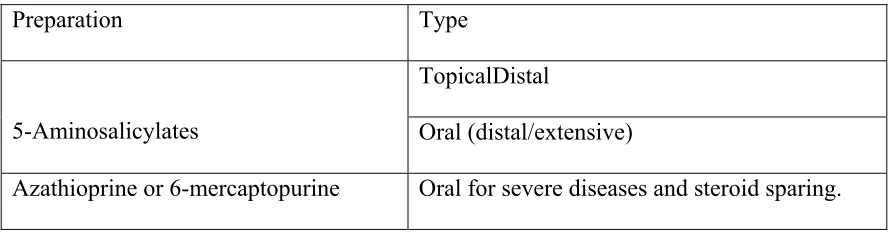
Table -2 -- Maintenance Therapy for Ulcerative Colitis
Preparation Type TopicalDistal
5-Aminosalicylates Oral (distal/extensive)
[image:33.612.84.531.536.655.2]DEFENSINS
Defensins are small cationic antimicrobial peptides that form an important part of the innate immune system and protect the intestinal mucosa against bacterial infection. 86 and play an important role in host immune system to some infectious diseases, immune diseases and skin disease Hexamers of defensins create voltage-dependent ion-channels in the target cell membrane causing permeabilization and ultimately, cell death
It has been suggested that deficient defensin expression may underlie the chronic inflammation of Crohn’s disease. The DNA copy number of the β-defensin gene cluster on chromosome 8p23.1 is highly polymorphic within the healthy population, which suggests that the defective β -defensin induction in colonic CD could be due to low β -defensin-gene copy number (89).
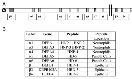
Defensins’ are two types alpha(α) and Beta(β)-defensin. The α - defensins are the first group discovered and until now six human α-defensins were detected (90). α -defensins are strongly expressed in neutrophils and are also present in certain epithelia, such as gut wall, endocervix and vagina. β -defensins are expressed in a variety of epithelia of the respiratory air ways (91). α - defensins are closely related to the β
-defensins, differing in the spacing pattern of conserved cytosine residues and the linkage arrangements of the distal fide bands. These genes have been mapped to 8p22-p23 (Figure 1). Several types of beta–defensin are noted. DEBF4 is effective against Escherichia coli and prevention of Pseudomonas aeruginosa infection at micromolar concentration. The anti microbial properties of DEFB4 were expressed in leukocytes and acts as a chemokine for cells of the adaptive immune response (102).Recent studies
-defensin 3 (DEFB103) and human β defensin 4 (DEBF104) showed variation in copy numbers. In Crohn’s disease, reduced Paneth cell production of a-defensins in the ileum is considered to be a significant factor in pathogenesis of the disease. In ulcerative colitis, studies have shown that epithelial cells and plasma cells in the colon have increased expression of human β -defensin 2 (or DEFB4) mRNA. This increased expression of the defensins may be due to increased bacterial load in the lumen. Alternatively it may be genetically regulated, by the copy number polymorphisms of the DEFB4 gene. This variation may have an impact on gene expression levels. There has been no study of copy number polymorphisms of DEFB4 and ulcerative colitis, so the present studies were designed to measure the DEFB4 gene copy numbers in patients with ulcerative colitis (116 – 120)
AIMS OF THE STUDY
To perform a case control study in patients with ulcerative colitis and matched controls:
1. To determine whether there are specific associations of ulcerative colitis with factors connected with childhood hygiene, and effect of appendectomy or tobacco usage on incidence of ulcerative colitis.
MATERIALS
SUBJECTS
CASES
:
Confirmed cases of ulcerative colitis diagnosed by clinical history,colonoscopic findings and histology. The clinical history consisted of history of blood and mucus diarrhoea presently or in the past. Colonoscopic findings of presence of symmetrical and continuous inflammation that began in the rectum and extended
proximally without interruption with features of loss of normal vascular pattern, erythema and edema of mucosa with or without granularity, friability with presence of yellow brown mucopurulent exudates associated with mucosal ulceration which can be punctate, annular, linear or serpigenous and +/- presence of pseudopolyps. Histology showing cryptitis or crypt abscesses and absence of granulomas. Histologically the disease was graded as quiescent, mild, moderate and severe depending on the activity.Quiescent colitis was characterized bypresence of architectural alterations with or without the presence fibrosis with no features of neutrophils infiltration, no edema no ulcers. Mild, moderate, severe disease were divided depending on the
1) Degree of neutrophil infiltration of the crypts and depth of mucosal infiltration 2) Amount of mucin depletion
3) Amount of edema and congestion with or without presence of the ulcers. Grade increases if the above features increase in severity.
METHODS
The study was designed as a prospective case control study. It was presented to the institutional review board and the protocol and the consent form were approved.
All participants were investigated only as clinically indicated. In the case of patients with ulcerative colitis, this included colonoscopy and biopsy and tests to
establish the general severity of the disease and complications. Informed written consent was obtained from all participants or the guardian. A detailed questionnaire including risk factors of ulcerative colitis was read out to the patients and controls and their responses were recorded in the form. All the responses were later entered in to a computer
spreadsheet for analysis using a statistical program.
Questions regarding risk factors concerned with hygiene were demarcated as during “childhood” and “current”. The reason was to determine whether specific associations existed with childhood hygiene or with current state of hygiene. These included (1) residence in urban area (or) village, (2) history of intestinal parasitic infestation, (3) drinking water source for the household and (4) availability of toilets. Questionnaires also included a detailed evaluation of socioeconomic status based on the modified Kuppuswamy scale, a past history of appendicectomy, and a history of
consumption of tobacco in any form.
DNA Extraction and Polymorphic Analysis:
DNA isolation from blood
Materials required:
1. EDTA coated vacutainer tube 2. Centrifuge
3. Plastic pasteur pipette (sterile)
4. 15 ml self standing centrifuge tube (sterile) 5. RBC lysis buffer
6. WBC lysis buffer 7. Proteinase K 8. 10% SDS 9. Saturated NaCl 10. Absolute alcohol
11. Eppendorf tubes (sterile) 12. 70% ethanol
13. TE buffer
Reagent preparation:
RBC lysis buffer:
WBC lysis buffer:
Mix 25ml of 0.5 M EDTA and 2.19 g of NaCl in 250ml of water and adjust the pH to 8.0 and make up the volume to 500 ml with water. Sterilize by autoclaving. Can be stored at room temperature for 3 months.
Tris-EDTA buffer (10 mM Tris & 0.1 mM EDTA):
Mix 1 ml of 1 M Tris (pH 8.0) and 400µl of 0.25 M EDTA (pH 8.0) and make up the volume to 100 ml with water. Sterilize by autoclaving. Can be stored at room
temperature for 1 year.
10% SDS:
Dissolve 10 g of SDS in 100 ml of water and heat the solution at 65° C to assist dissolution. Sterilization is not required. Can be stored at room temperature for 6 months.
Proteinase K:
Dissolve the lyophilized Proteinase K (Cat no.PK1, genei) in 10 ml of sterile water to obtain a concentration of 10mg/ml. Aliquot 500µl volume into Eppendorf tubes. Can be stored at –20° C for a long period.
Saturated NaCl:
Dissolve 36.05 g of NaCl in 100 ml of water and is saturated or 6M NaCl solution. Sterilize by autoclaving and store at room temperature for up to 1 year. (Note: Always use milliQ water for the reagents preparation)
Procedure:
3. Three layers will be visible: the top yellow layer is the plasma, the middle white layer is the buffy coat and the bottom red is the erythrocytes. Carefully remove the plasma and discard it and collect the buffy coat in a fresh tube.
4. Add ten volumes of RBC lysis buffer and incubate at room temperature for 10 min.
5. Then centrifuge the tube at 4000 rpm for 10 min. Discard the supernatant and a white pellet will be seen.
6. Repeat the wash until a clear white pellet is obtained.
7. Then to the pellet add 4.5 ml of WBC lysis buffer and make a homogenous solution. Add 250 µl of 10% SDS and 100 µl of Proteinase K and mix them well and incubate at 37° C for overnight or at 55° C for two hours.
8. Check the lysis, if the lysis is not complete, then add 50 µl of Proteinase K and continue the digestion.
9. If the lysis is complete, add 1.5 ml of saturated NaCl solution and mix well till the solution turns milky white.
10.Centrifuge the solution at 4000 rpm for 15 min and collect the supernatant in a fresh tube.
11.To the supernatant add two volumes of absolute ethanol and mix gently, a white thread like structure will appear and will reach the top of the solution.
12.Collect the thread like structure in a fresh eppendorf tube containing 1ml of 70% ethanol, using a sterile pipet tip.
15.When the precipitate turns transparent, transfer it into an eppendorf tube containing 200-300 µl of TE buffer.
16.The DNA can be dissolved by incubating at 37° C for 2 hr or 65° C for 30 min. 17.Store the DNA in deep freezer (-20 is adequate) for long-term storage or in the
fridge for short term.
COPY NUMBER POLYMORPHISM ANALYSIS
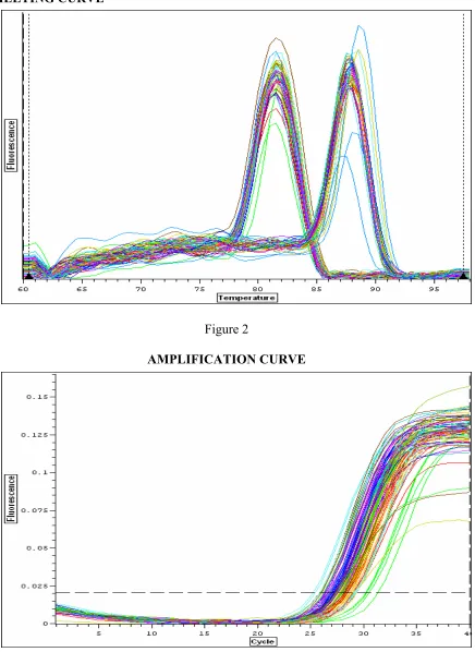
DEBF4 copy number was assessed by quantitative real time polymerase chain reaction using SYBR Green method. PCR was carried out on a Chromo4 (MJ Research, USA) thermal cycler equipped with a continuous fluorescence detector and the PCR protocol was an initial denaturation step for 1 min at 95° C followed by 40 cycles
MELTING CURVE
[image:43.612.98.534.81.679.2]Figure 2
AMPLIFICATION CURVE
DISCUSSION
[image:43.612.100.532.86.366.2]SAMPLE SIZE DETERMINATION:
As there was no Indian data regarding the frequency of defensin copy number
polymorphisms in the general population, we calculated sample size assuming arbitrary frequencies of 5% for the general population and 25% for patients with ulcerative colitis. We calculated that a sample size for this study of 59 cases and 59 controls would detect the above difference with a power of 80% and an alpha value of 0.05. Patients with irritable bowel syndrome or functional bowel disorder were chosen as the control subjects because by definition these patients do not have a structural disorder of the bowel, and they are drawn from the same population as the cases.
STATISTICAL ANALYSIS:
RESULTS
Demographic characteristics
:
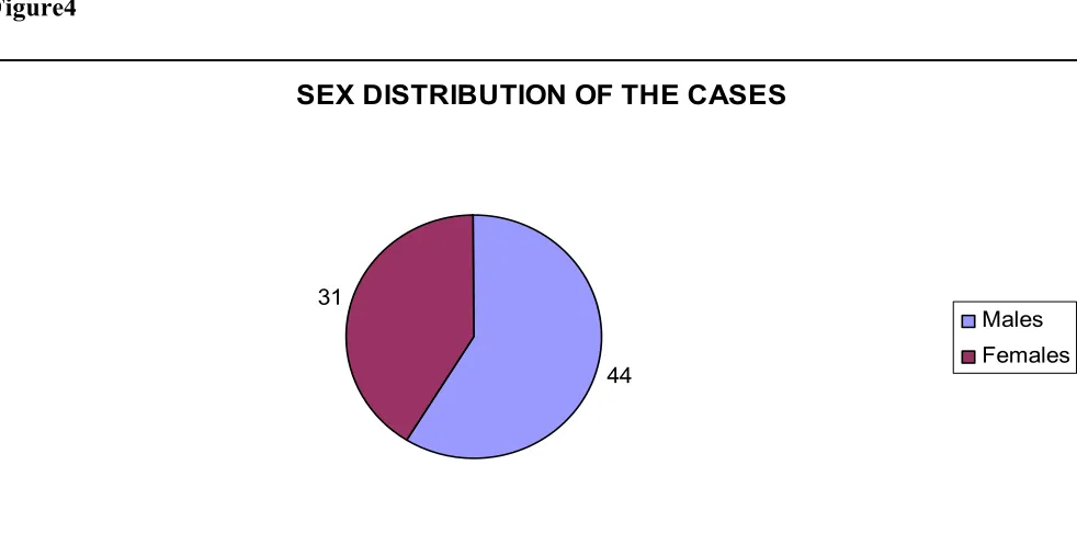
Cases: 75 cases of chronic ulcerative colitis were included in this study with mean age being 37 years (range 15–70 years). 44 were male and 31 were female (Figure 4). They hailed from various states like Tamil Nadu, West Bengal, Jharkhand, Andhra Pradesh and others. Their body mass index mean (SD) was 20.7 (4.3).
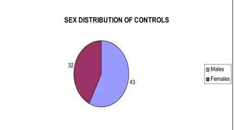
[image:45.612.95.586.398.641.2]Controls: 75 age, sex and religion matched controls were studied for the epidemiological associations and for study of defensin gene copy number polymorphism. The mean age of controls was 38 years (range 18-66 yrs) of which 43 were male and 32 were female (Figure 3). Their state of origin corresponded to the cases. Their body mass index was 21.3 (4.8).
Figure4
SEX DISTRIBUTION OF THE CASES
44 31
Figure5
SEX DISTRIBUTION OF CONTROLS
43 32
Males Females
Epidemiological associations and risk factors:
TABLE- 2
RISK
FACTORS
ULCERATIVE
COLITIS
CONTROLS RR 95% CI P
Childhood Residence Urban Village 31 44 30 45
1.05 0.56 – 2.02 1.00
Current Residence Urban Village 39 36 43 32
0.80 0.42-1.53 0.62
Childhood Water source Protected Unprotected 10 65 6 69
1.76 0.608 -5.144 0.42
Current water source Protected Unprotected 41 34 29 46
1.91 0.998 –
3.664
0.07
Childhood toilet Closed
Open 44
31
36 39
1.53 0.806 –
2.932 0.252 Current toilet Closed Open 68 7 66 9
1.32 0.46 – 3.76 0.79
* Fisher exact test was used to compare these dichotomous variables, none of the differences was statistically significant.
b) Personal habits and diagnosis of ulcerative colitis:
TABLE 3
RISK FACTORS ULCERATIVE
COLITIS
CONTROLS RR 95% CC P
Toothpaste use in childhood Users Non users 38 37 31 44
1.45 0.76 –
2.78
0.32
Toothpaste usage in Current Users Non users 72 3 66 9
3.27 0.85
-12.6
0.13
Worm infestation in childhood
Present
Absent 31
44
41 34
0.584 0.30 –
1.11
0.071
Exposure to Pets in childhood
Yes
No 27
48 21 54 1.45 0.72 – 2.89 0.38
Exposure to Pets now Yes
No 14
61
9 66
1.68 0.68 –
4.17
0.37
Exposures to Cows / Poultry In childhood
Yes
No 39
36
38 37
1.05 0.56 –
2.001
1.00
Exposures to Cows / Poultry Now Yes No 17 58 13 62
1.39 0.62 –
3.13 0.54 Breast Fed Yes No 72 3 73 2
0.65 0.10 –
4.05
1.00
Separate Bedroom in childhood Yes No 12 63 3 72
4.571 1.234 –
16.935
0.02
the household, having a separate bedroom in childhood and breast feeding in infancy between cases and controls.
In this study we did not find any significant difference in any of these putative risk factors except that the availability of a separate bedroom for the person in childhood was significantly associated with ulcerative colitis, with a relative risk of 4 (CI 1.23 – 16.93) (P=0.02). A separate bedroom in childhood probably signifies higher socioeconomic status and may be a surrogate marker of higher hygiene status in childhood.
Socio Economic Status:
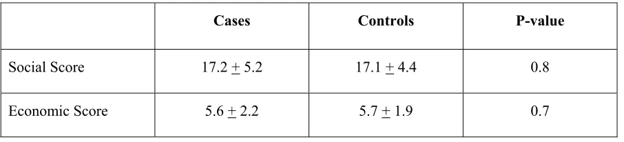
[image:49.612.84.537.521.624.2]Socio economic status of the cases and controls was also calculated (current status) to assess whether it has any effect on incidence of ulcerative colitis and also to see if it is an indirect marker of sanitation and health. The modified Kuppuswamy scale has been in wide use since 2 decades, and this was used for calculation of social economic status of subjects in this study. This scale is a composite score of per capita monthly income, education of head of family and profession of the head. It gives maximum score of 29 and lowest score of 3. There was no significant difference in social or economic score.
Table- 4. Socio-economic Status:
Cases Controls P-value
Social Score 17.2 + 5.2 17.1 + 4.4 0.8
Risk Factors Associated with Past Medical, Family and Personal
History:
In the past medical and surgical history, we collected the data regarding the history of appendectomy, diabetes mellitus, hypertension and other abdominal surgeries (Table 4). In the family history we collected history of inflammatory bowel disease and
[image:50.612.85.531.329.680.2]consanguinity (Table 5). In the personal history we obtained history regarding smoking habits, alcohol consumption (Table 6) and types of food consumed, vegetarian or non vegetarian (beef, fish, pork, meat) (Table 7).
Table-5. Past Medical and Family History in Cases and Controls:
RISK FACTORS ULCERATIVE
COLITIS
CONTROLS RR 95% CI P
Appendiecectomy Done
Not done 1
74
3 72
0.32 0.33 – 3.19 0.62
Had abdominal Surgery Yes No 10 65 16 59
0.56 0.23-1.34 0.28
Diabetes Mellitus Present
Absent 5
70
7 68
0.694 0.21 – 2.29 0.76
Hypertension Present
Absent 5
70
4 71
1.27 0.32 – 4.91 1.00
Family history of IBD
Present Absent
2 73
0 2.027 1.72 – 2.38 0.248
Consanguinity Present Absent 8 67 8 67
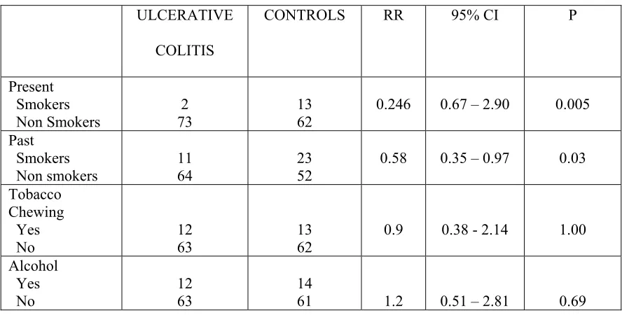
Table-6. Personal Habits
ULCERATIVE
COLITIS
CONTROLS RR 95% CI P
Present Smokers Non Smokers 2 73 13 62
0.246 0.67 – 2.90 0.005
Past Smokers Non smokers 11 64 23 52
0.58 0.35 – 0.97 0.03
Tobacco Chewing Yes No 12 63 13 62
0.9 0.38 - 2.14 1.00
Alcohol Yes No 12 63 14
61 1.2 0.51 – 2.81 0.69
Table-7. Food Habits:
ULCERATIVE
COLITIS
CONTROLS RR 95% CC P
Vegetarian Yes No 13 62 5 70
2.93 0.99 – 8.7 0.07
Beef Yes No 17 58 9 66
0.465 0.19 – 1.12 0.13
Fish Yes No 62 13 67 8
1.7 0.68 – 4.52 0.34
Pork Yes No 4 71 2 73
0.486 0.86 – 2.74 0.68
Meat Yes No 61 14 65 10
1.49 0.61 – 3.60 0.5
[image:51.612.87.528.366.616.2]ulcerative colitis. Past smokers had a relative risk of developing ulcerative colitis of 0.58 (P=0.03), indicating a protective effect of 42%. However, tobacco chewing did not show any significant association with ulcerative colitis. Previous studies have shown that alcohol consumption, western type of diet consumption especially like ham, meat were associated with increased incidence and increased relapse of ulcerative colitis. However, in this study, dietary factors did not appear to significantly influence the development of ulcerative colitis. In this study, we did not get any significant family history of IBD or history of consanguinity in the cases or controls.
CLINICAL FEATURES
OF ULCERATIVE COLITIS
[image:52.612.85.533.392.575.2]PATIENTS:
Table-8. Age of onset of the disease
Age range of Incidence of cases
Number of cases Percentage
1 – 10 1 1.34
11 – 20 8 10.67
21 – 30 22 29.34
31 – 40 28 37.34
41 – 50 9 12
51 – 60 6 8
61 – 70 1 1.34
other studies in which incidence was in the same decade as mentioned above but the second peak which was mentioned in the 6th decade was not seen in our study (93).
Table-9. Symptoms of ulcerative colitis patients:
Number of Cases Percentage
Abdominal Pain 54 73.3
Diarrhoea 73 97.3
Blood in stool 73 97.3
Urgency 70 93.3
Tenesmus 59 78.7
Incontinence 44 58.7
Tiredness 64 85.3
Fever 21 28
Loss of weight 58 77.3
Arthritis 55 73.3
Most common symptom in ulcerative colitis was diarrhea with blood and mucus, and stools associated with urgency. These symptoms were present in 93 to 97% of the cases. Other symptoms like abdominal pain, tenesmus were little less around 70 to 75% and fecal incontinence was present in 58% of the cases. Arthritis was the only
complication which was common extra intestinal manifestation in our study which was present around 73% of the cases. The high incidence of arthritis and of fecal
Table-10. Extent of colitis:
Colon Extent Number of Cases Percentage
Pan colitis 44 58.7
Left sided colitis 17 22.7
Proctitis 14 18.7
Table- 11. Clinical course of the disease in ulcerative colitis patients:
Number of Cases Percentage
Intermittent flare with complete resolution
42 56
Mostly continuous illness 23 30.7
Asymptomatic after initial attack
7 9.3
First Episode 3 4
[image:55.612.82.533.526.699.2]Most of the cases included in this study were already on treatment instituted here or elsewhere. During the study, only three cases presented for the first time. As seen in Table 11, the majority of our patients, 56%, had intermittent flares of disease with complete remission between flares, which is also common presentation in other studies (94). Mostly continuous disease was seen in 30.7% cases. Quiescent disease after the first attack was seen in 9.3% cases.
Table 12. Grade of Ulcerative Colitis (Histology):
Grade Number of Cases Percentage
Mild 19 25.34
Moderate 43 57.34
Severe 12 16.00
Table 13. Laboratory Parameters
Mean + SD
Hemoglobin g% 10.9 + 2.3
ESR mm/h 41.8 + 28.19
Albumin g% 3.76 + 0.71
Most of our patients were on treatment by the time of presentation. So, Hb, albumin and ESR were within the normal range. Hb. is being 10.9 + 2.3gm%, albumin 3.76 + 0.7 and ESR 41.8 + 28.1.
The Ulcerative Colitis Disease Activity Index (UCDAI) is the sum of scores of 4 components, stool frequency, rectal bleeding, sigmoidoscopic findings and physician’s global assessment. This disease activity index ranges from 0 to 12 with a higher total score representing more severe disease. Disease activity index in our study was 7.11 + 3.67 which indicates most of these patients had mild or moderate activity, most probably secondary to the treatment.
The Seo’s disease activity index(94) was also used for quantification of the disease activity. The activity index (AI) is calculated using the formula:AI =
Table-14. Seo’s Activity Index
Mild ( < 150) 31 41.34
Moderate (150 – 220) 27 36.0
Severe (> 220) 17 22.67
In this study, around 40% patients had mild disease and 36% patients had moderate disease and around 22% patients had severe disease as per the Seo’s activity index which was in correlation with the ulcerative colitis activity index.
Table -15. Treatment
DRUG HISTORY NUMBER OF
PATIENTS
PERCENTAGE
H/O PAST AMINO SALICYLATE CONSUMPTION YES NO 55 20 73.3 26.7 PAST ORAL STEROID CONSUMPTION
YES NO 41 34 54.7 45.3 PAST RECTAL STEROID USAGE
YES NO 20 55 26.7 73.3 PAST H/O IV STEROIDS
YES NO 9 66 12 88 PAST H/O IMMUNOSUPRESSION
YES NO 16 59 21.3 78.6 PRESENTLY ON SALICYLATES
YES NO 50 25 66.7 33.3 PRESENTLY ON STREOIDS
YES NO 21 54 28 72 PRESENTLY ON IMMUNOSUPPRESSION
[image:57.612.88.524.355.700.2]Most of the patients mostly on salicylates treatment is around 66.7% only few of them work on steroids 28% only few needed immunosuppresion that is around 14.7%.
HUMAN BETA-DEFENSIN 2 COPY NUMBER
POLYMORPHISMS
The human beta-defensin gene 2 (also known as DEFB4) copy number
[image:58.612.91.521.419.669.2]polymorphism results are shown in the Figure 6. The majority of the subjects had less than 4 copies of DEFB4 gene but significant number of the cases had 4 or more copies when compared to controls which were statistically significant (p- 0.005). Subjects with defensin gene copy numbers > 4 had relative risk of 2.85 times to develop UC. Thus the presence of four or more copies of this gene may be one of the causes of the pathogenesis of the ulcerative colitis.
Figure 6
0 10 20 30 40 50 60
1 2 3 4 5 6 7
Copy numbers
F
re
quency
o
f s
u
bj
ect
s
CTRL UC
TABLE 16
COPY NUMBER
POLYMORPHISMS
CASES CONTROLS P value* RR OF CNP
<4 60 59
>4 26 7
0.005 2.85(1.32– 6.16)
DISCUSSION
Ulcerative colitis is a disease which was described mostly in the developed countries. There are only a few studies from developing countries like India regarding incidence of ulcerative colitis which was increasing in present days and to see the effect of hygiene on the incidence and course of IBD. A study from the Punjab showed that prevalence of UC was nearly similar in India and in Western countries (115).
The hygiene hypothesis has been developed to indicate why certain immune diseases are increasing in the modern world. If ulcerative colitis is increasing in India, perhaps it could be due to recent changes in the social practices, diet patterns and hygiene in India. Therefore this was undertaken as a case-control study to examine the effect of these parameters on the diagnosis of ulcerative colitis.
The incidence of the ulcerative colitis in our population was approximately in the same age group as described by Yang SK et al (93) and Probert C .S et al (94) dscribed in 2000 and 1996 respectively, that incidence was common in the 2nd and 3rd decades. As our study was done at a point of when patients presented to our institution but were notfollowed the 2nd peak as described by Yang SK et al was not observed. Equal sex incidence in our study was almost keeping with the two other studies described above.
The body mass index of this study group was within the range as described by Kuganthosan.S. et al (97) and Jabpsen et al (98).
AE. Gent et al (99) suggested that there was no effect of the environmental factors and childhood hygiene as the outcome of ulcerative colitis except that study by Gent et al found that usage of protected water in childhood was associated with increased incidence of the ulcerative colitis. This may be due to immune conditioning of the gastrointestinal tract.
In this study, we also found that there was no significant difference between the cases and controls in factors like childhood environmental conditions such as residence in village (or) urban area, usage of closed toilet (or) history of worm infestation. Like the study by Gent et al who found that hot water taps were more common in houses of subjects with IBD, in this study also people who consumed protected water had a relative risk of 1.96 when compared to people not consuming the protected water and increased risk may be secondary to the immune condition of the GI tract by the GI infections.
Around 10 cases (13%) were not sure of the worm infestation in childhood, and these were considered as not having the infection.
Tooth paste which contains micro particles could also serve as proxy measures of hygiene conditions (or) have an impact on the microbial flora of the gut. However observational studies did not show any association implicating tooth paste as an independent risk factor which was shown in our study also which was consistent with the study by Bussel M.G. Et al in 1998 (103).
In our study we studied the relation of pets (or) exposure to partly which did not show any significance which was the same in other studies by Benotein et al in 2006 (104).
Breast feeding appears to be protective and its effect on ulcerative colitis was evidenced by various studies and a meta-analysis was done by Klement E et al(105) in 2004 which revealed odds ratio ranged from 0.56 to 0.77 depending on the quality of study. Even in our study the odds ratio was 0.65 indicating that breast feeding was possibly associated with protection from the ulcerative colitis.
Having a separate bedroom in childhood is the one factor in our study highly significant with a p value of 0.02 and relative risk of 4.57 (1.23-16.03) indicating that having a separate room from childhood indicating higher socio economic studies and also may reflect various psychosocial effects.
Human appendix has been considered as a vestigial remnant, many case control studies suggest that previous appendectomy is rare in ulcerative colitis. In the appendix of the ulcerative colitis patients, the CD4/CD8 ratio is significantly increased and the proportion of CD4 + CD69 + (early activation antigen) T cells, but not of CD4+HLA-DR4 (mature activation antigen). T Cells is also significantly increased. These findings suggest that the appendix may be primary site in development of ulcerative colitis (106).In our study we found that OR of 0.32 for not developing the ulcerative colitis (CI 0.3-3.19) but this was not statistically significant. It was a little high when compared to study by Desanssure petat (107) who found OR of 0.1 (95% CI 0.05-0.21) (107). We did not find any much difference in the incidence of ulcerative colitis in patient has had previous abdomen surgeries between the cases and controls.
We found that family history of IBD in the first degree relative had an increased risk by 2, seen in 2 patients (2.06 of patients). There are consistent with the study done by park JB et al in 2006 who studied in Korean population found to have incidence of ulcerative colitis was around (2.01%) (108).
development of ulcerative colitis when compared with controls withOR-0.58 (95%CI 0.45-0.75).
In one study we found that relative risk was increased 7 times (CI 1.67-35.2; P-0.005) in nonsmoker when compared to present smokers. We also found that past smokers were protected 2.58 times (95 % CI 1.14-5.76) when compared to non-smokers with P value of 0.03. But we did not find any protective effect of tobacco chewing (or) alcohol consumption.
Various studies were done in different geographic areas with various types of dietary habits and the results varied. In our study there was significant association
between the various types of food consumed with OR ranging from 0.486 for having pork to 2.93 being a vegetarian. It appears that strict vegetarians were at a risk of 2.93
compared to non-vegetarians although this did not reach statistical significance.
Zuinbliene et al (111) also studied various dietary habits but were not able to establish any specific association with any food habits.
Table 17
Clinical features Park SH et al Out Study
M : F 0.94 : 1 1.59 : 1
Median Age 40 (12 – 72 yrs) 38 (15 – 20 yrs)
Proctitis 134 pts (44.1%) 14 pts (18.7%)
Left sided colitis 69 pts ( 22.7%) 17 pts (22.7%)
Pan colitis 101 pts (33.2%) 44 pts (58.6%)
Mild 149 pts (49%) 19 pts (25.34%)
Moderate 125 pts (41.1%) 43 (57.34%)
Severe 26 pts (86%) 12 (16%)
[image:65.612.80.543.461.633.2]Quiscent -- 1 (1.3%)
Table 18
Clinical Course Park SH et al Cases (percentage)
First episode 100 % 3 ( 4%)
Asymptomatic after initial attack
97.4% 7 (9.3%)
Mostly continuous illness - 23 (30%)
Complete resolution 42 (56%)
Paneth cells(114) may play important roles in the maintenance of intestinal immune homeostasis.(116) Rahaman et al(120) demonstrated that lamina propria in colon from UC patients, Crohn's colitis patients, and controls contain cells that express hBD-2. They proposed that plasma cells were 2-3 times more abundant in UC colon than in control colon and Crohn's colitis. Moreover, plasma cells in UC colon expressed hBD-3 and hBD-4 mRNA. Additionally, hBD-2 mRNA expression was demonstrated in 3 out of 4 well-characterized plasma cell lines and they concluded that mature colonic plasma cells can express multiple β-defensins. In UC, defensin production by plasma cells is probably clinically relevant since plasma cells accumulate in large numbers between the distorted crypts and muscularis mucosa.(117) A study by Fellermann(119) who tested the genome wide DNA copy number profiling by array-based comparative genomic hybridization and quantitative polymerase-chain-reaction analysis of the human β -defensin 2 (HBD-2) gene they that healthy individuals, as well as patients with ulcerative colitis, have a median of 4 (range 2-10) HBD-2 gene copies per genome. In a surgical cohort with ileal or colonic CD and in a second large cohort with inflammatory bowel diseases, those with ileal resections/disease exhibited a normal median HBD-2 copy number of 4, whereas those with colonic CD had a median of only 3 copies per genome (P=.008 for the surgical cohort; P=.032 for the second cohort). Individuals with < or = 3 copies have a