Copyrightq1996, American Society for Microbiology
The Human Immunodeficiency Virus Type 1 5
9
Packaging Signal
Structure Affects Translation but Does Not Function
as an Internal Ribosome Entry Site Structure
GINO MIELE,
1ANDREW MOULAND,
2GEOFFREY P. HARRISON,
1E
´ RIC COHEN,
2ANDANDREW M. L. LEVER
1*
University of Cambridge Department of Medicine, Addenbrooke’s Hospital, Cambridge CB2 2QQ,
United Kingdom,
1and Laboratoire de re´trovirologie humaine, De´partement de microbiologie
et immunologie, Universite´ de Montre´al, Montre´al, Canada H3C 3J7
2Received 28 August 1995/Accepted 7 November 1995
The role of the RNA secondary structure in the 5
*
packaging signal region of human immunodeficiency virus
type 1 (HIV-1) in initiating translation of
gag
mRNA has been investigated both in vitro and in the presence
of cellular cofactors in vivo. Heat denaturation of the structure and mutagenic deletion both lead to an increase
in levels of translated products, indicating that the structure is a significant inhibitor of translation. The
proximity of the
gag
AUG to the packaging signal structure suggested that it might function as an internal
ribosome entry site. However, in both a cell-free system and eukaryotic cells, translation will initiate at a novel
upstream initiation codon introduced within the 5
*
noncoding region. This codon is utilized exclusively,
resulting in
gag
protein products with an extra 11 amino acids at the amino terminus, which, when expressed
in T lymphocytes, are confined intracellularly, probably because of the lack of an N-terminal glycine
myris-toylation signal. Deletion of the secondary structure abolishes
gag
production even in the presence of
tat
and
rev
in
trans
. Using dicistronic constructs containing the HIV-1 5
*
leader cloned between two heterologous open
reading frames, we were unable to detect any significant expression of the second open reading frame that
would have been supportive of an internal ribosome entry site mechanism. Using mutant proviruses either
lacking the entire packaging signal structure region or containing the introduced upstream initiation codon in
long-term replication studies, we were unable to detect reverse transcriptase activity in culture supernatants.
The 5
*
packaging signal structure of HIV-1 does not serve as an internal ribosome entry site. The translation
of
gag
is consistent with ribosomal scanning. However, the packaging signal structure causes significant
translational inhibition.
The 5
9
leader sequence of human immunodeficiency virus
type 1 (HIV-1) has multiple cis-acting functions which are
essential in the virus life cycle. These include transcriptional
control through the tat-responsive element (TAR) (5, 45, 56,
61, 65), binding of the tRNA primer (31, 53), dimerization of
the genomic RNA (11), and splicing (13). A signal important
to genomic RNA encapsidation in HIV-1 has been identified in
this region (3, 10, 30, 39). In COS cell transfection studies a
deletion within the 5
9
end of the gag open reading frame has
also been suggested to affect packaging (40).
We and others have demonstrated that a stable secondary
structure exists in the region between the primer binding site
and the gag AUG (4, 21, 22, 58). Deletions in this region which
reduce the efficiency of packaging would also disrupt this
pre-dicted secondary structure. Secondary structure predictions
vary somewhat among investigators; however, there are some
regions which are identified by all investigators. In particular,
the stem-loop from bases 763 to 780 is consistently predicted.
Expression of many eukaryotic genes is regulated at the level
of translation (64, 66). A scanning hypothesis of translation
initiation has been proposed (35), but the mechanism is not yet
fully elucidated. According to this model, after the 25-kDa cap
recognition protein (known as p25 eIF-4E or CBP1) has bound
to the mRNA cap, the 40S subunit of the ribosome (in
com-bination with Met-tRNA and other factors) binds somewhere
along the 5
9
end of the untranslated sequence and then
mi-grates along the RNA, unwinding the RNA secondary
struc-ture (dependent on ATP), until an initiator AUG is
encoun-tered (32, 35, 66). For efficient translation initiation, this AUG
should be in an appropriate sequence context (RccAUGG),
the purine (R) at position
2
3 being particularly important in
determining the strength of initiation together with, to a lesser
extent, the G at position
1
4 (33). Ribosomal scanning is
known to be inhibited by strong secondary structures (16, 17,
19, 20, 23, 32, 34, 36, 43, 44, 69). Therefore, if translation of
HIV-1 gag is initiated by the ribosomal scanning mechanism,
the predicted secondary structure in this region would be
ex-pected to be inhibitory.
Internal ribosome entry has been demonstrated in a number
of prokaryotic and eukaryotic systems, including
picornavi-ruses (1, 24–28, 50, 63), a plant potyvirus (8), cowpea mosaic
virus (67), the immunoglobulin (Ig) heavy chain-binding
pro-tein (41), homeotic gene antennapedia mRNA (48), and
hep-atitis C virus (68, 70). Most recently, internal initiation has
been shown with murine leukemia virus-based constructs in
which competing AUG and CUG initiation codons exist (6). In
HIV-1 the proximity of the predicted secondary structure to
the gag gene raised the possibility that it might also act as an
internal ribosome entry site (IRES).
In this study we have investigated how the secondary
struc-ture in the HIV-1 5
9
untranslated region affects translation of
the gag and gag/pol open reading frames.
* Corresponding author. Mailing address: University of Cambridge Department of Medicine, Addenbrooke’s Hospital, Hills Rd., Cam-bridge CB2 2QQ, United Kingdom. Phone: (1223) 336844. Fax: (1223) 336846.
944
on November 9, 2019 by guest
http://jvi.asm.org/
Mutation A4.The construction of the mutation A4 generated a new initiation codon 33 bases upstream of the native gag initiation codon, which is at position 790. The G at position 763 was mutated to C in order to remove a nonsense codon, so that the use of the new AUG would add an extra 11 amino acids at the 59end of the gag open reading frame. These 11 amino acids are MVSLAEAR-RRE, with a total predicted molecular mass of 1,498 Da. To create a favorable Kozak consensus, the C at position 753 was mutated to an A and the U at position 759 was mutated to a G (Fig. 1B).
Mutation A14.The construction of mutation A14 generated a deletion from bases 677G to 790A, removing the entire secondary structure, including the major splice donor signal. The resulting leader sequence retains a translation context identical to that of the wild type (Fig. 1C).
Mutation D2m.The construction of mutation D2m resulted from a mismatch between the mutagenic template and oligo D2. An AUG initiation codon at position 767 was created out of frame with the native AUG but in frame with a stop codon at position 776 (Fig. 1D).
The RNA cap site for the gag mRNA is at position 454, just 59of the BglII site at position 474, which was used to excise the packaging signal region for mu-tagenesis. To generate mRNAs which included the RNA cap site, a ScaI frag-ment (positions 313 to 2751) from the mutated proviral clones was subcloned into the EcoRV site of pBluescript KS1in the orientation of the T7 promoter. These clones are referred to as the pKSScaI series. All proviral clones were propagated in Escherichia coli DH5a, and plasmids were prepared by cesium chloride purification (42). pBluescript clones which were required for in vitro transcription (in the uncoupled system) were propagated in dam(2) Epicurian-coli SCS110 cells (Stratagene) to allow linearization with the methylation-sensi-tive restriction endonuclease BclI, which cleaves at HIV-1 position 2429. The linearized plasmid therefore contains the first 178 bases of the pol gene.
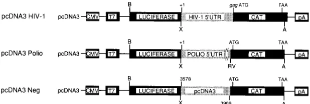
Dicistronic constructs.Dicistronic constructs (Fig. 2) were prepared to exam-ine directly internal ribosome entry on HIV-1 mRNAs. The luciferase gene was excised from pGEM-luc (Promega) with BamHI and XhoI and cloned into the mammalian expression vector pcDNA3 (Invitrogen) to generate pcDNA3-luc (kindly provided by Nahum Sonenberg, McGill University). Recombinant PCR was used to generate HIV (molecular clone pSVC21), poliovirus (type II Lans-ing), or control intercistronic sequences upstream of chloramphenicol acetyl transferase (CAT) sequences with 59oligonucleotides that contain an XhoI site and a 39oligonucleotide containing an ApaI site in CAT 10 nucleotides (nt) downstream of the stop codon. These were directionally cloned into pcDNA3-luc such that gag mRNA, including the cap (at position11) up to an including the gag initiation codon, a poliovirus leader sequence (positions11 to 735), or a negative control sequence from the pcDNA vector (nt13578 to13909) was placed upstream of CAT. In a similar manner, dicistronic constructs were made with the env leader as the test sequence, and the effect of the gag coding sequence on gag mRNA translation initiation was evaluated by including 100 and 200 nt of the gag coding sequence in addition to the gag mRNA leader. The following PCR primers were used.
pcDNA3 HIV-1: XhoIHIV59, 59-ATCACTCGAGGGGTCTCTCTGGTTAG AC-39; and HIV-CAT (antisense), 59-TCCAGTGATTTTTTTCTCCATCTCT CTCCTTCTAGCCTC-39
pcDNA3 Polio: XhoIP255, 59 -ATCACTCGAGTTAAAACAGCTCTGGGGT-39; and P25-CAT (antisense), 59-GCTTCCTTAGCTCCTGAAAAGATATCTT AACAATGAGGT-39
pcDNA3 Neg: XhoIpcDNA359, 59-ATCACTCGAGTCAAAGGCGGTAATA CGG-39; and pcDNA3-CAT (antisense), 59-GCTTCCTTAGCTCCTGAAAA CTGAGATACCTACAGCGTG-39
pcDNA3-env: HIV285-env 5525 (antisense), 59-TTCGTCGCTGTCTCCGCTT CAGTCGCCGCCCCTCGCC-39; and HIV 5525 (sense), 59-AAGCGGAGACA GCGACGAA-39
HIV 5525-CAT (antisense): 59-TCCAGTGATTTTTTTCTCCATTGCCACTG TCTTCTGCT-39
dicistronic construct containing the poliovirus leader sequence; pCDNA3 Neg, a dicistronic construct containing a random sequence; pCDNA3 env, a dicistronic construct containing the HIV-1 env leader sequence; pCDNA3gag100, a dicis-tronic construct containing the HIV-1 gag leader sequence plus the first 100 nucleotides of the gag coding region; pCDNA3gag200, a dicistronic construct containing the HIV-1 gag leader sequence plus the first 200 nucleotides of the gag coding region.
In vitro transcription and translation.One microgram of supercoiled plasmid DNA from the pKSBglII WT and pKSBglII A4 clones was transcribed and translated in a coupled rabbit reticulocyte lysate system (Promega) according to the manufacturer’s instructions with T3 RNA polymerase and 0.8 mCi of trans-lation-grade [35S]methionine (Amersham) per ml. One-tenth of the reaction
product was boiled in sample buffer (62.5 mM Tris-HCl [pH 6.8], 2% sodium dodecyl sulfate [SDS], 10% glycerol, 5%b-mercaptoethanol, 0.02% bromophe-nol blue) at 1008C for 4 min and loaded onto a denaturing polyacrylamide gel. Following electrophoresis, the gel was fixed, soaked in Amplify (Amersham) for 30 min, and dried and autoradiography was performed.
Since it is not known whether the wild-type RNA secondary structure can be fully formed prior to translation in the coupled transcription and translation reticulocyte system, transcription and translation reactions were also performed in an uncoupled system in which these processes were temporally separated.
Synthesis of capped RNA.Completely linearized pKSScaI plasmids (5mg) were transcribed in 50ml of 40 mM Tris-HCl (pH 7.5)–6 mM MgCl2–2 mM
spermidine–10 mM NaCl–10 mM dithiothreitol–0.5 mM each recombinant nu-cleoside triphosphate (except GTP, which was used at a final concentration of 0.05 mM)–0.5 mM m7
G (59) ppp (59) G (Boehringer Mannheim) with 50 U of RNasin (Promega) and 40 U of T7 RNA polymerase (Promega). Following DNase treatment (RQ1 DNase; at 378C for 10 min), the RNA was extracted with phenol-chloroform, precipitated with ethanol, and then redissolved in double-distilled autoclaved water (diethyl pyrocarbonate treated). To quantify the tran-scription products, aliquots were analyzed by agarose gel electrophoresis and with DNA dipsticks (Invitrogen).
Two micrograms of each of the test-capped mRNA samples was used to program micrococcal nuclease-treated rabbit reticulocyte lysate (Promega). Translation reactions were carried out at 308C for 1.5 h in the presence of 0.8 mCi of translation grade [35S]methionine (Amersham) per ml. The RNA
sec-ondary structure was disrupted by heating the RNA samples to 678C for 10 min. After denaturation, the RNAs were either translated immediately (to translate an unstructured mRNA template) or allowed to cool to room temperature and then to 08C over a period of 1 h to translate a reannealed mRNA template. Portions equivalent to one-tenth of the translation reaction mixtures were heated in sample buffer at 1008C for 4 min and loaded onto a denaturing polyacrylamide gel. Following electrophoresis, the gel was fixed, soaked in Amplify (Amersham) for 30 min, and dried and autoradiography was performed. Bands were quanti-tated by densitometry.
Size differentiation of gag proteins was performed by SDS-polyacrylamide gel electrophoresis with linear gels of 12.5% or gradient gels of 7.5 to 15% (42).
In vitro transcription and translation of dicistronic constructs were performed after linearizing with ApaI. The reaction buffer consisted of 40 mM Tris-HCl, 6 mM MgCl2, 2 mM spermidine, 100mg of bovine serum albumin, 5 mM (each)
ATP, CTP, and UTP and 100 mM GTP, 1 mM m7GpppG, 100 mM
dithiothrei-tol, 0.4 U of RNasin perml, and 0.5ml of T7 RNA polymerase (New England Biolabs). [3
H]cytidine (10 nCi) was added to monitor incorporation. Reaction mixtures were treated with RQ1 DNase and then passed through a G-50 spin column. The eluate was precipitated with ethanol. Equal numbers of counts per minute from each reaction were used to program protein synthesis in the pres-ence of [35
S]methionine in a rabbit reticulocyte lysate (Promega) (52). Because CAT was not identifiable by gel electrophoresis because of background, an aliquot of the reaction mixture was used in a standard CAT assay (42). Both a blank (reticulocyte lysate alone) and a positive CAT expressor (pSP6 [46]) were used.
Cell culture.Cells were transfected with DEAE dextran (62) for monolayer and suspension cells. COS-1 cells were cultured in Eagle’s minimal essential medium, and Jurkat-tat T lymphocytes (55) were grown in RPMI 1640. Cell lines
on November 9, 2019 by guest
FIG. 1. Schematic of previously proposed HIV-1 packaging signal structure, where panel A illustrates the wild-type sequence and panels B, C, and D illustrate the positions of mutations A4, A14, and D2m, respectively. Mutation A4 inserts an upstream initiation codon in the Kozak consensus, which if utilized, results in a gag protein product of circa 56.5 kDa that is also myristoylation defective. Mutation A14 deletes the entire structure, leaving the native gag AUG and the AUB in the Kozak consensus intact. Mutation D2m creates an upstream AUG which is out of frame with gag and also contains a nearby stop codon.
on November 9, 2019 by guest
http://jvi.asm.org/
were provided by the Medical Research Council (MRC) AIDS Directed Pro-gramme. Cell culture media were supplemented with 10% fetal calf serum, 105
IU of penicillin per ml, and 100mg of streptomycin per ml.
COS-1 cells were harvested after 72 h and heated to 1008C in sample buffer for 4 min to extract and denature cellular proteins, and equal quantities were loaded onto gradient SDS-polyacrylamide gel. After electrophoresis, proteins were transferred overnight at 250 mA onto Hybond-C extra nitrocellulose paper (Amersham). The filters were probed with a monoclonal antibody (ADP315 provided by the MRC) to the p17 matrix protein at a concentration of 30mg/ml for 2 h at room temperature. Bands were visualized by anti-mouse Ig horseradish peroxidase-linked whole antibody (from sheep) and enhanced chemilumines-cence Western detection reagents (Amersham) according to the manufacturer’s instructions. At 72 h posttransfection sufficient Jurkat-tat T cells were harvested to enable loading protein equivalent to 650,000 cells per lane on an 12.5% SDS–polyacrylamide gel. The cultures were maintained to constitute the long-term replication studies. Virions were prepared by incubating the culture super-natant overnight with a half volume of 30% polyethylene glycol 8000 in 0.4 M NaCl. The precipitate was collected by centrifugation at 2,000 rpm for 45 min at 48C in an MSE 43124-129 rotor and resuspended in 0.5 ml of TNE (10 mM Tris-HCl, 150 mM NaCl, 1 mM EDTA [pH 7.5]). This material was layered over equal volumes of TNE containing 20% sucrose and centrifuged at 98,0003g for 2 h at 48C. Virion preparations and cellular samples were loaded onto the same gel. Transfer and probing were as described above, except that a monoclonal antibody (ADP313, provided by the MRC) to the p24 capsid (CA) protein was used at a concentration of 3mg/ml for 1 h at room temperature.
HeLa and COS-1 cells were transiently transfected with the dicistronic con-structs by lipofection (46), and the CAT gene product was assayed by the standard thin-layer chromatography assay (42). Luciferase was assayed according to the manufacturer’s instructions (Promega).
To study the long-term replication kinetics of the mutant proviral clones, the transfected Jurkat-tat cells were cultured for a period of 21 days and split on the basis of cell concentration (approximately 1:10) to maintain equivalent cell densities in the cultures. Prior to splitting, reverse transcriptase activity in the culture supernatants was determined by the Potts method (51).
RESULTS
Translation in a coupled transcription and translation
re-ticulocyte system.
Coupled transcription and translation of
plasmid DNA from the (cap site-negative) pKSBglII series
gives gag precursor proteins of the appropriate size (Fig. 3).
These are not full length, as the BglII site at position 2096 used
for subcloning the gene is within the coding region of gag. In
pKSBglII A4 it is clear that translation has been initiated from
the novel upstream AUG. The lack of an RNA cap site in these
clones does not appear to impair translation.
The use of the novel upstream AUG might have been
arti-factual, resulting from the use of the coupled system in which
the secondary structure necessary for IRES activity may not
have had time to form. To resolve this problem, we separated
the transcription and translation reactions (programming
rab-bit reticulocyte lysate with in vitro-synthesized capped RNA)
to allow the RNA secondary structure to form prior to
trans-lation.
Uncoupled transcription and translation.
The translation of
in vitro-transcribed capped mRNA clearly demonstrated that
with some constructs the gag products were more abundant
when mRNAs were heat denatured and translated
imme-diately (Fig. 4). This is particularly evident when comparing
the pKSScaI WT p55 translation products. In both sets of
samples, the production of the precursor protein was greater
with pKSScaI A14 RNA than with pKSScaI A4, which was
also greater than the pKSScaI WT gag protein. Heat
denatur-ation of the mRNAs immediately prior to transldenatur-ation increased
the amounts of translation product from the wild-type RNA
and the pKSScaI A4 by approximately 25 and 36%,
respec-tively, and increased that from pKSScaI A14 by less than 15%.
The differences in the quantities of the products of
transla-tion likely relate to the lower level of secondary structure in
pKSScaI A14, with additional heat disruption of the secondary
structure in all three mRNA templates being most marked in
pKSScaI A4 and pKSScaI WT. RNA was quantitated after
denaturation and cooling or denaturation alone to ensure that
degradation of the message had not occurred, which would
account for the reduced quantities of protein production in the
former (data not shown). pKSScaI A4 produced a greater
quantity of gag protein than pKSScaI WT, probably because
less RNA structure needed to be unwound before the
ribo-some encountered the initiation codon. Minor decreases in
levels of stability caused by the downstream mutations to
re-move nonsense codons may also have contributed to this effect.
Since the leader sequence of HIV-1 is believed to contain
the dimer linkage site (11), it was possible that the introduction
of a mutant sequence such as A4 affected dimerization and
thus the ability of the RNA to be translated. However, in vitro
analysis of the RNA prior to translation confirmed that in all
cases the vast majority of the RNA was monomeric (data not
shown).
[image:4.612.150.459.73.177.2]promoter; T7, T7 RNA polymerase promoter; B, BamHI; X, XhoI; A, ApaI; pA, growth hormone polyadenylation signal of mammalian expression vector pcDNA3; ATG and TAA, initiation and stop codons of CAT mRNA.
FIG. 3. One microgram of plasmid DNA from the pKSBglII series was used to program a coupled transcription and translation system according to the manufacturer’s instructions (Promega). One-tenth of the reaction product was boiled in sample buffer for 4 min at 1008C and then electrophoresed on an SDS–12.5% polyacrylamide gel at 50 mA for 4 h. The positions of molecular mass markers (Amersham) are shown. Mock, no template.
on November 9, 2019 by guest
Translation initiation in intact cells.
A cellular factor known
as the polypyrimidine tract-binding protein has been shown to
be essential for internal ribosomal initiation in
encephalomyo-carditis virus (29). To assay the phenotype of the proviral
clones in the presence of cellular factors, we transfected the
pSVC21 A4 and pSVC21 WT proviral clones into COS-1 cells
and the pSVC21 A4, pSVC21 A14, and pSVC21 WT clones
into Jurkat-tat T lymphocytes.
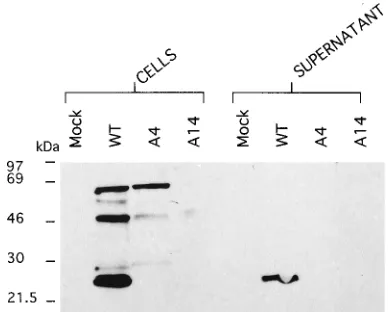
Transfection of the mutant A4 proviral plasmid into COS-1
cells gave rise to an 18.5-kDa p17 matrix cleavage product of
the gag precursor protein (Fig. 5). Transfection of the same
pSVC21 A4 mutant provirus into T cells resulted in the
pro-duction of a gag precursor protein larger than that of the wild
type (visible after further electrophoresis; data not shown)
which did not appear to be proteolytically cleaved (Fig. 6). In
the T-cell transfections of pSVC21 A4, gag could not be
de-tected in the supernatants. Transfection of the pSVC21 A14
mutant proviral clone resulted in no detectable expression of
gag, intra- or extracellularly. This could have been due to the
lack of spliced viral products such as tat and rev since the major
splice donor is absent in this construct. To investigate this, the
A14 mutant was transfected into a cell line stably expressing tat
and rev from a plasmid HVCP, described previously (54), and
probed with a monoclonal antibody to gag p55/p24. Even with
tat and rev expressed in trans, there was no expression of gag
from pSVC21 A14 (Fig. 7). RNase protection data
demon-strated the presence of the cytoplasmic A14 message (data not
shown); therefore, RNA instability was not the reason for the
absence of gag.
Confirmation of the lethality of either removal of the
struc-ture or introduction of the upstream AUG was obtained in
replication studies in which neither mutant A4 nor mutant A14
was shown to be replication competent (data not shown).
Transfection of the proviral pSVC21 D2m construct resulted
in the expression of trace amounts of gag (data not shown),
consistent with scanning of the first-encountered AUG
de-creasing usage of the second (native) initiation codon.
Translation of dicistronic constructs.
Data from the A4 and
D2m mutants strongly favor the ribosomal scanning model for
translation of HIV-1. It is possible, however, that the
second-ary structure upstream of the novel A4 AUG is itself capable
of conferring internal ribosome entry. To exclude this
possi-bility, the 5
9
leader sequence was cloned into a dicistronic
construct in which significant translation of the downstream
FIG. 4. Two micrograms of in vitro-synthesized capped mRNA wastrans-lated at 308C for 1.5 h, incorporating [35
[image:5.612.69.285.72.269.2]S]methionine. Secondary structure dis-ruption prior to translation was effected by heating the mRNA samples to 678C for 10 min. The samples were then either translated immediately or allowed to cool slowly to room temperature and then to 48C (to reform the structure). One-tenth of the translation reaction product was denatured by boiling it in sample buffer for 4 min at 1008C, and then it was electrophoresed on a 7.5 to 15% denaturing polyacrylamide gradient gel at 50 mA for 4 h. The positions of molecular mass markers (Amersham) are shown. Quantitation relative to the A14 mutant heat disrupted immediately prior to translation (100%): disrupted, WT, 25%; A4, 90%; A14, 100%; disrupted and reannealed, WT, 19%; A4, 66%; A14, 83%.
[image:5.612.335.529.475.631.2]FIG. 5. Western blot (immunoblot) of transiently transfected COS-1 cells. Cells were harvested after 72 h, and equal numbers were loaded per track (650,000) following boiling in sample buffer for 4 min at 1008C to extract cellular protein. Electrophoresis was carried out on a 7.5 to 15% denaturing polyacryl-amide gel at 50 mA for 4 h. Proteins were transferred overnight at 250 mA onto a Hybond-C extra nitrocellulose filter (Amersham) which was subsequently probed with a monoclonal antibody to the p17 cleavage product of HIV-1 gag at a concentration of 30mg/ml for 2 h at room temperature. Bands were visualized with anti-mouse Ig horseradish peroxidase-linked whole antibody (from sheep) and the ECL Western detection reagents. The positions of molecular mass markers (Amersham) are shown.
FIG. 6. Western blot of transfected CD41Jurkat-tat cells (corresponds to day 3 of the long-term study). Cells were harvested after 72 h, and equal numbers were loaded per track (650,000) following boiling in sample buffer for 4 min at 1008C to extract cellular protein. Electrophoresis was carried out on a 12.5% denaturing polyacrylamide gel at 50 mA for 4 h. Proteins were transferred overnight at 250 mA onto a Hybond-C extra nitrocellulose filter which was subsequently probed with a monoclonal antibody to the p24 cleavage product of HIV-1 gag at a concentration of 3mg/ml. Bands were visualized with anti-mouse Ig horseradish peroxidase-linked whole antibody (from sheep) and the ECL Western detection reagents (Amersham). The positions of molecular mass mark-ers (Ammark-ersham) are shown.
on November 9, 2019 by guest
http://jvi.asm.org/
exon would imply internal ribosomal entry. With a positive
control poliovirus IRES structure, translation of the
down-stream CAT exon could be detected easily in COS and HeLa
cell transfections (Fig. 8A), but as expected, the level of
ex-pression was less with reticulocyte lysates (Fig. 8B). By
con-trast, there was no evidence of CAT activity when the HIV-1
leader sequence was present. Identical findings occurred with
MT4 cells, and the addition of 100 or 200 bases of gag sequence
had no effect (data not shown). An env sequence was likewise
negative. The failure of the HIV-1 leader to act as an IRES
might be attributable to the use of the splice donor to skip the
downstream exon. This was shown not to be the case with a
T7/vaccinia virus expression system, in which nuclear processes
would not come into play and in which Northern (RNA)
anal-ysis confirmed the presence of a single transcript of correct size
in all cells, indicating no exon skipping had occurred (data not
shown).
DISCUSSION
The evidence presented here clearly documents
transla-tional inhibition by the intact 5
9
leader region of HIV-1. The
heat sensitivity of this effect and the increased quantity of
translation products when the structure is deleted support the
concept that, in vitro, this area significantly slows translation.
Indeed, the observation of translational inhibitory effects of the
HIV-1 leader is in accordance with the findings of several other
groups (12, 18, 49, 61). Dimerization of the RNA in our studies
was not responsible for the effects of or for the differences
among the constructs. The degree of translational inhibition is
clearly related to the extent of secondary structure the
ribo-some encounters immediately upstream of the first AUG, as
shown by the difference in inhibition levels between mutant A4
and the wild type.
Although the formation of the RNA secondary structure is
believed to be very rapid, our studies suggest that it does not
occur immediately after in vitro transcription in the coupled
system. This is in accordance with our previous studies of RNA
structure using single- and double-stranded probes in which
the presence and stability of a structure were to some extent
time dependent (21). It has been reported that the structural
properties of the TAR region of the HIV-1 leader influence
novel initiation codon also demonstrates that ribosome
shunt-ing, a third mechanism of translation by which the scanning
complex shunts from an upstream shunt site to a downstream
acceptor site without scanning the intervening region (as with
cauliflower mosaic virus [15]), does not occur. Introduction of
a novel out-of-frame AUG immediately upstream of the native
codon leads to a significant reduction in gag translation. Both
leaky scanning and reinitiation of translation have previously
been shown to occur with HIV-1 transcripts (40a, 60, 60a).
As a definitive test for the presence of an IRES, a dicistronic
construct containing two assayable reporter genes with the
sequence of interest cloned between them was used (in a
cel-lular environment). The results with such a dicistronic
con-struct, in transient transfections, unequivocally support the fact
that the HIV-1 packaging signal structure does not function as
extract cellular protein. Electrophoresis was carried out on a 12.5% denaturingpolyacrylamide gel at 50 mA for 4 h. Proteins were transferred overnight at 250 mA onto a Hybond-C extra nitrocellulose filter which was then subsequently probed with a monoclonal antibody to the p24 cleavage product of HIV-1 gag at a concentration of 3mg/ml. Bands were visualized with anti-mouse Ig horseradish peroxidase-linked whole antibody (from sheep) and the ECL Western detection reagents (Amersham). The positions of molecular mass markers (Amersham) are shown.
FIG. 8. The poliovirus mRNA leader can mediate internal ribosome entry but the gag leader cannot. Dicistronic constructs were as described in the legend to Fig. 2. Luciferase and CAT assays were performed as described in Materials and Methods. Plasmids were transfected into HeLa cells, and equal numbers of luciferase units were used for CAT assays. (A) CAT assay with HeLa cells (similar results were obtained with COS and MT4 cells). CAT activity was not detectable with pcDNA3gag 100 and pcDNA3gag 200 or pcDNA3env constructs (data not shown). Percent acetylation, reading from left to right (duplicate assays): pcDNA3 HIV-1, 2 and 1.3%; pcDNA3 Polio, 21 and 23%; pcDNA Neg, 1.2 and 1.2%. (B) CAT assay of reticulocyte lysates. Internal ribosome entry is achieved with the pcDNA3 Polio construct (30 and 33% acetylation). By con-trast, virtually no CAT activity was detectable in duplicate assays with the pcDNA3 HIV-1 (7.6 and 9.7% acetylation) and pcDNA3 Neg (4.5 and 6.0% acetylation) control lanes. pSP64CAT (46) was transcribed in vitro with SP6 RNA polymerase, and the RNA was translated as described above and used as a positive CAT control (97% acetylation). (2) indicates the control lane con-taining reticulocyte lysate alone (5.6% acetylation).
on November 9, 2019 by guest
an IRES. Using these in vitro methods, we would have been
able to detect an IRES effect of a magnitude at least 10-fold
lower than that demonstrated by the poliovirus leader had one
existed.
The inability to detect any intra- or extracellular gag
follow-ing transfection of T cells with the pSVC21 A14 proviral clone
is evidently a direct consequence of the profound effect of
deleting the entire 5
9
packaging signal structure. Since in vitro
translation with this region was actually enhanced by the
de-letion, the result implicates the 5
9
packaging signal structure as
having an important role in the translation of gag mRNA. The
absence of intracellular p24
gagcleavage products in T cells
transfected with pSVC21 A4 may reflect the effect of the extra
11 amino acids (initiating from the upstream AUG) on
pro-teolytic processing. The observation that A4 gag is present
intracellularly in T cells but not in the supernatant is further
evidence that the upstream novel initiation codon is being
utilized, since translation from the upstream AUG would
re-sult in a myristoylation-defective protein that would be
ex-pected to be intracellularly confined. In light of the evidence
from the T-cell transfections and the 21-day replication
stud-ies, it is clear that the A4 provirus is able to generate gag
protein intracellularly yet extracellular export is completely
abolished. The A14 provirus is, however, unable to sustain
protein synthesis in cells, as evidenced by the lack of gag p55,
of intracellular cleavage products, or of extracellular reverse
transcriptase activity. However, whether this is a direct result
of the inability of the provirus to integrate into the host cell
genome or due to the deletions of a packaging signal and splice
donor site requires further elucidation. The difference in the
abilities of COS and Jurkat cells to cleave pSVC21 A4 gag
precursor is striking and emphasizes the cellular dependence
of many viral processes and how results obtained with COS
cells may not be relevant to events occurring in cells for which
the virus is tropic.
The packaging signal structure of HIV-1 is present only in
unspliced RNAs. Translation of spliced RNAs, including those
of the regulatory proteins Tat and Rev, would thus not be
subject to translational inhibition as exemplified by the A4
mutant. It is possible that this inhibition is merely an
unavoid-able consequence of the necessity for some form of RNA
structure to allow packaging and splicing of genomic RNA.
Alternatively, the packaging signal structure may actually
con-tribute to the switch from early to late gene expression.
Trans-lation of unspliced RNA is likely to be inhibited in favor of
spliced products (even if nuclear export does occur), and the
accumulation of regulatory proteins to a high level before
structural protein synthesis may contribute to the explosive
burst of viral production of which HIV is capable. The
struc-ture may also affect encapsidation by acting as a negative
influence on translation, thus aiding in the sequestration of
full-length genomic RNA molecules for packaging in the
bud-ding particle. It has been suggested that the structure is a
favored site for interaction with the viral nucleocapsid protein
(11, 58). It is possible that after translation of some full-length
RNA the newly synthesized gag protein will then bind to this
region, inhibiting translation of a proportion of the unspliced
RNAs, thus ensuring the availability of the full-length message
for packaging.
The 5
9
leader region of HIV-1 may not necessarily be
exclu-sively responsible for translational regulation, and it should be
noted that translation suppressors have been discovered in the
3
9
untranslated regions of many RNAs (2, 7, 9, 37). Despite the
location of these suppressors at the 3
9
end of the genome, they
affect the 5
9
leader by an as-yet-undefined mechanism.
In this communication we have clearly shown that the 5
9
HIV-1 packaging signal structure inhibits the translation of gag
in vitro yet is essential in vivo and that initiation occurs via the
scanning model of translational initiation and not by internal
ribosome entry.
Further elucidation of the characteristics of the HIV-1
pack-aging signal structure and the effect of disruptive,
compensa-tory, and deletion mutations is currently under study in order
to determine the relevance of this phenomenon to the viral life
cycle.
ACKNOWLEDGMENTS
This work was supported by the MRC (United Kingdom) AIDS Directed Programme and the Sykes Trust. Part of this work was sup-ported by grants from the MRC of Canada and the National Health Research Development Program (NHRDP) of Canada to E.C., who is a recipient of a Career Award from the NHRDP. A.J.M. is a fellow of the Fonds pour la Recherche en Sante´ du Que´bec.
We thank R. B. Ferns for donating the p55/p24 and p55/p17 mono-clonal antibodies, through the MRC ADP, and Richard Jackson for helpful advice throughout the work. We are indebted to Nahum Sonenberg, Andrew Craig, and Yuri Svitkin for plasmids and advice.
REFERENCES
1. Agol, V. I. 1991. The 59untranslated region of picornaviral genomes. Adv. Virus Res. 40:103–108.
2. Ahringer, J., and J. Kimble. 1991. Control of the sperm-oocyte switch in Caenorhabditis elegans hermaphrodites by the fem-3 39untranslated region. Nature (London) 349:346–348.
3. Aldovini, A., and R. A. Young. 1990. Mutations of RNA and protein se-quences involved in human immunodeficiency virus type 1 packaging result in production of noninfectious virus. J. Virol. 64:1920–1926.
4. Baudin, F., R. Marquet, C. Isel, J. L. Darlix, B. Ehresmann, and C. Ehres-mann.1993. Functional sites in the 59region of human immunodeficiency virus type 1 RNA from defined structural regions. J. Mol. Biol. 229:382–397. 5. Berkhout, B., R. H. Silverman, and K. T. Jeang. 1989. Tat transactivates the human immunodeficiency virus through a nascent RNA target. Cell 59: 273–282.
6. Berlioz, C., and J.-L. Darlix. 1995. An internal ribosomal entry mechanism promotes translation of murine leukemia virus gag polyprotein precursors. J. Virol. 69:2214–2222.
7. Braun, R. E. 1990. Temporal translational regulation of the protamine 1 gene during mouse spermatogenesis. Enzyme (Basel) 44:120–128. 8. Carrington, J. C., and D. D. Freed. 1990. Cap-independent enhancement of
translation by a plant potyvirus 59nontranslated region. J. Virol. 64:1590– 1597.
9. Ch’ng, J., C. Lai, D. L. Shoemaker, P. Schimmel, and E. W. Holmes. 1990. Reversal of creatine kinase translational repression by 39untranslated se-quences. Science 248:1003–1006.
10. Clavel, F., and J. M. Orenstein. 1990. A mutant of human immunodeficiency virus with reduced RNA packaging and abnormal particle morphology. J. Virol. 64:5230–5234.
11. Darlix, J. L., C. Gabus, M. T. Nugeyre, F. Clavel, and F. Barre-Sinoussi. 1990. Cis elements and trans-acting factors involved in the RNA dimerisation of the human immunodeficiency virus HIV-1. J. Mol. Biol. 216:689–699. 12. Edery, I., R. Petershyn, and N. Sonenberg. 1989. Activation of
double-stranded RNA-dependent kinase (DSL) by the TAR region of HIV-1 mRNA: a novel translational control mechanism. Cell 56:303–312. 13. Feinberg, M. B., R. F. Jarrett, A. Aldovini, R. C. Gallo, and F. Wong-Staal.
1986. HTLV-III expression and production involve complex regulation at the levels of splicing and translation of viral RNA. Cell 46:807–817.
14. Fisher, A. G., E. Collati, L. Ratner, R. C. Gallo, and F. Wong-Staal. 1985. A molecular clone of HTLV-III with biological activity. Nature (London) 316: 262–265.
15. Fulterer, J., Z. Kisslaszio, and T. Hohn. 1993. Non-linear ribosome migra-tion on cauliflower mosaic virus 35S RNA. Cell 73:789–802.
16. Ganoza, M. C., E. C. Kofoid, P. Marliere, and B. G. Louis. 1987. Potential secondary structure at translation initiation sites. Nucleic Acids Res. 15: 345–360.
17. Garrel, C. M., A. R. McKenzie, M. M. Patino, W. E. Walden, and E. C. Theil. 1991. Ferritin mRNA: interactions of iron regulatory element with transla-tional regulator protein P-90 and the effect on base paired flanking regions. Proc. Natl. Acad. Sci. USA 88:4166–4170.
18. Geballe, A. P., and M. K. Gray. 1992. Variable inhibition of cell-free trans-lation by HIV-1 transcript leader sequences. Nucleic Acids Res. 20:4291– 4297.
19. Gold, L. 1988. Post-transcriptional regulatory mechanisms in Escherichia coli. Annu. Rev. Biochem. 57:199–233.
on November 9, 2019 by guest
http://jvi.asm.org/
berg, and E. Wimmer.1988. A segment of the 59nontranslated region of encephalomyocarditis virus RNA directs internal entry of ribosomes during in vitro translation. J. Virol. 62:2636–2643.
28. Kaminski, A., M. T. Howell, and R. J. Jackson. 1990. Initiation of encepha-lomyocarditis virus RNA translation: the authentic initiation site is not selected by a scanning mechanism. EMBO J. 9:3753–3759.
29. Kaminsky, A., S. L. Hunt, and R. J. Jackson. Personal communication. 30. Kim, H. J., K. Lee, and J. J. O’Rear. 1994. A short sequence upstream of the
59major splice site is important for encapsidation of HIV-1 genomic RNA. Virology 198:336–340.
31. Kleiman, L., S. Gaudry, F. Boulerice, M. A. Wainberg, and M. A. Parniak. 1991. Incorporation of tRNA into normal and mutant HIV-1. Biochem. Biophys. Res. Commun. 174:1272–1280.
32. Kozak, M. 1986. Influences of mRNA secondary structure on initiation by eukaryotic ribosomes. Proc. Natl. Acad. Sci. USA 83:2850–2854. 33. Kozak, M. 1986. Point mutations define a sequence flanking the AUG
initiation codon that modulates translation by eukaryotic ribosomes. Cell 44: 283–292.
34. Kozak, M. 1988. Leader length and secondary structure modulate mRNA function under conditions of stress. Mol. Cell. Biol. 8:2737–2744. 35. Kozak, M. 1989. The scanning model for translation: an update. J. Cell Biol.
108:229–241.
36. Kozak, M. 1989. Circumstances and mechanisms of inhibition of translation by secondary structure in eukaryotic mRNAs. Mol. Cell. Biol. 9:5134–5142. 37. Kruys, V., B. Beutler, and G. Huez. 1990. Translational control mediated by
UA-rich sequences. Enzyme (Basel) 44:193–202.
38. Kunkel, T. A., J. D. Roberts, and R. A. Zakour. 1987. Rapid and efficient site-directed mutagenesis without phenotypic selection. Methods Enzymol. 154:367–382.
39. Lever, A. M. L., H. Gottlinger, W. Haseltine, and J. Sodroski. 1989. Identi-fication of a sequence required for efficient packaging of human immuno-deficiency virus type 1 into virions. J. Virol. 63:4085–4087.
40. Luban, J., and S. P. Goff. 1994. Mutational analysis of cis-acting packaging signals in human immunodeficiency virus type 1 RNA. J. Virol. 68:3784– 3793.
40a.Luukonan, B. G. M., W. Tan, and S. Schwartz. 1995. Efficiency of reinitiation on human immunodeficiency virus type 1 mRNAs is determined by the length of the upstream open reading frame and intercistronic distance. J. Virol. 69:4086–4094.
41. Macejak, D. G., and P. Sarnow. 1991. Internal initiation of translation me-diated by the 59leader of a cellular RNA. Nature (London) 353:90–94. 42. Maniatis, T., E. F. Fritsch, and J. Sambrook. 1982. Molecular cloning: a
laboratory manual. Cold Spring Harbor Laboratory, Cold Spring Harbor, N.Y.
43. Manzella, J. M., and P. J. Blackshear. 1990. Regulation of rat ornithine decarboxylase mRNA translation by its 59 untranslated region. J. Biol. Chem. 265:11817–11822.
44. Manzella, J. M., W. Rychlik, R. E. Rhoads, J. W. B. Hershey, and P. J. Blackshear.1991. Insulin induction of ornithine decarboxylase: importance of mRNA secondary structure and phosphorylation of eukaryotic initiation factors eIF-4B and eIF-4E. J. Biol. Chem. 266:2383–2389.
45. Meusing, M. A., D. H. Smith, and D. J. Capon. 1987. Regulation of mRNA accumulation by a human immunodeficiency virus trans-activator protein. Cell 48:691–701.
ison, Wis.
53. Rhim, H., J. Park, and C. D. Morrow. 1991. Deletions in the tRNA (Lys) primer binding site of human immunodeficiency virus type 1 identify essen-tial regions for reverse transcription. J. Virol. 65:4555–4564.
54. Richardson, J. H., L. A. Child, and A. M. L. Lever. 1993. Packaging of human immunodeficiency virus type 1 RNA requires cis-acting sequences outside the 59leader region. J. Virol. 67:3997–4005.
55. Rosen, C. A. 1991. Regulation of HIV gene expression by RNA-protein interactions. Trends Genet. 7:9–14.
56. Rosen, C. A., J. G. Sodroski, K. Campbell, and W. A. Haseltine. 1986. Construction of recombinant murine retroviruses that express the human T-cell lymphotrophic virus type III transactivator genes. J. Virol. 57:379–384. 57. Roy, S., M. Agy, A. G. Hovanessian, N. Sonenberg, and M. G. Katze. 1991. The integrity of the stem structure of human immunodeficiency virus type 1 Tat-responsive sequence RNA is required for interaction with the interfer-on-induced 68,000-Mrprotein kinase. J. Virol. 65:632–640.
58. Sakaguchi, K., N. Zambrano, E. T. Baldwin, B. A. Shapiro, J. W. Erickson, J. G. Omichinski, G. M. Clore, A. M. Gronenborn, and E. Apella.1993. Identification of a binding site for human immunodeficiency virus type 1 nucleocapsid protein. Proc. Natl. Acad. Sci. USA 90:5219–5223.
59. Sanger, F., S. Nicklen, and A. R. Coulsen. 1977. DNA sequencing with chain-terminating inhibitors. Proc. Natl. Acad. Sci. USA 74:5463–5467. 60. Schwartz, S., B. K. Felber, E.-M. Fenyo¨, and G. N. Pavlakis.1990. Env and
Vpu proteins of human immunodeficiency virus type 1 are produced from multiple bicistronic mRNAs. J. Virol. 64:5448–5456.
60a.Schwartz, S. B., B. K. Felber, and G. N. Pavlakis. 1992. Mechanism of translation of monocistronic and multicistronic human immunodeficiency virus type 1 mRNAs. Mol. Cell. Biol. 12:207–219.
61. Sengupta, D. N., B. Berkhout, A. Gatinol, A. Zhou, and R. H. Silverman. 1990. Direct evidence for translational regulation by leader RNA and Tat protein of human immunodeficiency virus type 1. Proc. Natl. Acad. Sci. USA 87:7492–7496.
62. Sodroski, J., W. C. Goh, and C. Rosen. 1986. Replicative and cytopathic potential of HTLV-III/LAV with sor gene deletions. Science 321:1549–1553. 63. Sonenberg, N. 1990. Picornavirus RNA translation continues to surprise.
Trends Genet. 7:105–106.
64. Standart, N., and T. Hunt. 1990. Control of translation of masked RNAs in clam oocytes. Enzyme (Basel) 44:106–119.
65. Steffy, K., and F. Wong-Staal. 1991. Genetic regulation of human immuno-deficiency virus. Microbiol. Rev. 55:193–205.
66. Thach, R. E. 1992. Cap recap: the involvement of eIF-4F in regulating gene expression. Cell 68:177–180.
67. Thomas, A. A. M., E. Ter Haar, J. Wellink, and H. O. Voorma. 1991. Cowpea mosaic virus middle component RNA contains a sequence that allows inter-nal binding of ribosomes and that requires eukaryotic initiation factor 4F for optimal translation. J. Virol. 65:2953–2959.
68. Tsukiyamakohara, K., N. Iizuka, M. Kohara, and A. Nomoto. 1992. Internal ribosome entry site within hepatitis C virus RNA. J. Virol. 66:1476–1483. 69. Van De Guchte, M., T. Van Der Lende, J. Kok, and G. Venema. 1991. A
possible contribution of mRNA secondary sructure to translation initiation efficiency in Lactococcus lactis. FEMS Microbiol. Lett. 81:201–208. 70. Wang, C., P. Sarnow, and A. Siddiqui. 1993. Translation of human hepatitis
C virus RNA in cultured cells is mediated by an internal ribosome-binding mechanism. J. Virol. 67:3338–3344.