3
Introduction
Iodination is considered the best therapeutic option in regions endemic for iodine deficiency goiter. A program of iodine supplementation using salt as the carrier was instituted in Sri Lanka in 1995. Such programs are usually monitored carefully for their effects over a period of time. The Sri Lankan program has been evaluated five yearly on three occasions1,2,3. The evaluation has been mainly on the availability and the composition of iodized salt, the biochemical effects of iodination on urine and the regression of the goiter. We wish to present another concern brought about by
iodination, namely the changes to thyroid function by making iodine available to “iodine hungry goiters”. The aim of the study was to determine if there was an increase in the prevalence of hyperthyroidism after iodination, as has been reported in other international series 4,5.
Materials and Methods
A descriptive study was conducted on 408 patients with hyperthyroidism seen over a period of thirty one years (1981- 2011) in a General Surgical Clinic (GSC) of the University Surgical Unit at Teaching Hospital Peradeniya, Sri Lanka. The patient
IODINATION AND ITS EFFECT ON HYPERTHYROIDISM
P.C.A. Ratnatunga1, K. B. Galketiya1, R. Dassanayake1, A.M. Kumari1, G.B.Agalawatte1, N.V.I Ratnatunge2, C.N. Ratnatunga3
1
Department of Surgery, Faculty of Medicine, University of Peradeniya, Sri Lanka 2
Department of Pathology, Faculty of Medicine, University of Peradeniya, Sri Lanka 3
Department of Microbiology, Faculty of Medicine, University of Peradeniya, Sri Lanka
Corresponding Author:Dr. P.C.A Ratnatunga, Email:[email protected]
Doi: http://dx.doi.org/10.4038/sljm.v25i2.19
_____________________________________________________________________________ Abstract
Background
Iodine supplementation with salt as a carrier was introduced in Sri Lanka in 1995. Literature from elsewhere suggest that there is an increase in hyperthyroidism after the initiation of such programmes.
Objectives
To identify weather iodination precipitated an exacerbation of hyperthyroidism in the Central Province of Sri Lanka
Methods
Four-hundred-and-eight consecutive patients with hyperthyroidism seen over a period of thirty one years in a General Surgical Clinic at Teaching Hospital, Peradeniya were reviewed.
Results
The prevalence of secondary hyperthyroidism increased after iodination. No change in the demography and presentation of simple diffuse toxic goiter was observed.
Conclusions
Physicians must be aware of this increased incidence of hyperthyroidism after iodination and need to be vigilant of the need to control it, in view of its complications.
Keywords: iodination, hyperthyroidism
4 details were recorded from the clinic notes to a digitized format and entered on a spread sheet frequently throughout this period. The diagnosis of hyperthyroidism was made clinically and was reinforced always by serology. Autonomous activity though recorded was not taken into account. The occasional case with the suggestion of a hot nodule in a toxic patient was further studied by isotope studies to tailor surgery to the patient’s needs. Fine needle aspiration (FNA) cytology was not done unless thyroiditis or cancer was suspected. All patients with multi nodular goiter (MNG) with secondary toxicity were operated on, after control of their toxic state, unless contraindications existed or the patient refused to give consent. Histological diagnosis was made on all patients operated on.
The toxic patients were separated into two groups. Those presenting before iodination (Pre) (1981-1995), (n=129) and those presenting after (Post) (1996-2011) (n= 258). Hence the Pre was seen over 15 years and post, over 16 years.
The diagnosis of Graves’ disease was on clinical grounds -i.e. mostly as a simple diffuse goiter (SDE) with toxicity in a young adult, being usually vascular, and with possible associated features of opthhalmopathy. Finding acropachy, myopathy or dermopathy was unusual.
Assessment of serum Thyroid receptor antibody (TRAb) was not done. No ultrasound assessment for sub-clinical nodules was done in the early years and only selectively done on suspicious nodules in the late pre-iodination and post-iodination period. Hence it was not taken into account. Ultrasound was done in some patients, where toxicity was present (biochemical) but whose thyroid was not clinically palpable. These patients together with toxic patients where no definite clinical diagnosis
was possible were grouped together and not taken into the analysis (n=19).
It is well known that in endemic regions there are patients with toxic multi-nodular goiters with co-existing ophhthalmopathy. These were included in the Graves’ group. Throughout this period, data on the age structured number of MNGs which were benign and not toxic were recorded, as was the age structured General Surgical Clinic admissions, to calculate the prevalence. The size of the goiter was recorded on the basis of the WHO classification.
The statistical analysis was done using a SPSS 20 package. Ethical clearance was obtained from the Faculty of Medicine University of Peradeniya.
Results
Clinical hyperthyroidism was detected in 408 patients presenting to the GSC. Secondary thyrotoxicosis due to autonomous nodules and MNGs were the dominant cause of hyperthyroidism, in the study cohort (Table 1).
Table 1: Pattern of clinical
hyperthyroidism in the Central Province
Simple diffuse toxic goiter
Demographic data of SDTG (? Graves’ disease) is shown in Table 2, and Table 3 shows the age structured data related to
Type Pre
n=121 Post n= 287
Total n=408 SDE Toxic Goitre (?Graves’
disease)
Graves’/SDE/(opthalmopathy+) Graves’/MNG/(opthalmopathy+)
38 9 1
49 6 4
87 15 5 IIry toxic MNG and *Toxic Hot
Nodule
61 (*6)
218 (*30)
279 (*36)
Thyroiditis (Hashitoxicosis) 0 3 3
Uncertain 12 7 19
* Value in brackets indicate number of toxic hot nodules
5 SDTG (? Graves’ disease) before and after iodination.
Table 2: Demography of SDTG (?Graves’disease), pre and post iodination
Table 3: Age structure and prevalence of SDTG (? Graves’ Disease )
Age group 0 – 10 yrs were not seen in the GSC as they attend the Pediatric clinic.
No significant difference of SDTG (? Graves’ disease), in its demography and in its prevalence related to GSC admissions was seen in any age cohort between the pre and post iodination periods.
Figure 1, highlights the presentation of secondary toxic goiter and it’s gender prevalence throughout the period of the study. The gender prevalence remains the same (P= 0.123) unaffected by iodination, as seen in Table 4.
On comparing the prevalence of toxic multi-nodular goiter in the pre and post iodination periods, a significant to highly significant increase in toxic patients after iodination
Figure 1: Percentage prevalence of secondary toxicity
Table 4: Demography of MNG with secondary toxicity, pre and post iodination
was observed among all cohorts of the age structured GSC admissions (which is thought to represent population of the drainage area ) ( Table 5 and Figure 2).
The odds ratios with 95% confidence intervals for each age group is depicted in Figure 3.
Table 5: Age structure and prevalence of toxic MNG
Pre (n=48)
Post n=(59)
Significance
M : F ratio 6:42 15:44
P= 0.09 (chi square test) Mean age
(SD) yrs:
30.06 (10.15)
32.42 (12.34)
P=0.28 (t test) Age range
yrs 11-60 17-81
Pre –Pre- iodination period; 1981-1995, Post – Post-iodination period; 1996 - 2011
Age Pre Post P = test of 2
proportions
Range yrs N= % Prev: N= % Prev:
11-20 6 0.19 5 0.15 0.84
21-30 21 0.42 31 0.61 0.19
31-40 13 0.31 9 0.21 0.35
41-50 5 o.17 9 0.23 0.58
51-60 3 0.12 4 0.13 0.93
61-70 0 0.00 0 0.00 -
71-80 0 0.00 1 0.07 -
Total 48 59
Pre –Pre- iodination period; 1981-1995, Post – Post-iodination period; 1996 - 2011
1984 -198
7 1988
-199 1 1992
-199 5 1996
-199 9 2000
-200 3 2004
-200 7 2008
-201 1
0.00 0.25 0.50 0.75 1.00 1.25 1.50
%
P
r
e
v
a
le
n
c
e
Female Male
Pre ( n =61)
Post ( n=218 )
Significance
M : F ratio 10:51 17:20
P=0.12
(chi square test) Mean age
(SD)
45.08 (9.96)
47.0 (12.1)
P=0.23 (t test)
Age range yrs 26-70 16 -80
Pre –Pre- iodination period; 1981-1995, Post – Post-iodination period; 1996 - 2011
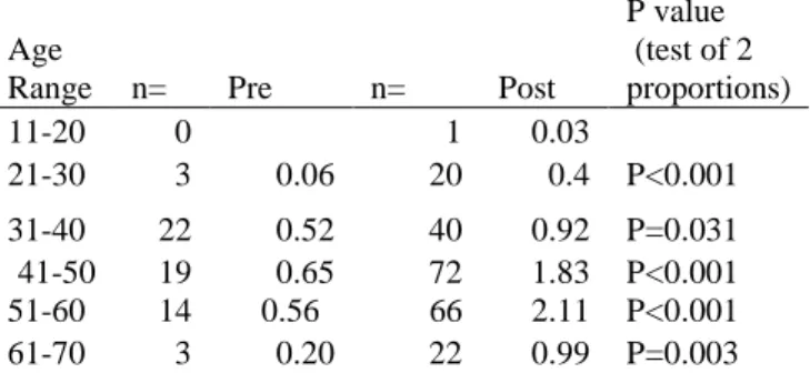
Age
Range n= Pre n= Post
P value (test of 2 proportions)
11-20 0 1 0.03
21-30 3 0.06 20 0.4 P<0.001
31-40 22 0.52 40 0.92 P=0.031
41-50 19 0.65 72 1.83 P<0.001
51-60 14 0.56 66 2.11 P<0.001
61-70 3 0.20 22 0.99 P=0.003
6
Figure 2: Age structured prevalence of secondary toxic goiters
Figure 3: Age structured odds ratio for the occurrence of secondary toxic goiter for post iodination vs pre-iodination periods
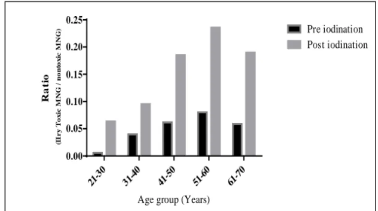
Secondary toxicity in multi-nodular goiters and it’s prevalence in all multi-nodular goiters
The prevalence of toxic goiter, pre and post iodination is expressed as a ratio to the
eu-thyroid multi nodular goiter as per age group is shown in Figure 4.
Figure 4: Age structured ratio of secondary toxic MNG to non-toxic MNG patients
The odds ratio with the 95% confidence limits per age group pre and post iodination is depicted in Figure 5.
In those with MNGs, the older the patient, the greater the likelihood of the MNG being toxic, this was more marked after iodination.
Figure 5:Odds ratio of post iodination vs pre iodination occurrence of toxic MNG compared to non-toxic NMG
Prevalence of toxicity according to the grade of the multi-nodular goiter
11-2 0 21-3 0 31-4 0 41-5 0 51-6 0 61-7 0 71-8 0 0.0 0.5 1.0 1.5 2.0 2.5
Age group (Years)
% P r e v a le n c e Pre iodination Post iodination 0 5 10 15 20 Age group O d d s ra ti o ( 9 5 % C I)
21-30 31-40 41-50 51-60 61-70 1 21-3 0 31-4 0 41-5 0 51-6 0 61-7 0 0.00 0.05 0.10 0.15 0.20 0.25
Age group (Years)
R a ti o (I I r y T o x ic M N G / n o n to x ic M N G
) Pre iodination
Post iodination 0 5 10 15 20 25 30 35 Age group O dd s r at io (9 5% C I)
21-30 31-40 41-50 51-60 61-70
7 The larger the MNG, the greater the prevalence of toxicity (Figure 6 and Table 6).
Pre –Pre- iodination period; 1981-1995, Post – Post-iodination period; 1996 - 2011
Figure 6: Prevalence of graded toxic goiters in the pre vs post iodination periods
Table 6: Percentage prevalence of size of toxic goiter in relation to iodination
Admissions with complications (other than obstructive states due to size), caused by pure toxicity were not frequently seen in this study (they go to the respective Medical, Psychiatric and Orthopaedic wards and clinics), but were seen in a few instances. A few patients presented with cardiac complications, two in heart failure and two others with toxic cardiomyopathy. None had
fractures or were affected by neuropsychiatric disorders.
Careful control of the toxic state, followed by surgery on those with no contraindications, gave good results with no mortality and internationally acceptable morbidity.
Discussion
In our study simple diffuse toxic goiter (? Graves’ disease) shows no demographic or prevalence change related to iodination. Graves’ disease being diagnosed without serology (TRAb) and mostly on its complications, leaves a possibility of under-diagnosis. This is a limitation of the study. Thyroiditis was detected on clinically suspicious cases by FNAC of the thyroid. Thyroid antibodies both the microsomal and the thyroglobulin were only available in the last decade, though done, were not uniformly done due to the prohibitive cost factor. Hashitoxicosis, the transient form of hyperthyroidism was only seen in three of our patients, a prevalence of 1.12%, which fits in with the relative rarity (<5%) of the disease 6.
Our results feature a highly significant increase in secondary toxicity, post iodination, an increase in the percentage of toxicity in MNG specially seen in the elderly,
This possible “Jod-Basedow" effect is attributed to the sudden availability of iodine to iodine starved endemic goiter, which is autonomously active7,8. Thyroid follicles are not suppressed by the excess iodine, or the low levels of thyroid stimulating hormone (TSH) that exist after iodination. The pre iodination TSH induced thyroid bulk is also thought to contribute 9,10. The fact that this may not be the only cause is highlighted by occasional evidence of iodine induced
P r e io d in a tio n P o s t io d in a tio n
Pre Post
Grade
% Prev
%
Prev P value
I 0.00 5.55 -
II 1.59 6.84 <0.001
III 4.59 7.46 0.020
IV 3.333 26.09 0.019
Pre –Pre- iodination period; 1981-1995, Post – Post-iodination period; 1996 - 2011
8 hyperthyroidism in iodine sufficient areas10,
11 12
.
In Tasmania, known for its endemicity for goiters, iodization of bread resulted within months, the doubling of the prevalence of toxicity4. They too take it as a Jod-Basedow effect.
However, only physiological doses of less than 300 µg per day were used. This is alarming because it was contended that such small doses were innocuous. In Sri Lanka country-wide compulsory iodination of the carrier i.e. salt, highlights the possible risks of Jod-Basedow effects on an entire population7. Caution has also been highlighted by the finding of high age related prevalence of TgAb in the population, though the sample was small, implying a possible florid immune response
13,14
.
Imposing such effects on the unsuspecting elderly in the population whose access to effective medical care is doubtful because of personal beliefs, ayuruvedic medication ,or simply sub clinical nature of the symptoms ,is worrying , to say the least. Sri Lankan national policy makers, conscious no doubt of the gravity of the need to minimise such effects reduced the amount of iodine in the salt from twenty five to fifteen parts per million at the consumer end, during this period 3,14. This reduction seems to have had the desired effect on the prevalence of toxicosis (Figure 1). It is claimed that the increase in the prevalence is said to be present for about four to five years post iodination 9,14,15, but that is not conclusive ,as is seen when one scans Figure1.
Sub-clinical toxicity has been shown to be present after iodination in a larger population of elderly patients in areas endemic for goiter, than overt secondary clinical toxicity 16.
Both groups have an associated morbidity and hence require, clinical vigilance for, cardiac dysfunction i.e. atrial fibrillation (AF), and heart failure. Also associated are osteoporosis and neuropsychiatric disorders in those with secondary toxicity 16.
Our study confirms the internationally accepted fact that those affected are often in the older age groups. Females are affected more because goiter is more common among them. It affects them in their post menopausal years when cardiac disease and osteoporotic fractures are not uncommon. Secondary toxicosis will no doubt increase such risks of AF which is found in 2 – 30 % of patients presenting with toxicity especially in those above 60 years with the attendant risks of cardiac sourced embolism17.
It is thought that AF caused by thyrotoxicosis is more liable to embolism and the central nervous system is thought to be more often involved18. Anti-coagulation now is considered mandatory in such circumstances, however has its own risks at this age.
Even though AF and heart failure are documented, even in patients with subclinical toxicity, osteoporotic fractures, are well recognised in toxic patients 19, have as yet not shown to be a feature of subclinical toxicity, though bone mineral depletion is seen in the latter group20. A greater predilection to Dementia and Alzheimer’s disease has also been documented in those with toxicity 21.
Clinical features of toxicity are less prominent in the elderly and the failure to diagnose, exposes them to the risks of such morbidity which increases after iodination. Hence it becomes mandatory that clinicians country-wide recognize these at risk groups
9 especially in the current post iodination period .
In this study the percentage prevalence of toxicity in MNGs, based on size, are respectively, grade II [ 6.8%], III [ 7.5 % } and IV [ 26%] after iodination. Hence it is imperative that clinicians are vigilant of “old ladies with a large goiter” and if there are no contra-indication they should be offered to have it removed after control of the toxicosis.
The obstructive effects of grade IV goiters do not regress with radioactive iodine therapy. The latter therapy may not detect an incidental cancer in the gland and is not encouraged unless strong contraindications exist for surgery.
Many of the Grade IV glands are often retro-tracheal or retro–sternal and need to be surgically removed. In a thyroid or endocrine surgical unit, the mortality is almost nonexistent and the morbidity very low.
Another point that should be emphasized is that, secondary toxicity on MNG’s is the commonest cause of toxicity at present in Sri Lanka and as such should be given due consideration.
As it is a mild form of toxicity, giving the patient Lugol’s iodine pre surgery to evoke the Wolfe – Chaikoff effect and reduce the “vascularity”, as is done in Graves’ disease is both unnecessary 6,7 and maybe downright risky by provoking enhanced toxicity.
Conclusions
We conclude that this study done on patients presenting to a surgical clinic in the Central Province of Sri Lanka shows an increase in the prevalence of toxicity in multinodular goiters after the program of iodination of salt.
Acknowledgements
We thank Mr. Chandana Jayasundera for helping with the sorting of the enormous amount of data that this study entailed.
Conflict of interest and financial disclosure: None
References
1. Jayatissa R. Iodine deficiency status of children in Sri lanka 2000-2001, Report of the Medical Research Institute Sri Lanka, Department of Health Services Sri Lanka in collaboration with the UNICEF.
2. Jayatissa R. Iodine nutrition status in Sri Lanka, 2005. Report of the Medical Research Institute, Sri Lanka, Department of Healthcare and Nutrition 2006, in collaboration with the UNICEF.
3. Jayatissa R. Gunatilleke M.M. Third national survey on Iodine Deficiency status in Sri Lanka 2010. Medical Research Institute, Department of Health care and Nutrition, Ministry of Health and the UNICEF.
4. Connolly R.J. Vidor G.I., Stewart J.C. Increase in thyrotoxicosis in endemic goiter area after iodination of bread.
Lancet 1970 1:500-502. Doi:
10.1016/S0140-6736(70)91582-5
5. Kohn L.A. The Midwestern American “epidemic” of iodine induced hyperthyroidism in the1920’s. Bulletin of the New York Academy of Medicine
1976, 52: 770.
6. Unikrishnan A.G., Hashitoxicosis: A clinical perspective. Thyroid Research and Practice, 2013. 10 55-56. Doi:
10.4103/0973-0354.106803
7. Ermans A.M., Camus M. Modifications of thyroid function induced by chronic administration of iodide in the presence
10 of autonomous thyroid tissue. Acta Endocrinologica 1972, 70, 463-475. Doi: 10.1530/acta.0.0700463
8. Miller J.M., Horn R.C., Block M.A. The evolution of toxic nodular goitre.
Archives of Internal Medicine 1964, 113,72-88.Doi:
10.1001/archinte.1964.00280070074014
9. Martins M.C., Lima N., Knobel M., Natural course of iodine induced thyrotoxicosis [JodBasedow] in an endemic area: a five year follow up.
Journal of Endocrinological Investigation, 1989, 12, 239-44. Doi:
10.1007/BF03349973
10. Saffran, M., Terri L.P. Roti E. Braverman L.E., Environmental factors affecting autoimmune thyroid disease
Endocrinology and Metabolism Clinics of North America 1987, 16:327.
11. Vagenakis A.G., Wang C., Burger A., Maloof F., Braverman L.E., Ingbar S.H. Iodide induced thyrotoxicosis in Boston. New England Journal of Medicine 1972, 287, 523-527. Doi:
10.1056/NEJM197209142871101
12. Eggertsen R., Petersen K., Lundberg P.A., Nystrom Lindstedt G. Screening for thyroid disease in a primary 12 care unit with a thyroid stimulating hormone assay with a low detection unit British Medical Journal , 1988, 297, 1586-1592. Doi: 10.1136/bmj.297.6663.1586
13. Premawardhana L.D.K.E. Iodine excretion, goiter and thyroid autoimmunity in Sri Lanka .The current status and lessons for the future.
Journal of the Ceylon College of Physcians 2000, 32, 11-13.
14. Wickremanayake T.W. Iodised salt and hyperthyroidism. Ceylon Medical Journal 2000, 45,173-174.
15. Weiping T., Zhonggyan S. Xiochun T. et al. Effect of Iodine intake on Thyroid Diseases in China. New England Journal of Medicine 2006, 354 2783 – 2793. Doi: 10.1056/NEJMoa054022
16. Boelaert K, Franklyn J.A. Thyroid hormone in health and disease. Journal of Endocrinology, 2005, 187, 1-15. Doi: 10.1677/joe.1.06131
17. Sarwin C.T., Geller A.,Wolf P.A., Belanger A.J., Baker E., Bacharach P., Wilson, Benjamin E.J. and D’Augastino R.B. Low serum thyrotropin concentrations as a risk factor for Atrial fibrillation in older persons. New England Journal of Medicine 1994, 331 1249-1252. Doi:
10.1056/NEJM199411103311901
18. Presti C.F., Hart R.G., Thyrotoxicosis atrial fibrillation and embolism revisited. American Heart Journal
1989, 117,976-977. Doi: 10.1016/0002-8703(89)90642-X
19. Fraser S.A, Anderson J.B., Sith D.A., Wilson G.M. Osteoporosis and fractures following thyrotoxicosis.
Lancet 1971 I, 981-983. Doi:
10.1016/S0140-6736(71)91383-3
20. Muddle A.H., Reijinders F.J.L., Kruseman A.C. Peripheral bone density in women with untreated multinodular goitre. Clinical Endocrinolology (Oxf)
1992, 37, 35-39. Doi: 10.1111/j.1365-2265.1992.tb02280.x
21. Kalmin S., Mehta K.M., Pols H.A.P., Hofman A., Drexhage H.A., Breteler M.B. Subclinical hyperthyroidism and the risk of dementia, The Rotterdam study. Clinical Endocrinology 2000, 53,733-737. Doi: 10.1046/j.1365-2265.2000.01146.x