femoral central venous catheters to detect deep venous
thrombosis and prevent pulmonary embolism among the
critically ill-a cost-effectiveness analysis
Christopher E. Cox
A Master's Paper submitted to the faculty of the University of North
Carolina at Chapel Hill in partial fulfillment of the requirements for the
degree of Master of Public Health in Health Care and Prevention in the
School of Public Health
Chapel Hill
2002
ABSTRACT
Background: Femoral central venous catheter (CVC) use is complicated by a
high risk of proximal deep venous thrombosis (DVT). DVTs may result in
potentially harmful pulmonary embolism (PE). Performing routine ultrasound after
removal of femoral catheters to detect DVT and thus prevent pulmonary
embolism (PE) has been suggested, but the potential cost-effectiveness of this
strategy has not been examined.
Objective: To evaluate the potential cost-effectiveness of routine unilateral
duplex Doppler ultrasound versus no imaging study in averting PE-associated
deaths associated with femoral CVCs among the critically ill.
Design: Decision model.
Data sources: Probabilities for clinical outcomes were obtained from
meta-analyses of clinical trials and literature syntheses of other prospective studies.
Cost estimates were derived from average physician and Medicare
reimbursements, institutional costs, and other sources.
Target population: Mechanically ventilated 60-year old patients with femoral
CVCs treated in a medical intensive care unit (ICU) for acute respiratory failure.
Time horizon: Duration of hospitalization.
Intervention: Unilateral duplex ultrasound examination of the proximal veins of
the lower extremity performed after removal of a femoral CVC versus no
ultrasound examination.
"--Outcome Measures: Costs, costs per PE-associated death and per PE averted,
and incremental cost-effectiveness ratios.
Results of Base-Case Analysis: A strategy of screening ultrasound cost
$14,350 per PE death and $1 ,969 per PE averted compared with no imaging
test.
Results of Sensitivity Analyses: In one-way sensitivity analyses, the
ultrasound strategy ranged from cost-saving to costs of $151,703 per PE death
prevented after varying prevalence of CVC-associated DVT, probability of PE
associated with DVT, sensitivity and specificity of ultrasound for DVT, and
probability of eligibility to receive low-molecular weight heparin versus an Inferior
vena cava {IVC) filter.
Conclusions: A strategy of performing ultrasound after removing femoral CVCs
appears to be a cost-effective modality in reducing both PE and PE deaths
associated with CVCs among the critically ill. If further study confirms the benefits
of preventing catheter-associated venous thromboembolic disease among the
critically ill, then this study suggests that these improved outcomes come at
INTRODUCTION
Almost half of the 31 million intensive care unit (leU) patients in the US receive
central venous catheters (eves) annually 1• These catheters provide reliable
intravascular access for life-sustaining medications and allow measurement of
hemodynamic variables. Femoral vein placement of eves is popular and has
advantages relative to subclavian and internal jugular vein placement including
the comparative ease of insertion, lack of need for confirmatory imaging tests
after insertion, and very low risk of potentially life-threatening acute complications
such as pneumothorax.
Despite their convenience, femoral eves are associated with significant
complications such as catheter-related bloodstream infection and venous
thrombosis . The incidence of ipsilateral proximal deep venous thrombosis (DVT)
may be as great as 38% 2• Because more than 50% of proximal DVTs may result in pulmonary embolism (PE) 3·5, the burden of catheter-associated venous
thromboembolic disease is quite high among critically ill patients who require
mechanical ventilation for respiratory failure and who have already limited
cardiopulmonary reserve 6.
Although cost-effective therapies exist for the treating DVT and DVT-associated
complications such as PE 7, the best strategy for detecting eve-associated DVT
and thus preventing eve-related pulmonary embolism PE is not clear. Physical
exam alone is unreliable in this population 8, and patients often are unable to
verbalize suggestive symptoms because of endotracheal intubation or sedation.
•
L
F
I
The reference standard, contrast venography, is rarely used because of its
invasive nature and potential for nephrotoxicity 9•
A more acceptable test to detect eve-related DVT may be lower extremity
ultrasound, a test already used by most physicians managing leU patients as
their primary DVT imaging modality. Ultrasound is relatively inexpensive,
noninvasive, can be performed at the bedside without need for patient transport
outside the leU, and is highly accurate for detecting symptomatic proximal DVT
10
• However, it is less sensitive for detecting asymptomatic DVT 11 as is the
typical case among leU patients. Therefore, it is uncertain if treatment based on
ultrasound's lower positive predictive value would lead to excessive costly
complications from anticoagulation or inferior vena cava filter placement in those
falsely diagnosed with DVT.
We developed a decision model to assess the potential cost-effectiveness of
routine lower extremity Doppler ultrasound in preventing PE and PE-related
death when performed after removal of femoral eves among critically ill patients
requiring mechanical ventilation in a medical leu. We hypothesized that a
strategy of screening ultrasound would result in acceptable costs relative to no
ultrasound examination. Our patient population included those at high risk for
DVT. These patients are the group most likely to experience poor outcomes from
PE, and are therefore the most likely to be affected by such a screening strategy.
We included sensitivity analyses to address the potential variability in baseline
probabilities, outcomes incidences, as well as the accuracy of ultrasound testing
METHODS
We used a decision model to compare outcomes of ultrasound screening relative
to no screening among the critically ill with femoral CVCs. Additionally, we
incorporated two treatment strategies for positive tests on ultrasound,
low-molecular weight heparin (LMWH) and inferior vena cava interruption filters
(IVCFs). The probabilities and costs used in the decision model are based on our
judgement about the best estimates from the medical literature. For values that
were not well-supported by the literature, we relied on the opinions of experts in
critical care medicine and venous thromboembolic disease.
Decision Model
We performed a cost-effectiveness analysis by adopting a decision tree
modelling approach and incorporating the recommendations of the Panel on
Cost-Effectiveness in Health and Medicine in our research design and analysis
12
. Figure 1 presents our full decision model in which two approaches to
detection of catheter-associated DVT can be made: screening ultrasound after
removal of femoral CVC versus no ultrasound. The ultrasound strategy used
unilateral duplex Doppler examination of the proximal veins of the lower extremity
catheterized by the femoral CVC. Patients with proximal DVT by ultrasound were
treated with either low molecular weight heparin (LMWH) or an inferior vena cava
(IVC) filter. The management of patients without proximal DVT by ultrasound or
who did not receive screening ultrasound changed as clinically evident
complications arose such as PE. In our model we assumed that outcomes are
only those that clinicians could directly observe. By taking this approach, we
'
r
were only able to assess PEs detected clinically among those who did received
no ultrasound. We also assumed that some DVTs and PEs among these
severely ill, mechanically ventilated patients were clinically undetected and
therefore were not associated with greater costs.
Patients and Baseline Assumptions
We analyzed treatment costs and important clinical outcomes relevant to a
60-year old male patient with a femoral eve who received mechanical ventilation for
acute respiratory failure. This demographic profile best represented the critically
ill patients described in a recent international epidemiologic study of 5,183
patients requiring mechanical ventilation, whose mean age was 59.2 and of
whom 61.3% were male 13. Patients were assumed to receive mechanical ventilation for 7 days, stay in the leU for 8 days, remain on the hospital ward for
an additional 7 days, and have a SAPS II score of 44 13. Although we did not take into account patients' underlying severity of illness in mortality calculations, we
assumed the mortality rate would be approximately 33% during hospitalization 14•
The base-case patient was assumed to receive DVT prophylaxis with sequential
compression devices while in the leU. Femoral eves were assumed to have
three lumens, were not heparin- or antibiotic-impregnated, and were left in place
for at least 1 week. We defined DVT as a partial or complete occlusion of the
proximal veins of the lower extremity on the ipsilateral side of the femoral eve.
Time Horizon and Perspective
The period of initial hospitalization was examined in our analysis. Therefore, we
did not incorporate values for health states (utilities) to calculate quality-adjusted
!___
t
r
I
tlife years (QAL Ys). We took the perspective of the health care payer in our cost
analyses.
Diagnostic and Treatment Alternatives
In our model, a screening unilateral lower extremity ultrasound could be
performed after removal of a femoral eve or not. If ultrasound was chosen, we
assumed that all eligible patients found to have proximal DVT would be treated
with LMWH because of its superior cost-effectiveness to unfractionated heparin 7 •
In our analysis, enoxaparin given 1 mg/kg twice a day was used for LMWH
calculations, assuming patients weighed 70kg. An IVeF was placed in those with
contraindications to anticoagulation. If no DVT was detected by ultrasound, no
therapy was given. We assumed that DVT was not clinically obvious in patients 8.
Probability Estimates
The probabilities of clinical events associated with femoral eve-related DVT
used in our decision model are shown in Table 1. We have based these
probabilities on review of the relevant literature including a MEDLINE search
(1966 to April 2002) as well as a bibliographic search of retrieved articles'
reference lists. Although data specific to critically ill medical ICU patients with
catheter-associated DVT and PE are limited, we have included studies of the
closest possible relevance to our target population and have utilized multiple
sensitivity analyses to investigate the potential variability in variables with
uncertain values.
I
Femoral eve-Associated DVT
The prevalence of eve-associated DVT ranges from 9% to 38% among the
critically ill and post-operative surgical patients 2· 15-22. Our base-case probability
of 21.5% for DVT is based on a large, randomized controlled trial comparing
complications of femoral eves with subclavian eves in critically ill adults 2• We
also performed a systematic review of femoral eve-associated venous
thrombosis (Appendix 1). Sensitivity analyses included the lowest and highest
rates from recent prospective studies of femoral eve-associated DVT 15-21 as
well as a study based on autopsy results for eve-associated thrombi 22•
Ultrasound
The sensitivity (62%) and specificity (94%) of ultrasound were based on a
meta-analysis of sixteen clinical trials performed among asymptomatic hospitalized
patients, most of whom were post-operative orthopedic patients in 1eus 11• We
examined ranges for both sensitivity (15-75%) and specificity (75-98%) in
sensitivity analyses based on the reported confidence intervals of this
meta-analysis. Such post-operative patients are likely to fairly approximate the
thromboembolic risk and logistic imaging difficulties (patient positioning, edema,
need for bedside testing) of medical leu patients.
Pulmonary Embolism
The 30% probability of PE resulting from DVT was derived from pooled rates
reported in three prospective studies 3· 5· 23 as well as from one large, randomized
trial of leU patients 2 that examined PE incidence among hospitalized patients.
Our sensitivity analyses reflect the likelihood that the critically ill may experience
a higher incidence than other less severely ill hospitalized patients because of
j
L
L
jtheir many risk factors for thromboembolic disease as well as the difficulty in
diagnosing this condition within such a group of sedated, mechanically ventilated
patients. We based the probability of PE while being treated with LMWH (1.9%)
on results of a meta-analysis comparing LMWH with unfractionated heparin for
the treatment of DVT 24• Sensitivity analyses include data from a large
randomized trial comparing IVC filters versus LMWH for prevention of PE among
those with DVTs 25• A mortality rate of 30% from untreated PE is based results of
a systematic review 26 and sensitivity analyses include the ranges of individual
studies' reported mortality rates (18%-38%). Finally, both the probability of PE
and death from PE after IVC filter placement was based on results of a
400-patient randomized trial of IVC filters versus low-molecular weight hepain for
prevention of PE in those with proximal deep vein thrombosis 25• This is the only
randomized trial evaluating outcomes of IVC filter placement to our knowledge.
Treatment Eligibility
Patients found to have proximal femoral vein DVT by ultrasound were treated
with LMWH if they had no contraindications to anticoagulation. Those who could
not receive LMWH for any reason received an IVC filter. We estimated that
approximately two-thirds of patients would be eligible to receive LMWH based on
institutional experience, though examined wide ranges of eligibility for
anticoagulation in our analyses.
Treatment Complications
We derived the probability of both major fatal (0.5%) and non-fatal bleeding
(1.1 %) due to LMWH from a meta-analysis of 11 randomized trials that examined
LMWH versus unfractionated heparin for the treatment of acute DVT 10•
I
!Thrombosis of the femoral vein or inferior vena cava is one of the most common
serious adverse effects of IVC filter placement 27 and was the sole major
complication considered in our modeL The 6% probability of this major
complication of IVC filter placement was based on a randomized clinical trial that
examined LMWH versus IVC filters 25• Systematic reviews of IVC filters have
reported similar results based on data from non-randomized trials 27• 28 and their
results were used for upper and lower limits of the sensitivity analyses. We did
not consider complications due to heparin-induced thrombocytopenia because of
the very low risk associated with LMWH and the infrequent occurrence of
clinically significant complications 29.
Because few data relevant to our population and this disease process exist, for
most therapy-related complications we assumed additional days of ICU and
hospital ward care that are estimates based on average lengths of stay of such
patients with these diagnoses at our institution. Because the increase in ICU
length of stay attributable to PE has not been evaluated to our knowledge, we
based our estimate of 4 extra ICU days on institutional experience and analyses
of other ICU complications including gastrointestinal bleeding (4 to 8 extra ICU
days) 30 and ventilator-associated pneumonia (3 to 10 extra ICU days) 31•
Generally however, complications prolonged ICU stay by 3 to 4 days and hospital
ward stay by 2 to 3 days. Costs related to death are based on a recent
cost-effectiveness model 7, though are included only in sensitivity analyses because
Costs
The costs used in our model are shown in Table 2.Total costs of each scenario
tested are also shown in Appendix 2. We incorporated a health care payer
perspective and included direct medical costs. Indirect costs such as days lost
from work by the patient or family members were not included in analyses. All
non-pharmaceutical medical costs were standardized to 2001 US dollars by
using the medical care component of the Consumer Price Index 32•
Costs of Initial Diagnosis and Treatment
In our calculations of the inpatient costs for those critically ill patients who
underwent ultrasound after removal of a femoral CVC, we utilized costs for
hospital care, diagnosis and treatment of DVT, as well as costs of potential
complications related to therapy for DVT or PE. We used the average institutional
costs per day of care for medical ICU patients in our baseline cost calculations.
These costs were calculated by multiplying average costs of all medical ICU
patients treated at UNC hospitals during ihe year 2001 by the institutional cost to
charge ratio (0.601) (Greco P, personal communication). These average charges
include hospital costs, physician fees, average costs of laboratory tests,
administrative costs, and electricity. Likewise, hospital ward costs in our analyses
reflect similar institutional costs. Pharmaceutical costs were based on average
wholesale prices for 2001 33• The costs of procedures included in our decision model such as ultrasound, IVC filters, esophagogastroduodenoscopy (EGO), and
ventilation-perfusion (V/Q) scans included physician interpretation. We derived
these costs from the
so'h
percentile of usual customary physician reimbursementrates 34 based on current procedural terminology (CPT) codes 35• Our sensitivity
Fee Scale reimbursement 34 and the upper bound the 90'" percentile of usual customary reimbursement.
Costs of Complications
Major complications of both LMWH treatment and IVC filter placement were
considered in our analyses. For costs associated with major non-fatal bleeding
secondary to use of LMWH, we assumed an EGD with intervention would be
pertormed and one unit of blood transfused, similar to past cost-effectiveness
analyses of LMWH 36• Costs for IVC filter thrombosis include an extra ultrasound examination. Extra days of ICU care and hospital ward care were assumed
based on the occurrence of complications such as PE, major bleeding 30, and IVC thrombosis and these costs based on institutional data.
Base-Case Analysis
We calculated incremental cost-effectiveness ratios (ICERs) by comparing
ultrasound versus no ultrasound in averting PE-associated death and overall PE.
Our best estimates of probabilities for all variables included in our decision model
were used in the base-case analysis and the uncertainty present in variables
assessed in one- and two-way sensitivity analyses. We used DATA 4.0 (TreeAge
Software, Williamstown, MA) and Excel (Microsoft Corporation, Redmond, WA)
software for analyses.
Sensitivity Analyses
Published data have provided wide ranges of probabilities and outcomes of
CVC-associated DVT and PE. Therefore, we pertormed one-way sensitivity analyses
by varying values of uncertain probabilities or costs within specified ranges
j
'
L
~
!
included in the decision model to investigate the effect of such differences on
incremental cost-effectiveness ratios. In these sensitivity analyses, we
incorporated either the greatest variability seen in medical literature, 95%
confidence intervals of meta-analyses, or added or subtracted half of probabilities
or costs. We also examined the probable costs associated with the worst case
scenario in which variables were all set to minimize the positive impact of
ultrasound.
Two-way sensitivity analyses were also performed to examine the effect of
changing two of the most important variables in our analysis simultaneously,
sensitivity (as well as specificity) of ultrasound and prevalence of
CVC-associated DVT. This was done to address the presence of ultrasound operator
differences between centers may affect overall outcomes. We also examined the
effect of varying probability of
PE
associated with CVC use and probability ofPE-associated death, both variables PE-associated with wide ranges of values in
published literature.
RESULTS
Base-Case Analysis
In the base-case analysis, performing screening ultrasound compared with no
imaging after femoral CVC removal resulted in an increase in overall costs from
$30,372 to $30,677 (Table 3). The incremental cost-effectiveness ratio, reflecting
costs per
PE
death averted by ultrasound screening followed by appropriatetherapy, was $14,350. In secondary analyses, use of ultrasound raised overall
costs frorn $30,092 to $30,199 and was associated with incremental costs of
$1 ,969 per CVC-related PE averted.
L
r
f
!
1
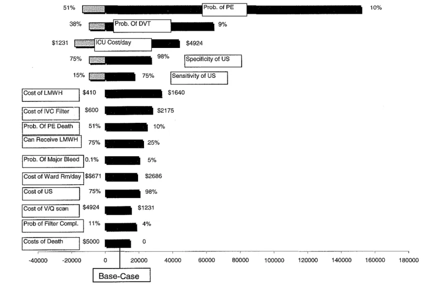
Sensitivity Analyses
Results of one-way sensitivity analyses comparing ultrasound versus no
ultrasound are shown in Figure 2. We varied the values of probabilities and costs
that were uncertain or of perceived interest those caring for the critically ill.
Overall, incremental cost-effectiveness ratios remained less than $50,000 per PE
t-death averted for most analyses, with the exception of probability of DVT and
F
'
probability of PE death.
Costs per PE death averted became significantly greater as we increased either
the sensitivity or specificity of ultrasound. For example, varying sensitivity from
15% to 75% changed incremental cost-effectiveness ratios from initial cost
savings to $17,082 per PE death averted. However, even at a sensitivity of 95%,
I
ultrasound cost $20,134 per PE death averted. Likewise, as specificity increasedfrom 75% to 98%, cost savings dominated up to a specificity of 86% after which
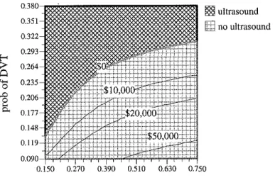
the ICER was $26,734. In two-way sensitivity analyses, we explored the effect of
ultrasound accuracy on ICERs further by simultaneously varying sensitivity and
specificity of ultrasound with the probability of PE (Figures 3 and 4). As both
sensitivity and specificity were improved, the incremental costs associated with
ultrasound in averting PE death increased. The trend toward increased costs with
improved accuracy of imaging represents the reduction in expenses associated
with treating ore expensive complications such as PE after '1alse negative" tests
and major bleeding events caused by anticoagulation given for '1alse positive"
tests.
The effect of varying costs of ICU and hospital ward care produced wide ranging
preventing each PE death cost nearly $26,000. Not until daily costs of leU care
reached $4,000 did the ultrasound strategy dominate. Similarly, doubling and
halving ward costs changed leERs by a nearly identical factor, with lower ward
costs associated with higher leERs.
When we varied the prevalence of femoral eve-associated DVT from 9% to
38%, a strategy of ultrasound screening ranged from costs of $63,805 per PE
death averted at a prevalence of 9% to cost savings at a prevalence of 30%.
Further, a greater incremental effect on PE death was observed at a prevalence
of 29%, with 3 deaths averted per 100 femoral eve DVTs. An exception to the
generally stable incremental cost-effectiveness ratios was variance of PE. When
the probability of developing PE from a eve-associated DVT was changed,
leERs ranged from $151,703 at a 10% probability to cost savings at 38%
probability. When we examined probability of receiving LMWH without
contraindications, the cost per PE prevented dropped from $22,136 at 25% to
$12,681 at 75% likelihood, consistent with an overall cost savings of LMWH
compared with IVC filter placement.
Varying the costs of treatments produced a wide range of leERs. For example,
when LMWH was available for $410, the leER was $5,031. However, when
pharmacy costs increased to $1640, incremental costs rose to nearly $33,000
per PE death averted. Similarly, varying IVe filter placement costs in our model
from $600 to $2175 changed increased the leER from $6,300 to $27,503.
Varying costs of most procedures and treatments such as ultrasound, V/Q scans,
IVe filters, and Uv1WH had little effect, typically changing leERs less than $1 ,000
r
L
t
r
from the base-case scenario per PE death averted. However, varying
probabilities of treatment-related death of both IVe filters and LMWH each
increased leERs nearly $5,000 from that of the base-case scenario.
~-In analyses evaluating the cost-effectiveness of ultrasound in averting overall PE,
varying the probability of PE with DVT was associated with costs of $60,614 per
PE averted at a probability of 9% to cost savings at probability of PE of 32%.
Likewise, prevalence of DVT varied leERs from $17,222 at 9% to cost savings at
24%.
DISCUSSION
The critically ill have an especially high incidence of DVT, likely as a result of
prolonged immobility, overall illness severity, and coagulation dysfunction 37• The
optimal strategy for the prevention, detection, and treatment of central venous
catheter-associated DVT and PE among the critically ill is unclear. We used
decision analysis modeling based on the probabilities most relevant to the
critically ill to examine the cost-effectiveness of performing ultrasound after
removing femoral eves in averting PE and death attributable to PE. Our results
suggest that an ultrasound-based screening strategy is associated with
acceptable potential costs of $14,350 per PE death averted and $1 ,969 per PE
1
averted. In the base-case analysis, we found that for every 100 ultrasound tests
performed, 5 non-fatal and 2 fatal PEs could be potentially averted by using
ultrasound screening. Our results were stable over the wide range of
probabilities, treatment strategies, and treatment costs we examined in the
CoxCE
Barriers in Decision Making
Decision-making regarding eve-associated DVT and PE management may be
challenging for physicians who manage critically ill medical patients. First, there
are few data on venous thromboembolic disease specific to this population.
Perhaps as a result, DVT and PE issues relevant to the critically ill have not been
specifically addressed in recent statements by consensus groups such as the
American Thoracic Society 38 and the American College of Chest Physicians 39• Also, the diagnosis of DVT and PE is difficult among ICU populations. Physical
examination is inaccurate for detecting DVT among these patients 8, who
themselves are usually unable to complain of symptoms associated with DVT
because they are intubated and sedated. Diagnosing PE among patients who
require mechanical ventilation is daunting, considering how commonly potential
confounding pathophysiology is present such as pulmonary edema, pneumonia,
and the acute respiratory distress syndrome. Although ultrasound testing
improves diagnostic accuracy, its sensitivity and specificity is lower among the
asymptomatic population relative to symptomatic outpatients . Nevertheless,
ultrasound is the most common test intensivists currently use to detect DVT
because the reference standard, contrast venography, is invasive, requires
patient transport to an imaging department, and is perceived as having a high
risk of nephrotoxicity. Our findings suggest that ultrasound may improve
outcomes at an acceptable cost per PE and PE death averted with either
anticoagulation or IVC interruption, supporting physicians' current practice.
The Potential Value of Ultrasound Screening
l
Not all DVTs and PEs are clinically important or require immediate
anticoagulation among the critically ill. However, considering that PE mortality is
strongly associated with hemodynamic status 40, the attributable morbidity and
mortality of PE within an leU population is likely to have been underappreciated.
For those who develop PE from DVT and survive, pathophysiologic impairments
may included increased work of breathing, increased dead space ventilation with
resultant hypercapnea and hypoxemia, and right ventricular overload 41• These
changes are likely to reduce weaning success and increase days of mechanical
ventilation. Increased time of mechanical ventilation is directly associated with
increased complications and costs 42•
The Potential Problems of Ultrasound Screening
Despite the potential benefits of ultrasound, there are many possible problems
with an ultrasound-based screening program. First, there is the concern for
bleeding related to heparin. However, even when we increased the probability of
major bleeding as high as 10%, over four times greater than that reported in a
recent clinical trial of LMWH for acute PE 43, the cost per PE death averted
remained less than $50,000. Recognizing that many leU patients have bleeding
diastheses, we found that if as many as 75% of patients with DVT received the
more expensive IVe filter for treatment rather than anticoagulation, costs
remained less than $23,000 per PE death averted. There are also overriding
questions of femoral catheter safety relative to upper extremity eves. Although
subclavian placement of eves was associated with nearly ten times fewer
thrombotic complications than femoral catheters in a recent large randomized
trial 2, other studies have reported vessel thrombosis in up to 4 7% of subclavian
and internal jugular vein catheters and PE in over 36% of these patients 44•
Therefore, choosing a site other than the femoral vein for eve placement will not
avoid thrombotic complications completely. Finally, the role of heparin flushes in
eves is important. Although the addition of daily heparin to eves reduce vessel
thromboses, rates of catheter-associated venous thrombosis are still troubling 45•
We believe that an ultrasound-based screening program of DVT detection and
treatment could address these concerns.
The Decision Model
The decision model used in our analyses has particular usefulness in addressing
eve-associated DVT and PE. First, the model accommodates individual
assessment of probabilties and institutional differences in costs in the
interpretation of ultrasound's cost-electiveness. This improves the generalizability
I
jf of our results. Our model is also able to compare the performance and costs
associated with two different therapies for eve-associated DVT, LMWH and IVe
filters, among the critically ill. Lastly, precisely because there is little data
applicable to the target patient population and relevant clinical trials have been
so difficult to perform, our model can help inform decision making by gathering
many of the best relevant estimates of disease probability, treatment benefits and
complications, and costs from methodologically strong clinical trials.
The probabilities used in our model were the best estimates available relevant to
our target population. However, some of the probability estimates we used in our
model are likely to be controvesial. For example, both the incidence of PE
associated with eves and the attributable mortality of these PEs is unknown.
Our analyses instead used PE data gathered from patients with DVT
any lower among those with eves, this is still conceivable. However, our
sensitivity analyses showed that costs per PE death averted do not exceed
traditional cost-effectiveness threshold values of $20,000 and $50,000 until PE
incidence drops below relatively conservative estimates of 27% and 20%,
respectively. Also, the probability of eve-associated DVT is not well-established
in clinical trials that have used a reference standard. Our estimate of 21.5% is
based on a large, methodologically strong randomized trial that used ultrasound
as the diagnostic standard for eve-associated DVT 2• However, this estimate is
likely an underestimate of the true prevalence of eve-associated DVT because
of the low sensitivity of ultrasound among the critically ill (ref) and asymptomatic
patients 11•
In sensitivity analyses, we were able to address concerns of physicians regarding
not only the potential benefits of ultrasound, but the possible adverse effects of
treatment based on ultrasound. We found that even when the incidence of major
bleeding related to LMWH was multiplied by a factor of four (to 1 0%), the cost
per PE death averted remained under $50,000. Likewise, increasing the IVe filter
thrombosis rate to 20% resulted in a cost increase to $24,555 per PE death
prevented. Because of higher cost of IVe filter placement and greater risk of
complications, LMWH therapy was associated with lower incremental costs.
However, institutional and operator-dependent differences may be important
when considering interventions. Overall, incremental cost-effectiveness ratios
remained less that $30,000 per PE death averted over the full ranges of
probabilities and costs examined, yet were sensitive to small changes from
base-case values.
r
r
Lt
The daily costs of leU and hospital ward care had a smaller effect than disease
probabilities on incremental cost-effectiveness ratios in our analyses. There are
many likely reasons why this was observed. First, because severity of illness and
length of stay was not considered in these data, the true costs of PE are likely
underestimated, because patients who require mechanical ventilation have
average leU and hospital stays that are significantly longer than those who do
not need respiratory support 46. Also, we have attempted to use conservative
cost estimates in our model in order to reduce potential bias in favor of
ultrasound. Further, we have not included costs of death in our model, unlike
some recent cost-effectiveness analyses 7, which would bias our analyses
against "standard of care".
The Course for Future Research
Our decision model is helpful in highlighting areas where more research is
needed to clarify the burden of venous thromboembolic disease among the
millions of leU patients who receive eves annually. Epidemiologic data are
needed on incidence of eve-associated DVT and PE. Further, imaging tests
need to be rigorously tested in this complicated population versus reference
standards of pulmonary angiography and contrast venography to better assess
their value relative to the same tests performed among symptomatic,
non-critically ill populations. The fact remains that physicians caring for leU patients
currently use tests to detect PE and DVT that have uncertain accuracy within this
population, including helical computerized tomography (eT) and ultrasound.
Also, the value of rapid assays for thrombosis such as D-dimer may provide
benefit to patients when their role is better defined. Finally, it is important to
better appreciate factors associated with vessel thrombosis such as relationship
l
l
!
l
I
.,
'
to time left in situ, affect of underlying disease process (sepsis, cancer), heparin
flushing dose and flushing frequency, and type of infusate (TPN versus other).
Overall, more research is needed to describe the attributable morbidity, mortality,
and costs of venous thromboembolic disease among the critically ill.
Limitations
It is important to consider the limitations of our model and analyses. First, few
data exist on the incidence of PE and PE mortality specific to the critically ill.
However, it is likely that such data may not become available soon for both
ethical and logistical reasons because many physicians may be reluctant to
enroll their severely ill patients in clinical trials examining PE incidence and
treatment because of concerns for contrast-induced nephrotoxicity associated
with angiography and venography, the reference standards for PE and DVT
diagnoses. Further, other diagnostic imaging modalities including helical
computerized tomography (eT) and V/Q scans, require transport of patients to
other areas of the hospital which might place patients at increased risk for
adverse outcomes and require additional staffing. Likewise, the true accuracy of
ultrasound pertormed on medical leU patients is unknown. However, our
sensitivity analyses accounted for potentially wide variability in both PE incidence
and ultrasound sensitivity and specificity.
We used the some of the most methodologically strong data available for
probabilities and costs in our model. Nevertheless, our model may underestimate
the true impact of eve-associated DVT and PE. First, we did not incorporate the
morbidity and mortality of eRBI, which has mortality rates ranging from 10-25%
and increases hospital costs significantly 47• Such catheter-associated
i
F
F
l
thrombosis is strongly related to eRBI among the critically ill 22• Also, mortality
attributable to PE among the critically ill is not known. Although we used rates
gathered from non-critically ill patients, it is probable that these too are
underestimates because of the association between hemodynamic status and
PE mortality . Also, we did not include costs associated with death, which would
have further biased the model toward ultrasound.
Conclusion
A good screening test is accurate, relatively inexpensive, and can improve
outcomes of an otherwise serious disease 48• Although we have shown that ultrasound screening may prevent potentially fatal PEs at acceptable costs to the
health care payer over a wide range of disease probabilties and costs of
treatment, it is premature to recommend routine ultrasound-based screening and
treatment of eve-associated DVT in the absence of relevant clinical trials. First,
the true attributable morbidity and mortality of DVT and PE must be determined
among those who are at greatest risk for poor outcomes-the critically ill.
However, in the absence of such definitive data, the potential burden of disease
associated with catheter-related DVT and PE among the critically ill is sobering:
even at a 10% prevalence of DVT, a 10% incidence of PE associated with DVT,
and a 10% probability of death from untreated PE, a group of 1000 critically ill
patients with femoral eves will experience 10 PEs and 1 PE-related death.
When multiplied by the millions who receive femoral eves annually, it becomes
apparent that there is likely room for improved outcomes from venous
thromboembolic disease among the critically ill.
L
'-1
1
1=
r
'
'
I
r
I
We have shown that screening ultrasound versus no ultrasound performed after
removal of femoral eves may be cost-effective in preventing both PE-associated
death and overall PE. This is the first cost-effectiveness study of screening
ultrasound to prevent CVC-associated PE, to our knowledge. In the absence of
randomized clinical trials examining detection and prevention of eve-associated
PE within the critically ill population, physicians should consider performing
screening ultrasound tests after removing femoral vein CVCs. The potentially
large burden of venous thromboembolic disease among the critically ill warrants
further study in areas of epidemiology, prevention, detection, and treatment. The
clinical outcomes of critically ill patients may be improved significantly through a
better understanding of DVT and PE.
REFERENCES
1. Mermel LA. Prevention of intravascular catheter-related infections. Ann
Intern Med. Mar 7 2000;132(5):391-402.
2. Merrer J, De Jonghe B, Golliot F, et al. Complications of femoral and subclavian venous catheterization in critically ill patients: a randomized controlled trial. Jama. Aug 8 2001 ;286(6):700-707.
3. Moser KM, Fedullo PF, LitteJohn JK, Crawford R. Frequent asymptomatic pulmonary embolism in patients with deep venous thrombosis. Jama. Jan 19 1994;271 (3):223-225.
4. Monreal M, Ruiz J, Olazabal A, Arias A, Roca J. Deep venous thrombosis and the risk of pulmonary embolism. A systematic study.
Chest. Sep 1992; 1 02(3) :677-681 .
5. Huisman MV, Buller HR, ten Gate JW, et al. Unexpected high
prevalence of silent pulmonary embolism in patients with deep venous thrombosis. Chest. Mar 1989;95(3):498-502.
6. Legere BM, Dweik RA, Arroliga AC. Venous thromboembolism in the intensive care unit. Clin Chest Med. Jun 1999;20(2):367-384, ix. 7. Gould MK, Dembitzer AD, Sanders GD, Garber AM.
Low-molecular-weight heparins compared with unfractionated heparin for treatment of acute deep venous thrombosis. A cost-effectiveness analysis. Ann
Intern Med. May 18 1999;130(10):789-799.
8. Geerts WH, Code Kl, Jay RM, Chen E. Szalai JP. A prospective study of venous thromboembolism after major trauma. N Eng/ J Med. Dec 15 1994;331 (24}:1601-1606.
~-[
r
I
r
9. Cook D, McMullin J, Hodder R, et al. Prevention and diagnosis of venous thromboembolism in critically ill patients: a Canadian survey.
Grit Care. Dec 2001 ;5(6):336-342.
10. Gould MK, Dembitzer AD, Doyle RL, Hastie TJ, Garber AM. Low-molecular-weight heparins compared with unfractionated heparin for treatment of acute deep venous thrombosis. A meta-analysis of randomized, controlled trials. Ann Intern Med. May 18
1999;130(10):800-809.
11. Kearon C, Julian JA, Newman TE, Ginsberg JS. Noninvasive diagnosis of deep venous thrombosis. McMaster Diagnostic Imaging Practice Guidelines Initiative. Ann Intern Med. Apr 15 1998;128(8):663-677.
12. Gold MR. Cost-effectiveness in health and medicine. New York: Oxford University Press; 1996.
13. Esteban A, Anzueto A, Frutos F, et al. Characteristics and outcomes in adult patients receiving mechanical ventilation: a 28-day international study. Jama. Jan 16 2002;287(3):345-355.
14. Le Gall JR, Lemeshow S, Saulnier F. A new Simplified Acute
Physiology Score (SAPS II) based on a European/North American multicenter study. Jama. Dec 22-29 1993;270(24):2957-2963.
15. Nolewajka AJ, Goddard MD, Brown TC. Temporary transvenous pacing and femoral vein thrombosis. Circulation. Sep 1980;62(3):646-650.
16. Meredith JW, Young JS, O'Neil EA, Snow DC, Hansen KJ. Femoral catheters and deep venous thrombosis: a prospective evaluation with venous duplex sonography. J Trauma. Aug 1993;35(2):187-190; discussion 190-181.
17. Mian NZ, Bayly R, Schreck DM, Besserman EB, Richmand D. Incidence of deep venous thrombosis associated with femoral venous
catheterization. Acad Emerg Med. Dec 1997;4(12):1118-1121.
18. Joynt GM, Kew J, Gomersall CD, Leung VY, Liu EK. Deep venous thrombosis caused by femoral venous catheters in critically ill adult patients. Chest. Jan 2000;117(1):178-183.
19. Durbec 0, Viviand X, Potie F, Vialet R, Albanese J, Martin C. A prospective evaluation of the use of femoral venous catheters in critically ill adults. Grit Care Med. Dec 1997;25(12):1986-1989.
20. Harris LM, Curl GR, Booth FV, Hassett JM, Jr., Leney G, Ricotta JJ. Screening for asymptomatic deep vein thrombosis in surgical intensive care patients. J Vase Surg. Nov 1997;26(5):764-769.
21. Trottier SJ, Veremakis C, O'Brien J, Auer AI. Femoral deep vein thrombosis associated with central venous catheterization: results from a prospective, randomized trial. Grit Care Med. Jan 1995;23(1 ):52-59.
22. Raad, II, Luna M, Khalil SA, Costerton JW, Lam C, Bodey GP. The relationship between the thrombotic and infectious complications of central venous catheters. Jama. Apr 6 1994;271 (13):1 014-1016. 23. Plate G, Ohlin P, Eklof B. Pulmonary embolism in acute iliofemoral
venous thrombosis. Br J Surg. Nov 1985;72(11 ):912-915.
24. Dolovich LR, Ginsberg JS, Douketis JD, Holbrook AM, Cheah G. A meta-analysis comparing low-molecular-weight heparins with
unfractionated heparin in the treatment of venous thromboembolism: examining some unanswered questions regarding location of treatment, product type, and dosing frequency. Arch Intern Med. Jan 24
2000;160(2):181-188.
;
i
L
I
r
i
F
25. Decousus H, Leizorovicz A, Parent F, et al. A clinical trial of vena caval ~
filters in the prevention of pulmonary embolism in patients with proximal deep-vein thrombosis. Prevention du Risque d'Embolie Pulmonaire par Interruption Cave Study Group. N Eng/ J Med. Feb 12
1998;338(7):409-415.
26. Dalen JE, Alpert JS. Natural history of pulmonary embolism. Prog Cardiovasc Dis. Jan-Feb 1975;17(4):259-270.
L
27. Becker DM, Philbrick JT, Selby JB. Inferior vena cava filters. Indications, safety, effectiveness. Arch Intern Med. Oct
1992;152(10):1985-1994. L
l
28. Streiff MB. Vena caval filters: a comprehensive review. Blood. Jun 15
2000;95(12):3669-3677. l
29. Warkentin TE, Levine MN, Hirsh J, et al. Heparin-induced
thrombocytopenia in patients treated with low-molecular-weight heparin or unfractionated heparin. N Eng! J Med. May 18
1995;332(20):1330-1335.
i
30. Cook DJ, Griffith LE, Walter SD, et al. The attributable mortality and
'
•length of intensive care unit stay of clinically important gastrointestinal
l
"
bleeding in critically ill patients. Grit Care. Dec 2001 ;5(6):368-375. f='
31. Heyland DK, Cook DJ, Griffith L, Keenan SP, Brun-Buisson C. The
' '
attributable morbidity and mortality of ventilator-associated pneumoniain the critically ill patient. The Canadian Critical Trials Group. Am J '
I
RespirCritCare Med. Apr 1999;159(4 Pt 1):1249-1256.
32. Consumer Price Indexes. Bureau of Labor Statistics. Available at:
http://www.bls.gov/cpi/home.htm. Accessed March 15, 2002.
33. Drug Topics Red Book. Montavale, NJ: Medical Economics; 2001. l
'
34. Physician Fees: A Comprehensive Guide for Fee Schedule Review and
Management. Los Angeles: Practice Management Information
Corporation; 1998.
35. Physician's Current Procedural Terminology. Chicago: American
i
Medical Association; 2001. j
36. Estrada CA, Mansfield CJ, Heudebert GR. Cost-effectiveness of low- ~ molecular-weight heparin in the treatment of proximal deep vein
f
thrombosis. J Genlntern Med. Feb 2000;15(2):1 08-115.37. Hirsch DR, Ingenito EP, Goldhaber SZ. Prevalence of deep venous thrombosis among patients in medical intensive care. Jama. Jul 26
1995;27 4(4):335-337.
i
38. Tapson VF, Carroll BA, Davidson BL, et al. The diagnostic approach to
1
acute vencus thromboembolism. Clinical practice guideline. American ,!
Thoracic Society. Am J Respir Grit Care Med. Sep 1999;160(3):1 043- if
t
1066.
39. Opinions regarding the diagnosis and management of venous
thromboembolic disease. ACCP Consensus Committee on Pulmonary Embolism. American College of Chest Physicians. Chest. Feb
1998; 113(2):499-504.
40. Kasper W, Konstantinides S, Geibel A, et al. Management strategies
and determinants of outcome in acute major pulmonary embolism:
L
results of a multicenter registry. JAm Col/ Cardia/. Nov 141. Wood KE. Major pulmonary embolism: review of a pathophysiologic
F
r
approach to the golden hour of hemodynamically significant pulmonary embolism. Chest. Mar 2002;121(3):877-905.
42. Ely EW, Baker AM, Dunagan DP, et al. Effect on the duration of mechanical ventilation of identifying patients capable of breathing spontaneously. N Eng/ J Med. Dec 191996;335(25):1864-1869.
43. Simonneau G, Sors H, Charbonnier B, et al. A comparison of
low-molecular-weight heparin with unfractionated heparin for acute
f--pulmonary embolism. The THESEE Study Group. Tinzaparine ou
L
Heparine Standard: Evaluations dans I'Embolie Pulmonaire. N Eng/ J
Med. Sep 4 1997;337(1 0):663-669. h l
44. Prandoni P, Polistena P, Bernardi E, et al. Upper-extremity deep vein thrombosis. Risk factors, diagnosis, and complications. Arch Intern
Med. Jan 131997;157(1):57-62.
45. Randolph AG, Cook OJ, Gonzales CA, Andrew M. Benefit of heparin in central venous and pulmonary artery catheters: a meta-analysis of
~
randomized controlled trials. Chest. Jan 1998;113(1):165-171.
:1-46. Ryskamp RP, Trottier SJ. Utilization of venous thromboembolism i prophylaxis in a medical-surgicaiiCU. Chest. Jan 1998;113(1 ):162-164.
r
47. Veenstra DL, Saint S, Sullivan SO. Cost-effectiveness of
antiseptic-l impregnated central venous catheters for the prevention of catheter- !
related bloodstream infection. Jama. Aug 11 1999;282(6):554-560. }
~
48. U.S. Preventive Services Task Force. Guide to Clinical Preventive ~
Services: Report of the U.S. Preventive Services Task Force. Vol 1996.
I
Washington, D.C.: Department of Health and Human Services.
"
49. Wells PS, Lensing AW, Davidson BL, Prins MH, Hirsh J. Accuracy of F !
ultrasound for the diagnosis of deep venous thrombosis in
asymptomatic patients after orthopedic surgery. A meta-analysis. Ann
Intern Med. Jan 11995;122(1):47-53.
50. Moser KM. Venous thromboembolism. Am Rev Respir Dis. Jan
1990;141 (1):235-249. i
51. Durbec 0, Viviand X, Petie F, Vialet R, Martin C. Lower extremity deep
¥-~'
vein thrombosis: a prospective, randomized, controlled trial in comatose
r
or sedated patients undergoing femoral vein catheterization. Grit CareMed. Dec 1997;25(12):1982-1985.
i
Base-Case
Variable Value (Range) Reference
Femoral eve-associated DVT 21.5 (9,38) 2,15-22
Sensitivity of ultrasound 62 (15-75) 11, 49
Specificity of ultrasound 94 (75-98) 11,49
DVT -associated PE 30 (1 0-51) 2, 3, 5, 23, 50
PE while on LMWH 1.9 24
PE death with LMWH 0.5 10
PE death without LMWH/IVe 30 (18-38) 26
PE alter IVe filter placement 1.1 25
No contraindications to LMWH 66 (25-75) Estimate
Major LMWH-related bleeding 1.1 (1-5) 10,43
Death from LMWH-related bleeding 0.5 (0.1-2.5) 10,43
IVe thrombosis due to 6 (4-11) 25, 27,28
IVe filter placement
27
Costs of initial hospitalization
Ultrasound with interpretation 235 (151-389) 34,35
LMWH for 10 day course 820 (410-1640) 33
ICU costs for 8 days 19,7'12 (9,856-39,424) Institutional
Hospital ward costs for 7 days 9,410 ( 4,705-18,820) Institutional
IVC filter placement 1,198 (600-2175) 34,35
Costs of major complications
Additional ICU day 2462 (1231-4924) Institutional
Additional ward day 1343 (671-2686) Institutional
Major bleeding episode 8626 (4193-16,780) 34-36 :j:
V /Q scan w/ interpretation 342 (133-541) 34,35
Major IVC filter complication 5383 (2, 724-1 0,685) 34,35 t
'
Death from any cause 2500 (0-5000) 12
·1n 2001 US dollars
t Assumes procedure-related DVT or lVC thrombosis. Includes two extra days of ICU care, repeat ultrasound
:t:Major bleeding included EGO w/ intervention, 3 extra ICU days30, 2 extra ward days, and transfusion of 1 unit of blood
Scenario Strategy Cost($) Effect Cost ($) Effect C/E Ratio
PE Death Averted no ultrasound 30,084 0.98
ultrasound 30,351 1.00 267 0.02 14,350
PE Averted no ultrasound 30,092 0.94
ultrasound 30,199 0.99 107 0.05 1,969
Table 3: Results of Cost-Effectiveness Analysis Comparing Screening Ultrasound Versus
Get ultrasound when removing femoral CVL?
ultrasound r
-~itive ultrasound ,-."
1negative ultrasound ~->
t~--,)
~---<
True Negative/Live ;j
False Negative ,,
Live DVT resulting in PE
no ultrasound ')~
''(
'·'-,.; '-"'Di~' --~;
No event/Live ,
Live -<]
PE :J~
True DVT ·J· Major complicatio~--r< Live ' , .•. '" Die <]
No problem/Live
-'-1 I
Live
\False DVT .)
Major complication
,--,<
'" DieNo problem/Live
•CJ I
Live---:::
PE
J~-<j
True Positives
----()
liv_, _ _ _ .,
Major complication ,--,~- '-..
'·'~---<
No problem/Live -<J 1
Live •
False Test Positives C
Major complication ,-·,( -J
'-' _Die
~----:
No problem/Live ·J I
DVT resulting in PE Live -···<)
-,)~
::)'~""="~'"~1/~L~ive"---~<: 1
51°/0
38% Prob. Of DVT 9%
$1231 $4924
Td><~<;: 9f %
fi>eCii;Ciiy
of us
11 5 % - 7 5 % !Sensitivity of US
I
ICostofLMWH 1$410 $1640
r:. ....
r.;osi OT tvG Filter
J
$GOO - $ 2 1 7 5 Prob. Of PE Death I 5 1 % - 1 0 %Can Receive LMWHl
7 5 % - 2 5 %
IProb. Of Major Bleect]0.1%
-
5%ICostofWard Rm/d~$$671 - $2686
!Cost of us
I 7 5 % - 9 8 %
ICostofV/Qscan I $4924 - $1231
IProbofFilterCompl.l11%- 4%
f....,~~~--~'"'--~'- I ~
40000 60000 80000
10%
100000 120000 140000 160000 180000
Figure 2: Results of one-way sensitivity analyses. The range of values examined is depicted on either side of each bar. Incremental cost
effectiveness ratios less than $0 indicate cost-savings; those greater than zero indicate additional costs of ultrasound screening.
Sensitivity Analysis on
sensitivity of ultrasound and prob of DVT
sensitivity of ultrasound
~ ultrasound
[;] no ultrasound
Figure 3: Two-way sensitivity analysis of probability of DVT and sensitivity of ultrasound. Incremental cost-effectiveness ratios are labelled with costs per PE death averted.
Sensitivity Analysis on
specificity of ultrasound and prob of DVT
specificity of ultrasound
Figure 4: Two-way sensitivity analysis of probability of DVT and specificity of ultrasound. Incremental cost-effectiveness ratios are shown. Incremental cost-effectiveness ratios are labelled with costs per PE death averted.
t
Appendix 1: Systematic Review of Femoral eve-Associated DVT
Literature Search Strategy
We systematically searched MEDLINE between January 1966 and March 2002
as well as the Cochrane Collaboration Database of Systematic Reviews and the
Database of Abstracts of Reviews for relevant manuscripts and abstracts. The
following medical subject heading (MeSH) and text words were used to search
for potentially relevant articles: ''femoral vein", "venous thrombosis",
"thromboembolism", "central venous catheterization", "peripheral venous
catheterization", "central line", and "deep vein thrombosis". We then
cross-referenced this search with a second that included "intensive care units", "critical
care", "critical illness", and "intensive care". Only articles written in English and
that included adult patients were reviewed for this analysis.
Selection of Articles for Inclusion
The computer-based literature search was done by the primary author (C.C.).
Articles were included in the systematic review if they fulfilled the following
criteria:
• randomized controlled or prospective cohort study design
• severely ill adult patients admitted to medical or surgical intensive care units
• femoral venous catheters used; and
• an objective imaging test was used to verify the presence of DVT such as
venous ultrasound, impedance plethysmography, fibrinogen scan, or
venography
We excluded studies that did not focus primarily on eve-associated
complications or did not specify relation of thrombosis to catheter location
(femoral, subclavian, or internal jugular).
Outcome Definitions
Catheter-associated DVT was defined as partial or complete thrombosis of the
proximal femoral vein on the ipsilateral side to a femoral eve, demonstrated by
an imaging test.
Data Extraction
The primary author abstracted data from each relevant article on study design,
patient characteristics, enrollment strategy (consecutive versus not), type of
imaging study performed, use of venography as a comparative gold standard
when ultrasound performed, frequency of DVT associated with catheter use,
location of catheter, duration of catheter use, blinding of outcomes assessment,
and completeness of follow-up. Because few randomized trials exist that are
relevant to this issue among the critically ill, we did not use an established validity
scale to rate the strength of study results. Instead, we included all prospective
studies, cohort or randomized, that used an imaging test to evaluate DVT in
patients with femoral eves.
Statistical Methods
The incidence of femoral eve-associated catheterization was evaluated using
odds ratios (ORs) and 95% confidence intervals (Cis) calculated for each study.
Summary ORs were calculated using Mantei-Haenszel methods under a
random-effects model. We examined a funnel plot of standard errors to
determine existence of evidence for publication bias. The Q statistic was
calculated to test for significant heterogeneity among studies. Analyses were
done including all studies (including prospective studies using the contralateral
femoral vein as a control) as well as for only the 3 randomized controlled trials.
We used Comprehensive Meta-Analysis software (Biostat, Englewood NJ) for
our analyses.
Results
Study Selection
A total of 232 articles were located from all sources using our search terms.
Many articles were case reports, addressed hemodialysis catheters, or focused
on oncology patients. We identified four prospective studies 16.19 and three
randomized clinical trials 2· 21• 51 that mei criteria for inclusion in the systematic
review. A summary of the eight studies is given in Appendix Table 1.
l
L
t
iI
I
t
L
~'
!
I
I
r
i
Study, Patient Imaging Catheter Total Total
year Design Population Test Duration,d (sd) Catheters DVT
Meredith, Cohort Trauma Duplex U/S 76 9
1993 76 2 (cFV)
Mian, Cohort Hospitalized Duplex U/S 42 11
1997 42 0 (cFV)
Durbec, Cohort MICU/SICU Venography 8.8 (4.4) 70 6
1997
Joynt, Cohort MICU/SICU Compr+Duplex 4 (2) 124 12
2000 5 (3) 124 2 (cFV)
Trottier, RCT MICU/SICU Duplex U/S 3.8 (1.5) 24 6
1995 4.2 (2.4) 21 0 (SCL)
Durbec, RCT MICU/SICU Venography 7.1 (4.6) 31 3
1997 9.9 (5.5) 30 1 (IJ)
Merrer, RCT MICU Doppler U/S 9.6 (6.3) 116 25
2001 11.3 (6.4) 107 2 (SCL)
Appendix Table 1: Studies reviewed for systematic review of eve-associated
venous thrombosis. ReT =randomized controlled trial, MleU=medical intensive care
unit, SleU=surgical intensivie care unit, U/S=ultrasound, cFV=contralateral femoral
vein, IJ=internal jugular vein, SeL=subclavian vein
Studies included a total of 641 patients with 483 femoral catheters. Most patients'
catheters were triple-lumen and the mean duration of femoral eve use ranged
from 3.8 to 9.6 days. Most patients were enrolled from medical or surgical JeUs.
The primary imaging modality was duplex ultrasound and only two studies used
the reference standard of venography 19• 51• Most patients in these studies were
receiving some type of DVT prophylaxis in the form of heparin or sequential
compression devices.
Overall, femoral eves were associated with a greater risk of venous thrombosis
compared to contralateral femoral vein controls or upper extremity eves (OR
8.55, 95% e;, 4.1, 17.9). When only randomized trials were included in the
analysis, the association between femoral eve use and venous thrombosis was
even greater than upper extremity eves (OR 10.0, 95% eJ, 3.2, 31.4). There
was not significant heterogeneity (0=2.94, p=0.82). All ORs are shown in
Appendix Table 3 and a Forrest plot of study results in Appendix Figure 1.
i
t--~
Venous Thromboses, no. (%)
Study, y Treatment Control OR (95% Cl)
Meredith, 9(11.8) 2 (2.6) 5.0 (1.0, 23.8)
1993
Mian, 11 (26.2) 0 (0) 31.0 (1.8, 546.6) 1997
Durbec, 6 (8.6) n/a 14.2 (0.8, 257.3)
1997
Joynt, 12 (9.7) 2 (1.6) 6.5 (1.4, 29.9)
2000
Trottier, 6 (25.0) 0 (0) 15.1 (0.8, 286.6)
1995
Durbec, 3 (9.7) 1 (3.3) 3.1 (0.3, 31.7)
1997
Merrer, 25 (21.6) 2 (1.9) 14.4 (3.3, 62.6) 2001
Appendix Table 3: Odds ratios for eve-associated venous thrombosis
Systematic Review
Citation Year Effect Lower Upper PValue 0.1 0.2 0.5 durbec 1 199714.209
durbec 2 1997 3.107 joynt 2000 6.536 meredith 1993 4.970 merrer 200114.423 mian 199731.032 trott!er 199515.108 RandomCombined (7) 8.554
.785!57.262 .305 31.680 1.431 29.846 1.037 23.828 3.325 62.565 1.762546.583 .796286.592 4.098 17.854
.020 .317 .006 .028 .000 .001 .023 .000 Control/Upper Extremity 2 Femoral
5 10
Appendix Figure 1: Odds ratios and Forrest plot of venous thrombosis associated with both upper extremity and femoral vein eves
Most trials enrolled small numbers of patients and did not use the reference
standard, venography, to confirm the presence of venous thrombosis. However,
considering that the sensitivity and specificity of ultrasound for detecting proximal
DVT among post-operative orthopedic patients (a group with similarities to
medical ICU patients) was found to be 75% and 98%, respectively, this suggests
that femoral DVT may have been underdiagnosed rather than overdiagnosed in
these studies. It is therefore possible that the true increased odds of DVT may
t-1 L
approach the upper bound of the 95% confidence intervals found in these
studies.
Conclusion
Femoral central venous catheters are associated with an increased odds of
venous thrombosis relative to upper extremity catheters or "control" femoral
veins. The most methodologically rigorous study examined demonstrated the
largest OR of 14.4 (95% Cis 3.3, 62.6). Femoral CVCs should be used sparingly
among the critically ill when proximal DVT-associated complications are a
concern.
Appendix 1: Total costs and cost-related data incorporated in the decision model by clinical scenario
Base-Case Value,
Scenario $(Range) Reference
DVT/LMWH/no complications 30,177 (15, 122-60,273)
34
' 35, CPT code 93971
Ultrasound with interpretation 235 (151-389)
LMWH (10 days) 820 (410-1640 33
ICU costs 19,712 (9,856-39,424) Institutional; 8 days Hospital ward costs 9,41 0 (4, 705-18,820) Institutional; 7 days
DVT/LMWH/major bleeding 41,203 (20,546-81 ,963)
33
LMWH 820 (410-1,640)
Ultrasound with interpretation 235 (151-389) 34' 35, CPT code 93971
ICU costs 27,104 (13,552-54,208) Institutional; 11 days Hospital ward costs 12,087 (6,044-24, 174) Institutional; 9 days
Transfusion 150 (75-300) Institutional
EGD/MD fees 807 (314-1 ,252) 34' 35, CPT code 43227
DVT/LMWH/PE 44,395 (22, 193-88,566)
Ultrasound with interpretation 235 (151-389) 34' 35, CPT code 93971
LMWH 820 (410-1,640) 33
ICU costs 29,568 (14,784-59, 136) Institutional; 12 days Hospital ward costs 13,430 (6,715-26,860) Institutional; 10 days V/Q scan w/ interpretation 342 (133-541) 34' 35, CPT code 78588
DVT/IVC Filter/no complications 30,555 (15,312-60,808)
34
' 35, CPT code 93971 Ultrasound with interpretation 235 (151-389)
ICU costs 19,712 (9856-39,424) Institutional; 8 days Hospital ward costs 9,410 (4,705-18,820) Institutional; 7 days IVC filter placement 1 '198 (600-2, 175) 34' 35, CPT code 75940
:t::t: Assumes procedure~related DVT or JVC thrombosis. Includes two extra days of ICU care, repeat ultrasound, lab testing, and extra physician visit