© 2018, IRJET | Impact Factor value: 6.171 | ISO 9001:2008 Certified Journal | Page 1675
Neuron Tracking and Segmentation in brain MRI images using
genetic algorithm
P.John thangavel
1, R.Nandhini
2, S.Monica
3,
B.Priyadharshini
41Professor,Dept. of Electronics and Communication Engineering, Jeppiaar SRR College, Tamilnadu, India 2,3,4 Student, Dept. of Electronics and Communication Engineering, Jeppiaar SRR College, Tamilnadu, India
---***---Abstract -Brain is the central nervous system of a human
being. One of the major causes of death among people is brain tumor. The present paper segments and classifies the MRI brain tumor image as benign or malignant. The methodology involves are Preprocessing, Segmentation, Feature Extraction and Classification. The present work segments the tumor using Genetic Algorithm and detects and classifies the tumor. This helps the doctor to analyze the tumor at earlier stages. The software package used is MATLAB version 13a platform.
Key Words: Brain Tumor, MRI Image, Genetic
Algorithm, Artificial Neural Networks.
I.INTRDUCTION
Brain is a complex organ since it contains more than 10 billion working brain cells .The damaged brain cells are diagnosed themselves by splitting to make more cells. This regeneration takes place in a controlled manner. If regeneration of the cells gets out of control the cells will continue to divide developing a lump which is called Tumor. Brain Tumor is a life threatening disease .The two major classification oftumor are Benign Tumor and Malignant Tumor. Benign Tumor is a non-cancerous cells. It does not causes death or serious injury. Moles are the example of benign tumor. Malignant Tumor is a cancerous cells. This malignant tumor tends to grow and spread in a rapid and uncontrolled way that can cause death and the Tumor are graded according to how aggressive.
They are as
A. Low Grade Tumor (Benign stage) B. High Grade Tumor (Malignant stage)
Some research shows that people affected by brain tumor died due to their inaccurate detection. Computed Tomography (CT), Magnetic Resonance Imaging (MRI), Positron Emission Tomography (PET), Single Positron Emission Computed Tomography (SPECT) are some of the imaging technique used majorly to identify diseases. Using these scanners doctors are able to easily visualize and locate the particular portion or area where the disease is being affected and finally to detect them.MRI is a diagnosing tool for detection of tumor in brain and it gives anatomical structure of brain. MRI uses magnetic field to capture image of brain instead of X-Rays.
The major drawback existing in this system of scan is misalignment may occur sometimes during locating the portion, as the image is rotated to 130 degree. Current
clinical methods that are used to differentiate the tumor from normal tissues, even after the injection of a contrast medium, may not detect the tumor in boundaries of the MRI brain image. The proposed system overcome such location of misalignment during rotation .
II.LITERATURE SURVEY:
A lot of research has been studied for brain tumor segmentation. some of the recent research methods are discussed here. Kamal Kant Hiran, RuchiDoshi[2] this paper outlines labwork using Artificial neural network for brain tumor detection using MRI images. This paper detects tumor area by darkening tumor portion and enhances the images for detection of brain tumor. The methods used are Image Acquisition, Preprocessing, Image Enhancement, Thresholding and Morphological operation. Medium filter is used in preprocessing to remove noise. A high pass filter is applied to digitized MRI image to get Enhanced image. The threshold segmentation is based on threshold value which converts Gray scale image into binary image. The purpose of morphological operator is to separate the tumor part of the image.
© 2018, IRJET | Impact Factor value: 6.171 | ISO 9001:2008 Certified Journal | Page 1676 and help radiologist to assist for correct detection of disease
in MRI.
III.
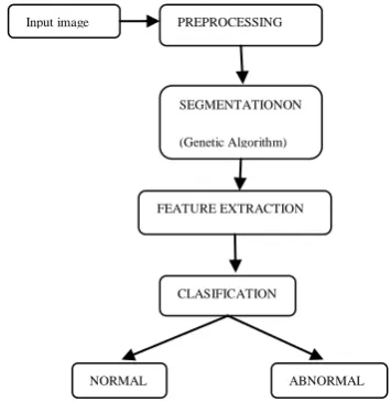
PROPOSED METHOD [image:2.595.50.228.188.376.2]The proposed method uses Genetic algorithm to segment the MRI brain tumor images. The architecture diagram of the proposed method is shown in Figure 1.
Figure 1. Architecture diagram
The proposed system involves the following modules
A. Preprocessing B. Segmentation C. Feature extraction D. Classification
A. PREPROCESSING:
Preprocessing is a technique which involves removal of noise or any distortion in an image. Wiener Filter is used in this phase to remove noise. Wiener filter is a 2-D adaptive noise removal filter and it uses pixel wise adaptive wiener method. Wiener estimates the local mean ( ) and variance ( ) around each pixel and it is shown in the following equation 1 and 2.
(2)
Where is the − by − local neighbourhood of each pixel in the image .Weiner then creates a pixel wise wiener filter using these estimates,
Where is the noise variance. If the noise variance is not
given, wiener uses the average of all the local estimated variances.
B. SEGMENTATION:
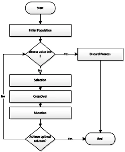
Segmentation is a process of partitioning a digital image into multiple regions. Genetic Algorithm is used in this method to segment the images.GA is based on the classical view of a chromosome as a string of genes.
Genetic algorithm is a natural inspired Meta heuristic algorithm. In GA each solution is represented as chromosome and each chromosome is built up from genes. . The best generated solutions will be added to the next iteration while the bad solutions will be rejected. While the algorithm iterates its solutions, these solutions are improved up to a point where a converge to near optimal solution is achieved.
In general, a GA has five stages: initialization of population, evaluation of fitness function, selection, crossover, mutation and termination. Initial population is created randomly, which can be done by setting genes to random values. After the initialization process, fitness function of each chromosome is evaluated.
• In the selection process, the fittest members in the current population are selected for reproducing the new solutions in crossover process two is chromosomes are selected and exchange genes by some point. In mutation process a gene is selected randomly and its value is changed. In last Termination of the iteration is done when a certain criteria is met. Generally termination is done by number of iterations.
Flowchart for Genetic Algorithm:
Figure 2: Genetic Algorithm
Input image PREPROCESSING
SEGMENTATIONON
(Genetic Algorithm)
FEATURE EXTRACTION
CLASIFICATION
[image:2.595.331.541.498.766.2]© 2018, IRJET | Impact Factor value: 6.171 | ISO 9001:2008 Certified Journal | Page 1677
C. FEATURE EXTRACTION:
Feature Extraction is a process of extracting the essential features of an Image in order to classify the various tumor stages. In this module Gray Level Co-occurrence Matrix (GLCM) is used to extract the feature of an image. It is a statistical method for examining the texture feature. The GLCM function characterize the texture of an image by calculating often pairs of pixel with specified value.
D. CLASSIFICATION:
[image:3.595.64.266.381.627.2]Genetic algorithm provides the optimal solution based on fitness function. Our proposed algorithm includes feeding the GA with an initial selection of population. These populations come from raw dataset of Micro array which represents the components of chromosome. The next step is calculating the fitness function. Based on output of these calculations; the higher fitness could be reserved and discarding the lower one. Third step is the crossover process, that responsible to yield good generation by combine the best component from different genetic. Finally, mutation process is implemented which will generate new gene structure with small random probability. Figure 1 illustrates the proposed system components.
Figure 3: Proposed Genetic Algorithm Components
The previous flowchart shows the general form of the algorithm that will be followed in this study and each stage of this flowchart have own technique. The first is to follow the flowchart so as to get satisfactory results regardless of the used technique. The below steps clarifies the different stages of the proposed GA.
Structural Feature
Artificial Neural Network (ANN) is one of the artificial neural networks, which has been proposed for segmenting
both gray-level and color images. The authors present the segmentation problem for gray-level images as minimizing a suitable energy function with ANN, it derived the network architecture from the energy function, and classify the sputum cells into nuclei, cytoplasm and background classes, where the input was the RGB component of the used images. In our work we used the ANN algorithm as our segmentation method. The ANN is very sensitive to intensity variation and it can detect the overlapping cytoplasm classes. ANN is considered as unsupervised learning. Therefore, the network classifies the feature space without teacher based on the compactness of each cluster calculated using the Euclidean distance measure between the pixel and the centroid of class l. The neural network structure consists of a grid of N M neurons with each column representing a cluster and each row representing a pixel. The network is designed to classify the image of N pixels of P features among M classes, such that the assignment of the pixels minimizes the criterion function
Where is considered as the Euclidean distance measure between the k th pixel and the centroid of class l, is the output of the neurons. The minimization is achieved using ANN and by solving the Motion equation satisfying
Where is as defined in a scalar positive function of time used to increase the convergence speed of the ANN. By applying the relation (5) to equation (4), we get a set of neural dynamics given by:
where and are the input and output of the neuron respectively. To assign a label to the pixel we use the input-output function given by:
The ANN segmentation algorithm can be summarized in the following steps:
1. Initialize the input of neurons to random values. 2. Apply the input-output relation given in (7) to obtain the new output value for each neuron, establishing the assignment of pixel to classes.
© 2018, IRJET | Impact Factor value: 6.171 | ISO 9001:2008 Certified Journal | Page 1678 Where is the number of pixels in class .
4. Solve the set of differential equation in (3) to update the input of each neuron:
5. Repeat from step 2 until convergence then terminate. We applied the ANN with the specification mentioned above to 1000 sputum color images and maintained the results for further processing in the following steps. Our algorithm could segment 97% of the images successfully in nuclei, cytoplasm regions and clear background. Furthermore, ANN took short time to achieve the desired results. By ex-periment, ANN needed less than 120 iterations to reach the desired segmentation result in 36 seconds.
IV.EXPERIMENTS AND RESULT
The proposed paper is initialized with Preprocessing step. In this step noise in an image is removed and quality of an image is improved using wiener filter successfully. The method is developed in MATLAB version 13a platform. The result of the proposed method is shown in Figure.
It can be seen from the segmented image that the proposed method segment the tumor part clearly form the given MRI brain image.
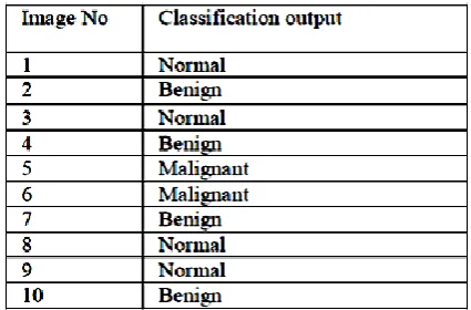
[image:4.595.327.540.143.283.2]The classification is analyzed for set of images collected from publicly available resources and it is shown in the following Table 1
Table 1:Classification analysis
It is seen from the table that the segmented image is correctly classified based on ANN classifier.
V. CONCLUSION:
The proposed method segments and classifies the MRI brain tumor images accurately. Brain tumor is curable if it is caught at earlier stages. This enables the doctors to grasp the exact progression of the disease state, which would help to make a decision about the appropriate treatment, surgery for radiologist and following-up for a series of disease control measures.
REFERENCES:
[1] GirjaSahu,Lalit Kumar P. Bhaiya.“A Survey Paper Based on the Classification of MRI Brain images using Soft Computing”, International Journal of Emerging Technology and Advanced Engineering Techniques” vol.4,pp.1-6,2014.
[2] Kamal Kant Hiran,RuchiDoshi, “An Artificial Neural Network Approach for brain tumor detection using Digital Image Segmentation” ,International Journal of Emerging Trends and Technology in Computer Science, vol. 2,pp.1-15,2013.
[3] AR.Kavitha, M.DivyaMeena, K.Gayathri,KP.Raghav “Brain Tumor Segmentation using Genetic Algorithm with Modified Region Growing Method”, International Journal of Emerging Technology in Computer Science and Electronics,vol.13,pp.1-7,2015.
[4] C.Logeshwaran,P.Bharathi,M.Gowthami“Brain Tumor Detection using Hybrid Techniques and Support Vector Machines” International Journal of Advanced Research in Computer Science and Software Engineering,vol .5,pp.1-8, May 2015.
© 2018, IRJET | Impact Factor value: 6.171 | ISO 9001:2008 Certified Journal | Page 1679 [6] U.Vanitha, P.PrabhuDeepak, N.PonNagesharan, R.
Sathappan. “Tumor Detection in Brain using Morphological Image Processing” Journal of Applied Science and Engineering Methodologies,vol.1,pp.1-6,2015.
[7] Michael O. Lam, Tim Disney, Daniela S. Raicu, Jacob Furst and David S. Channin, “BRISC-An Open Source Pulmonary Nodule Image Retrieval Framework", Journal of digital imaging, 2007.
[8] Arimura, S. Katsuragawa and K. Suzuki, “Computerized scheme for automated detection of lung nodules in low-dose computed tomography images for lung cancer screening”, Acad. Radiol., Vol. 11, pp. 617629, 2004.
[9] Ambrosini,S. Nicolini, P. Carolia, C. Nannia, A. Massarob, M.-C. Marzolab, D. Rubellob and S. Fantia, “PET/CT imaging in di_erent types of lung cancer: An overview”, European Journal of Radiology, Vol. 81, pp. 988-1001, 2013.