567
© 2018 by the Serbian Biological Society How to cite this article: Jurić-Lekić G, Bedrica Lj, Lončar D. Interscapular brown adipose tissue recruitment is hindered by a temperature environment of 33°C: Uncoupling protein-1 underexpression is not associated with obesity development in rats. Arch Biol Sci. 2018;70(3):567-79
Interscapular brown adipose tissue recruitment is hindered by a temperature
environment of 33°C: uncoupling protein-1 underexpression is not associated with
obesity development in rats
Gordana Jurić-Lekić1, Ljiljana Bedrica2 and Dragutin Lončar1,3,4,*
1Department of Histology and Embryology, School of Medicine, University of Zagreb, Salata 3, 10000 Zagreb, Croatia 2Clinic for Internal Diseases, Faculty of Veterinary Medicine, University of Zagreb, Heinzelova 55, 10000 Zagreb, Croatia 3Wenner-Gren Institute, The Arrhenius Laboratories F3, Stockholm University, S-106 91 Stockholm, Sweden
4Present address: University of North Carolina Chapel Hill, High Point Regional Hospital, Pediatrics 5th Floor, 601 N. Elm
St., High Point NC, 27262, USA
*Corresponding author: [email protected]
Received: December 15, 2017; Revised: March 7, 2018; Accepted: April 18, 2018; Published online: May14, 2018
Abstract: Brown adipose tissue (BAT) generates heat due to unique thermogenic UC-mitochondria, an event known as nonshivering thermogenesis. Cold, adrenergic agents, hormones, etc., activate nonshivering thermogenesis, resulting in lipid mobilization, an increase in the mitochondria and mitochondrial cristae, and increased uncoupling protein-1 (UCP1) expression and its incorporation into mitochondrial cristae. BAT precursor cells mature and contribute to BAT growth in a process known as BAT recruitment. For the first time, we herein report the effect of a thermoneutral environment of 33°C on interscapular BAT (IBAT) in rats delivered and raised at 33°C. The control animals were housed at 20°C. Thermoneu-tral IBAT was atrophic (73 mg vs. 191 mg) but with more adipocyte precursor cells; euthermia (37.6°C) was maintained without nonshivering thermogenesis. Although IBAT was inactive, the thermoneutral animals did not develop obesity, and on the contrary, the thermoneutral environment of 33°C hindered the rats’ growth, weight (65 gm vs. 139 gm), volume (67
gm vs.136 gm) and length (12 cm vs. 16 cm). The thermoneutral brown adipocytes were smaller (7234 µm3 vs. 9198 µm3)
with more lipids (4919 µm3 vs. 4507 µm3) and a smaller mitochondrial cristae area (52504 µm2 vs. 61288 µm2/adipocyte).
Lipoprotein lipase mRNA expression was 11% (vs. 58% in control) and UCP1 mRNA expression was 34% (vs. 93% control). UCP1 immunoelectron microscopic study detected 160 UCP1-gold particles (vs. 700 in control) per UC-mitochondrion;
thermoneutral brown adipocytes had 9-fold fewer UCP1-gold particles (0.34x10⁶ vs. 2.99x10⁶ UCP1-gold particles), and
thermoneutral UC-mitochondria developed specific intramitochondrial tubular inclusions.
Key words: brown adipose tissue; lipoprotein lipase; uncoupling protein; heat stress; mitochondria
INTRODUCTION
Brown adipose tissue (BAT) and its variant convert-ible adipose tissue (CAT, also referred to as beige or bright adipose tissue) generates heat and helps rewarm hibernating animals. BAT contributes to maintaining body temperature during cold exposure in mammals and, as recent data suggest, BAT may be involved in controlling obesity by burning excess calories [1-2]. Fatty acids imported from the circulation or from brown adipocyte lipid stores are oxidized inside the uncoupled brown adipocyte mitochondria (UC-mi-tochondria) to produce heat. The heat production is
heat instead of ATP, a process known as nonshivering thermogenesis [3-5].
At delivery, rat pups have two adipocyte popula-tions within their IBAT: mature adipocytes filled with lipids and specific UC-mitochondria, and another pop-ulation of adipocyte-precursor cells, described as endo-thelial cells, pericytes, preadipocytes, young adipocytes, etc. [3-4]. With delivery, as the newborn adjusts from intrauterine temperatures to an external, lower temper-ature milieu, their mtemper-ature brown adipocytes begin non-shivering thermogenesis. This process is accompanied by the growth and maturation of adipocyte precursor cells, which are filled with lipids and UC-mitochondria to join already mature adipocytes in nonshivering ther-mogenesis. This BAT activation process after delivery has been described as BAT recruitment and includes an up to 3-fold increase in BAT volume and weight, as well as increased adipocyte count, mitochondrial con-tent, mitochondrial cristae surface area, mitochondrial enzymes, including UCP1, and oxidative, lipolytic and lipogenic capacity [5-6]. Similarly, the increased expres-sion of UCP1 and increased nonshivering thermogen-esis have been postulated as a mechanism to prevent/ control obesity [1-2,5,7].
Since the discovery of nonshivering thermogenesis more than 50 years ago [8], substantial data have been generated regarding the effect of cold on the structure and function of brown adipocytes [3-7]. However, with the exception of a few sporadic studies that examined some aspects of young and adult animal behavior in a thermoneutral environment or BAT response in mature animals acclimated to a thermoneutral environment, there have been no detailed analyses of BAT in animals delivered and raised in a thermoneutral environment or in an elevated temperature environment that does not activate nonshivering thermogenesis. Our com-prehensive study fills that gap by analyzing BAT in rats delivered and raised in a temperature environment of 33°C during their first month of life.
The thermoneutral environment as an environ-ment in which the basic metabolic rate maintains euthermia without additional heat production by either shivering or nonshivering thermogenesis, has a minimal and constant metabolic rate, animals are inactive, and the elimination of CO2 is lowest, as is the core body temperature [5,9-10].
Historically, the thermoneutral environment, or thermoneutral zone (TNZ), delineated by lower criti-cal temperature (LCT) and upper criticriti-cal temperature (UCT), had been considered as the basic metabolic rate, and for rats it had been reported to be in the range of 28-34°C [9-10]. Considering the updated definition of thermoneutrality [11] where the core body tempera-ture is maintained through dry heat loss regulation, and after measuring such dry heat loss through the skin blood flow, the TNZ for 5 adult male rat strains had been reported to be in the range of 28-32°C [12].
The TNZ is species- and strain-dependent, influ-enced by the animal age, gender, nutritional status, caging and temperature conditions, pregnancy, lacta-tion, day-night circadian cycle, animal body tempera-ture, health conditions, etc. [9-10,12-17]. It has been reported that pregnant and lactating Sprague-Dawley rats tolerated the environmental temperature of 33°C better [18]. Therefore, in our search for the tempera-ture environment around the UCT of TNZ where nonshivering thermogenesis and BAT are inactive, we analyzed the effect of a temperature environment of 33°C on the postnatal development of IBAT.
MATERIALS AND METHODS
Animals
The care and use of animals were approved by the Animal Welfare Committee of the Faculty of Medi-cine, University of Zagreb. The review of literature [18], but also our pilot studies, showed that pregnant Sprague-Dawley rats and their delivered pups had good tolerance to a thermoneutral environment of 33°C. Therefore, the experimental rats were delivered and raised at this temperature, while the control group of animals was delivered and raised at a room tem-perature of 20°C, as had been already reported before by us and other authors [19-21].
The rats were housed at a 12-h light-dark cycle and had unlimited access to standard laboratory chow diet and tap water. On the 17th day of pregnancy,
six pregnant Sprague-Dawley rats were placed in a temperature environment of 33°C, and six pregnant animals were kept at room temperature (20°C). All pregnant animals were caged individually. The few-est number of pups delivered was 9, and so all litters were reduced to 9 pups per litter. After delivery, unless killed earlier, the young pups spent the first 20 days lactating and an additional 10 days feeding on their own. The experimental and control groups of animals were killed simultaneously at 7, 14 and 30 days of age. Six young animals from each age group were killed: 3 for morphology/electron microscopy studies and 3 for mRNA essays. At 30 days of age, 10 randomly selected animals from each group (experimental and control) were measured in detail as described in Table 1.
Electron microscopy (EM)
Three animals per age group in deep anesthesia were perfused transcardially, and the IBAT was fixed for EM analysis as previously described in detail [22]. We used Ringer’s solution warmed to 37°C and then a freshly prepared fixative (0.1 M phosphate buffer con-taining 2% glutaraldehyde and 1% paraformaldehyde at pH 7.4). After perfusion, IBAT adipose tissue was dissected, sliced into small samples and immersed in the fixative. Half of the IBAT sample was randomly selected to be stored in the fixative for 2 h and then processed for immunoelectron microscopy (IEM)
(see below). The other half of the IBAT sample was maintained in the fixative for 1-2 days, rinsed in sa-line solution and postfixed in 1% osmium tetroxide in cacodylate buffer for 1 h at 4°C. After dehydration in cold acetone and propylene oxide, the IBAT samples were embedded in Epon 812 and sectioned to 70 nm on a Reinchard Ultracut E ultramicrotome, stained with uranyl acetate and lead citrate and examined under a JEOL 100 S electron microscope.
UCP1 immunoelectron microscopy
IBAT for immunoelectron microscopic analysis of UCP1 was prepared as previously described in detail [20]. Perfused and freshly fixed IBAT was stored in the fixative for 2 h and then small samples of IBAT were washed in 0.1 M phosphate buffer and embedded in a mixture of 20% polyvinylpyrrolidone in 2 M sucrose. The samples were frozen in liquid nitrogen 2 h later and cut on a LKB Cryo Nova cryo-ultramicrotome.
Antibodies against rat UCP1 were prepared as de-scribed previously [23]. Preparation of colloidal gold markers measuring 10 nm in diameter and prepara-tion of protein A-gold complexes were carried out as per the method of Slot and Geuze [24]. The frozen tissue, in 70-nm-thick sections, was transferred to Formvar-covered carbon-coated copper grids and processed for staining. After washing the samples in a phosphate buffer containing 0.02 M glycine, the sections were treated with the diluted specific anti-body for 30 min. After washing in a phosphate buffer containing 0.1% bovine serum albumin, the sections were treated with protein A-gold for 30 min. Thin sections were negatively contrasted with 4% uranyl acetate for 5 min, embedded in methyl cellulose and analyzed by EM. To demonstrate the specificity of the labeling, control labeling was performed by omitting the incubation step with the primary antibody, or by blocking the primary antibody with purified UCP1 in the incubation solution as previously described [20].
Morphometry
sections were cut from each block and stained with toluidine blue. From these samples, sections were ran-domly selected for further study. To simplify the mea-surement of brown adipocytes’ diameter, we considered that the adipocytes had a spherical shape. From this data, 100 of the largest diameters were used to calculate the mean adipocyte diameter; from the data, 10% of the largest cells were identified as the largest adipocytes.
Following the procedures described by Weibel et al. [25], we measured the adipocytes’ cytoplasm occu-pied by lipids and/or mitochondria. The average mito-chondrial volume and mitomito-chondrial cristae area were determined as previously described in detail [26-27]. Volumetric densities of lipid droplets and mitochon-dria refer to the adipocyte cytoplasm excluding nuclei that occupied approximately 10% of the cell volume. All data were subjected to statistical analysis using a Student’s t test to assess the differences between the control animals and thermoneutral animals.
Determination of LPL mRNA and UCP1 mRNA
The amount of LPL mRNA and UCP1 mRNA was determined as previously described [28]. Three ani-mals per age group (7-, 14- and 30-day-old rats) were killed by cervical dislocation. The IBAT was excised and homogenized in guanidine extraction buffer. Total RNA was isolated according to Jakobsson et al. [29]. For the slot blots, an amount of the RNA preparation corresponding to 4 µg RNA was dissolved in 300 µL 10 x saline-sodium citrate buffer (SSC)/18% formal-dehyde with water to yield a total of 400 µl. After a 15-min incubation at 65°C, the solution was applied to a Zeta-Probe filter in a Minifold Slot-Blot apparatus, washed with 400 µl 10 x SCC and dried at room tem-perature. After prehybridization with salmon sperm DNA (Sigma) and a poly A/poly C mixture, the filter paper was hybridized with cDNA probes and nick translated with a Bethesda Research Laboratories kit. The UCP1 probe utilized was previously characterized by Jacobsson et al. [29], and the LPL probe was that characterized by Kirchgessner et al. [30]. The filter papers were washed, dried and exposed to Kodak X-OMAT AR film at -80°C, and the amount of darken-ing was evaluated with an LKB laser densitometer.
RESULTS
Animal growth and IBAT growth were retarded in the thermoneutral environment. Pregnant rats were housed in a temperature environment of 33°C from the 17th day of pregnancy, and their pups were
deliv-ered and raised at 33°C. The control group of animals was raised at 20°C. All animals had free access to food and water, the consumption of which was similar in both groups during the first 3 weeks. However, after 3 weeks, the consumption of water appeared to be higher in the animals caged at 33°C, although that could also have been the result of greater water evapo-ration at 33°C. At 20 days old, the pups were weaned, and the mother rats were removed from the cages.
We checked on our animals twice daily, then during feeding and bedding changes and during the conduc-tion of the experiments. We found that during the first 7-10 days of life, the animals in both groups behaved similarly. However, after 3 weeks of age, the pups raised at 33°C became less active in their cages, appeared to have slower growth rates, and their fur was shorter and thinner when compared to the animals raised at 20°C. At one month of age, both groups were euthermic, but the 33°C-animals weighed approximately half as much as the control group, their body volume was half and their length was only ¾ of the measurements of the control group (Table 1). After transcardial perfusion during which blood was washed from the tissue, IBAT appeared brown macroscopically in both groups, but the IBAT in the 33°C-animals was significantly smaller, had an atrophic-like appearance and its wet weight was 2.5-fold lower than the control animals (Table 1).
Basic BAT morphology is preserved in animals raised at 33°C
were similar in both groups. Both groups of animals had adipocyte precursor cells (pericytes and preadi-pocytes) and occasional macrophages and basophils near endothelial cells.
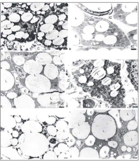
The size of the adipocytes increased with time in both groups (Table 2). The maximal adipocyte diam-eter grew with time and was approximately 33 μm at
1 month of age in both groups (Fig. 1, Table 2). In rats raised at 33°C, the average diameter was 24 μm at 30 days, i.e., slightly smaller than in the control animals, which resulted in a smaller adipocyte volume, so that at 1 month, their brown adipocyte volume was 7234 μm3 compared to 9198 μm3 in the control group (Fig.
1, Table 2).
As the size of brown adipocytes increased, the lipid amounts within the adipocytes also increased (Fig. 1, Table 2). The stored lipids in the 33°C brown adipocytes at 30 days of age occupied 2/3 of the brown adipocyte volume, while in the control adipocytes the lipids occupied approximately 1/2 of the brown adi-pocyte volume. Although the 33°C adiadi-pocytes had a slightly smaller diameter, their larger stored lipid vol-ume resulted in a larger volvol-ume of lipids per brown adipocyte compared to the control brown adipocytes (4919 μm3 vs. 4507 μm3) (Fig. 1f, Table 2). Such an
increase in the lipid volume in slightly smaller 33°C brown adipocytes resulted in a smaller volume of the brown adipocyte cytoplasm (Table 2).
Adipocyte precursor cells are more prevalent in IBAT of animals raised at 33°C
In animals raised at 33°C, the IBAT had small, pale cells with no lipids or a very small amount of lipids near capillaries. EM identified those cells as adipocyte precursor cells with less mitochondria and less mito-chondrial cristae: there were 2-3 adipocyte precursor cells per EM section, while the control IBAT had less than one adipocyte precursor cell per EM section (Fig. 1c and f, Table 2), because these cells were recruited and grew into mature brown adipocytes, thereby con-tributing significantly to the growth and thermogenic potential of control IBAT.
Table 1. The effect of temperature environment on 30-day-old rats delivered and raised at 33°C, as compared to rats delivered and raised at 20°C (control).
30-day-old rats Rectal body core
temperature (°C)a weight (mg)IBAT wet body weightmg IBAT/g Body weight (g) volume (mL)Body b excluding tail (cm)Body length
Control (20°C) (n-10) 37.3±0.3 191±21 1.73 139.4±6.9 136±4.2 16±0.3 Temperature of
33°C-rats (n=10) 37.6±0.4 73±9 1.11 65.5±7.5 65.5±7.5 12.3±0.6
aRectal digital thermometer was inserted up to 20 mm beyond the anal sphincter and the stable core temperature was recorded; bBody volume was measured by immersing the killed animals into a graduated cylinder.
The mean (±SD) represents the measurements of ten randomly selected animals from each group.
The mitochondrial cristae surface area is smaller in adipocytes of rats raised at 33°C
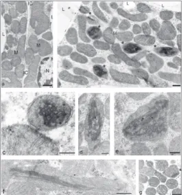
The average mitochondrial volume expanded with adipocyte growth in both groups (Fig 2a and b). At 30 days, the average mitochon-drial volume was approximately 20% bigger than the 7-day-old adipocyte mitochondria. Compared to the control at 30 days of age, the brown adipocytes in rats raised at 33°C had
Table 2. The effect of environmental temperature on the morphological parameters (±SD) in brown adipocytes of rats delivered and raised at 33°C, as compared to animals delivered and raised at 20°C (control).
Age
7-day-old 14-day-old 30-day-old
20°C (n-3) 33°C (n-3) 20°C (n-3) 33°C (n-3) 20°C (n-3) 33°C (n-3)
Mean maximal diameter of adipocytes (µm) 27±8 25±6 26±8 29±7 33±5 33+4
Brown adipocytes diameter (µm) 21±8 20±11 23±5 22±6 26±3 24±5
Brown adipocytes volume (µm3)a 4846 4186 6367 5572 9198 7234
Adipocyte volume occupied by lipids (%) 34±15 35 ± 16 37±16 36±19 49±14 68±8*** Volume of lipids per adipocyte (µm3) a 1647 1465 2355 2005 4507 4921
Adipocyte cytoplasm volume without nucleus (µm3) a 2897 2448 3610 3210 4221 2083
Preadipocytes per EM section <1 <1 <1 <1 <1 2-3
Adipocyte average mitochondrial volume (µm3) b 0.38±0.1 0.34±0.4 0.37±0.5 0.40±0.4 0.43±0.1 0.47±0.1
Volumetric density of mitochondria per cytoplasm (%) bc 45±9 40±12 51±7 46±4 44±3 60±2***
Volumetric density of mitochondria per adipocyte (µm3/
adipocyte) b 1296 979 1841 1477 1857 1249
Mitochondrial cristae per mitochondrion (µm2/µm3) b 27±4 31±6 29±7 33±8 33±7 42±3##
Mitochondrial cristae per cytoplasm (µm2/µm3) bc 12±3 12±4 15±5 15±7 14±3 25±2***
Mitochondrial cristae per adipocyte (µm2/adipocyte) d 34992 30366 53399 48727 61288 52504
Mitochondria with inclusions per EM adipocyte section (%) e <<1 <<1 <<1 <<1 <<1 1.3±6.9 aRecalculated from brown adipocyte diameters and postulated that the adipocyte is a sphere.
bFour randomly selected IBAT samples from each animal, and one hundred EM images per a sample were analyzed at magnifications of x8000 and
x20 000. Multipurpose test system M168 (for mitochondrial volume; volumetric density of mitochondria per cytoplasm) and M42 (for mitochondrial cristae per mitochondrion) were used for EM morphometric estimation (see reference 26).
cPer µm3 of adipocyte cytoplasm excluding the nucleus and lipid droplets (nucleus occupies 10% of the cytoplasm). dRecalculated data from the above measurement.
eSee Fig. 1.
***P<0.01, ##P<0.02;
Data are the mean±SD from three animals in each group.
mitochondria with a slightly larger volume (0.47 μm3
vs. 0.43 μm3) (Figs. 2a-b, 3a-b, Table 2). The larger
mitochondria packed in a smaller cytoplasmic volume of the 33°C brown adipocytes (likely “compressed” by an increased lipid volume) resulted in a relatively larger cytoplasmic volumetric density of mitochondria (60%) compared to control brown adipocytes (44%). However, the adipocytes raised at 33°C had a smaller cytoplasmic volume. This resulted in a smaller mito-chondrial mass per adipocyte during all three measur-ing points compared to the control group (Table 2).
Although the mitochondrial cristae area per mito-chondrion appeared to be slightly larger in the brown adipocytes of rats raised at 33°C at 7 and 14 days of age, the final measurement of the mitochondrial cris-tae area per 1 μm3 of cytoplasm remained stable
(12-15 μm2/μm3) (Table 2) during the first 2 weeks of life
in both groups. At 30 days of age, the control brown adipocytes had a mitochondrial cristae area per cyto-plasm of 14 μm2/μm3. However, the 30-day-old brown
adipocytes in rats raised at 33°C had a 30% larger mi-tochondrial cristae area per 1 μm3 of mitochondrion,
and a 60% larger mitochondrial cristae area per 1 μm3
of cytoplasm (Figs. 2a-b, 3a-b). The total mitochondri-al cristae area per adipocyte increased with age in both groups, and more so in the control group. At 7 and 14 days of age, the brown adipocytes of rats raised at 33°C had 10% less cristae area than the control group, while at 30 days of age, the adipocytes in the 33°C rats had 20% less mitochondrial cristae per adipocyte than the control group (52504 μm2/adipocyte vs. 61288 μm2/
adipocyte) (Table 2).
Intramitochondrial tubular inclusions are more frequent in adipocytes raised at 33°C
Intramitochondrial tubular inclusions were exception-ally rare findings in the brown adipocytes of the 7- and 14-day-old rats, but in rats raised at 33°C, 1-7% of the mitochondria per adipocyte electron microscope section had tubular inclusions (Fig. 2b-g, Table 2). The inclusions were comprised of uniform tubules. On transversal sections, each tubule appeared as a well-rounded cylinder with a dark peripheral osmo-philic wall and a clear central lumen (Fig. 2g-c). On longitudinal sections, the tubules had straight shapes with a dark osmophilic wall and a clear central lumen
(Fig. 2g-c). Each tubule had a diameter of about 50 Å, while the length of the tubule could span the entire mitochondrial length (i.e., up to 2 µm), and appeared to determine the shape of the mitochondria (Fig. 2f). Some intramitochondrial tubular inclusions were comprised of only a few tightly packed tubules, but most of the inclusions were assembled from several dozen tubules, reaching up to 100 plus tubules per in-clusion (Fig. 2c). Most inin-clusions were present inside mitochondria with very few or no intramitochondrial cristae (Fig. 2), although a few mitochondria with well-developed cristae also had inclusions (Fig. 2g).
LPL mRNA expression is significantly depressed in the IBAT of rats raised at 33°C
Table 3 shows that the expression of LPL mRNA de-clined with animal age in both the control and ani-mals raised at 33°C. At 14 days of age, the control IBAT expressed 78% and at 30 days of age, only 58% of LPL mRNA was expressed compared to LPL mRNA expressed in the 7-day-old control. In rats raised at 33°C, IBAT at 7 days of age expressed only 68% of the LPL mRNA, while at 14 and 30 days, the rates of LPL mRNA were 44% and 11%, respectively, com-pared with LPL mRNA expression in the 7-day-old control IBAT. By the time the animals were 1 month old, LPL mRNA expression was about 1/6 of the LPL mRNA expressed in the 7-day-old pups raised at 33°C (Table 3).
UCP1 mRNA expression is lower in brown adipocytes of rats raised at 33°C
Table 3 shows the effect of the temperature environ-ment of 33°C on the expression of UCP1 mRNA at 7, 14 and 30 days of age. During the first month of life, the UCP1 mRNA expression in the control group was the highest at 14 days (12% higher than at 7 days of life), while at 30 days, the UCP1 mRNA level de-creased to 93% of that in the 7-day-old pups.
in the 7-day-old control pups. The brown adipocytes of rats raised at 33°C had the highest expression of the UCP1 mRNA at 7 days of age and by the time the animals were one month old, UCP1 mRNA expres-sion was nearly halved (table 3).
The amount of UCP1 protein is
significantly reduced in thermoneutral UC-mitochondria
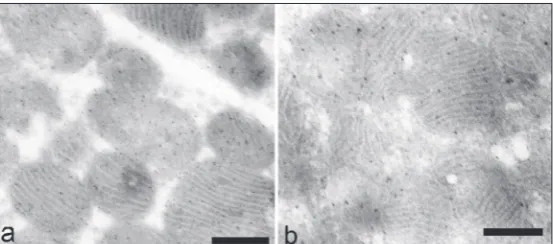
The qualitative and quantitative analysis of UCP1 protein by immunoelectron mi-croscopy in 30-day-old IBAT is detailed in Table 4 and in Fig. 3a-b. The distribu-tion of anti-UCP1 antibody protein A-gold complex particles (UCP1-gold particles) over the mitochondria follows the shape of the mitochondrial cristae (Fig. 3a-b).
The intermitochondrial membrane space and the mitochondrial matrix were not substantially labeled by the UCP1-gold particles; when they were labeled by UCP1-gold particles, they were labeled near the cristae, indicating that such labeling very likely rep-resented the incorporation of UCP1 protein into the cristae, or UCP1 protein en route to cristae incor-poration. Occasional UCP1-gold particle labeling was present throughout the cytoplasm of the brown adipocytes, particularly near the mitochondria, likely representing the UCP1 already synthesized en route to the mitochondria. The endothelial cell cytoplasm and their C mitochondria did not display any significant labeling by UCP1-gold particles.
In the control brown adipocytes, 85% of the UCP1-gold particles were distributed over the mitochondrial cristae, 11% over the intramembranous space and the mitochondrial matrix, and 4% over the outer
mitochon-drial membrane. With a 33-μm2 mitochondrial cristae
area per 1 μm3 of mitochondria (Table 2), there were
approximately 1400 UCP1-gold particles in the cristae per μm3 of mitochondria. With the UCP1-gold particles
over the mitochondrial intramembranous space, the mitochondrial matrix and over the outer mitochondrial membrane, it would appear that 1 μm3 of mitochondria
had about 1600 UCP1-gold particles (Table 4). Com-bining these data with an average control brown adi-pocyte mitochondrion volume of 0.43 μm3 (Table 2),
we established that one control UC-mitochondrion had about 700 UCP1-gold particles or, expressed through a control adipocyte, there were approximately 3x10⁶
UCP1-gold particles in the 30-day-old rats.
The mitochondria in the brown adipocytes of rats raised at 33°C exhibited a similar UCP1-gold particle labeling pattern as the control mitochondria (Fig. 3a-b), with the most UCP1-gold particle labeling (72%) observed over the mitochondrial cristae, 23%
Table 3. The effect of temperature environment on the expression of UCP1 mRNA and LPL mRNA in the interscapular BAT of rats delivered and raised at 33⁰C as compared to rats delivered and raised at 20°C (control).
Age
7-day-old 14-day-old 30-day-old
20°C (n-3) 33°C (n-3) 20°C (n-3) 33°C (n-3) 20°C (n-3) 33°C (n-3)
UCP1 mRNA (%) 100±17 64±12 112±23 52±16 93±11 34±9
LPL mRNA (%) 100±20 68±21 78±19 44±26 58±15 11±9
Interscapular BAT of the killed animals was dissected, total RNA was extracted and UCP1 mRNA and LPL mRNA levels were quantified by the Slot-Blot technique, as described in the Materials and Methods. UCP1 mRNA and LPL mRNA data were normalized and presented relative to the corresponding mean levels of the 7 day-old-pups housed at 20°C, with mean values set to 100 %.
Data are the mean±SD from three animals in each group.
over the intermembranous space and the mitochon-drial matrix, and 5% over the outer mitochonmitochon-drial membrane (Table 4). However, the total number of UCP1-gold particles per mitochondrion was drasti-cally reduced (Fig. 3b). There were only 5 UCP1-gold particles per μm2 of mitochondrial cristae, 1.6
UCP1-gold particles per μm2 of the intramembranous space
and the mitochondrial matrix, and 0.3 UCP1-gold particles per μm2 in the outer mitochondrial
mem-brane, totaling 6.8 UCP1-gold particles per μm2 of
mitochondria. With 42 μm2 of mitochondrial cristae
area per 1 μm3 of mitochondrion (Table 2), there were
about 200 UCP1-gold particles in the cristae per 1 μm3 of brown adipocyte mitochondria in rats raised
at 33°C. Adding to this the number of UCP1-gold particles present over the mitochondrial intermem-brane space and the mitochondrial matrix and the outer mitochondrial membrane, it appears that 1 μm3
of mitochondria contained about 270 UCP1-gold par-ticles (Table 4). As the average thermoneutral brown adipocyte mitochondrion had a volume of 0.60 μm3
(Table 2), there were approximately 160 UCP1-gold particles per mitochondrion. Thus, there were about 0.34 x 10⁶ UCP1-gold particles expressed per brown
adipocyte in the 30-day-old rats raised at 33°C, which was 9-fold fewer than in control brown adipocytes.
DISCUSSION
Control rat pups and pups delivered at 33°C were similar in appearance, lactation and behavior for 7-10 days post-delivery. In spite of being raised at 33°C from the time of delivery, the experimental group of pups behaved as poikilotherms and huddled with the mother in their nests until about 10 days of life when they began to leave the nests more often. After wean-ing at 20 days of age, the pups raised at 33°C moved in the cage less and spent more time out of the nest and away from each other, similarly as reported pre-viously [9-10,13-17,31], and at thirty days of age the rats were moving very little in the cages; they mostly lay down and appeared to be less interested in food. Their fur was sparse with short hair, and their bodies were significantly smaller, lighter and shorter than those of the control animals (Table 1).
Our animals raised at 33°C maintained normo-thermia, preventing hyperthermia by minimizing
mus-cle movements and maintaining low basal metabolism, which caused the global, retarded growth. It has been well documented that animals raised in a similar tem-perature environment have the lowest basal metabo-lism and maintain euthermia without an additional heat source, i.e., without shivering or nonshivering thermogenesis [9-10,12,18,31-32]. The findings that the IBAT of the animals raised at 33°C was atrophic, 2.5-fold lighter than the IBAT of control rats and more growth-arrested than the rest of the animals’ body (1.1 mg vs. 1.7 mg IBAT per 1 g of animal body weight) are similar to others [9,32]. In comparison, fully recruited IBAT in cold-acclimated rats had up to about 3.0 mg of IBAT per 1 g of animal body weight [33].
Since in the temperature environment around UCT any heat production by IBAT nonshivering ther-mogenesis would cause hyperthermia (a detrimental condition to the animals), their IBAT remained atro-phic, unrecruited. Such atroatro-phic, unrecruited IBAT in addition to mature developed adipocytes, had signifi-cantly more adipocyte precursor cells and young, un-der-developed adipocytes. They remained unrecruited, “dormant” in 1-month-old thermoneutral IBAT, con-tributing to a smaller, lighter and atrophic IBAT.
larger, with significantly more cristae per one µm3 of
mitochondrion, resulting in more mitochondrial cris-tae per 1 µm3 of adipocyte cytoplasm in rats reared
at 33°C (Table 2).
How brown adipocytes initiate and maintain a vast mitochondrial population and enormous mito-chondrial cristae surface area is not quite clear, but we know that stimulation of noradrenergic pathway proliferates the UC-mitochondria and its cristae in animals exposed to cold and in brown adipocytes cul-tured in vitro [5,35-39]. However, mitochondriogen-esis and the proliferation of UC-mitochondria also oc-curs before the pups are delivered and exposed to the cooler, external environment, suggesting that other, intrinsic mechanisms, likely hormonal, are involved in controlling mitochondriogenesis in brown adipocytes [1,3,5]. The animals raised at 33°C probably did not have any significant noradrenergic stimulation and therefore their extensive UC-mitochondriogenesis was probably the effects of intrinsic, hormonal stimuli.
LPL in BAT provide the fuel for nonshivering thermogenesis, and the expression of LPL mRNA and the activity of LPL enzyme in IBAT corroborate well. The expression of LPL mRNA starts early in conjunc-tion with lactaconjunc-tion and then declines slowly during the first few weeks following delivery, after which the expression of LPL mRNA declines slowly as there is less need for nonshivering thermogenesis [5-6,40,40-42]. In the control IBAT, the expression of LPL mRNA followed this pattern. Conversely, without the recruit-ment of IBAT in animals raised at 33°C, the expression of IBAT LPL mRNA expression declined sharply at the age of 1 month to only 11%.
One would expect that without LPL, fatty acid import from the capillaries would cease and the vol-ume of lipids within the brown adipocytes would de-cline or remain unchanged. However, morphometric analysis revealed that thermoneutral adipocytes had more stored lipids than control adipocytes. This may be a reflection of one of two processes: (i) even small, barely detectible amounts of LPL on the endothelial lumen of BAT capillaries could generate a small but continuous import of lipids into the inactive ther-moneutral adipocytes, or (ii) very low levels of LPL mRNA in 1-month-old thermoneutral adipocytes could originate not from adipocytes but from other
sources (such as macrophages frequently present in BAT) [42], and the accumulated lipids within the ther-moneutral brown adipocytes were thus due to a de novo lipid synthesis [42].
Ricquier’s team discovered that UCP was specifi-cally expressed in brown adipose tissue [43], while Loncar [4,28] showed that UCP can also be expressed in unique white-like-adipose tissue named convertible adipose tissue (CAT), currently referred to as beige or bright adipose tissue [1-2,6]. The amount of UCP1 mRNA in brown adipocytes corroborates well with the amount of UCP1 protein, BAT thermogenic capac-ity [1-2,5-6,43-44], with the ultrastructural features of brown and convertible adipocytes [4,20,28,45-46], with brown adipocytes in vitro [35], and with BAT-related tumors [21]. We showed that the IBAT of con-trol animals expressed UCP1 mRNA and LPL mRNA consistently during the first month of life. Although we had only 3 animals in each age group, our results corroborate well with the observation of other authors where 4 or more animals per age group were studied [5,47-48].
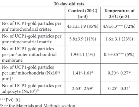
Table 4. Distribution of anti-UCP1-antibody-protein-A-gold complex particles (UCP1-gold particles)a in brown adipocytes of
30-day-old rats delivered and raised in a temperature environment of 33°C, as compared to animals delivered and raised at 20°C.
30-day-old rats Control (20°C)
(n-3) Temperature of 33°C (n-3)
No. of UCP1-gold particles per
μm2 mitochondrial cristae 43.1±11.9 (85%) 4.9±6.3*** (72%)
No. of UCP1-gold particles per
μm2 mitochondrial matrix 5.8±3.9 (11%) 1.6± 3.1 (23%)
No. of UCP1-gold particles per μm2 outer mitochondrial
membrane 1.9±1.1 (4%) 0.3±0.5*** (5%) No. of UCP1-gold particles
per μm3 mitochondria (Nx103/
μm3) b 1.41
c-1.61d 0.20 c- 0.27 d
No. of UCP1-gold particles per
adipocyte (Nx106) b 2.63c–2.99d 0.25c –0.34d
***P<0 .01
aSee the Materials and Methods section.
bRecalculated data from the above measurements and the adipocyte
morphometry data from Table 2.
cRecalculated data using the number of UCP1-gold particles per μm2 of
mitochondrial cristae.
dRecalculated data using the number of UCP1-gold particles per μm2
in the mitochondrial cristae plus the number of UCP1-gold particles per μm2 in the intermitochondrial space, mitochondrial matrix and the
outer mitochondrial membrane.
In contrast to the control rats, the IBAT of rats raised at 33°C and without recruitment, had lower UCP1 mRNA expression, starting from the first week of life and declining further by the age of 1 month (Table 3). At this point, it is unclear why IBAT at 33°C in 1-month-olds would expresses any UCP1. Although the UCP1 gene regulation data indicate a complex interplay of numerous transcription factors in the enhancer-promoter regulatory region of the UCP1 gene [44], we need more data to explain the mechanisms controlling UCP1 mRNA expression in 1-month-old unrecruited brown adipocytes.
Good correlation between UCP1 mRNA expres-sion and IEM quantification of the UCP1 described before [20-21,28,35,45] was confirmed, showing that UC-mitochondria at 33°C had approximately 5-fold less UCP1, and that in brown adipocytes at 33°C there
was about 9-fold less UCP1 protein as compared to the control brown adipocytes. However, in spite of sig-nificant underexpression of UCP1, and without BAT recruitment, rats raised at 33°C remained small and lean suggesting that to develop obe-sity [49], the animals should have had higher metabolism than rats delivered and raised at 33°C.
The finding of intramitochon-drial inclusions in adipocytes of 1-month-old rats raised at 33°C is significant. Such inclusions have been reported earlier by us and oth-ers [19,26, 33,34]; they were differ-ent and presdiffer-ent around the neonatal period or at the beginning of cold exposure, i.e. during periods of in-tensive mitochondriogenesis, con-ditions that were not present in rats reared at 33°C. Instead, the UC-mi-tochondria in adipocytes of animals raised at 33°C already had a crowded cytoplasm and well-developed mi-tochondrial cristae. However, the UC-mitochondria had low amounts of UCP1 protein (Fig. 3b, Table 4).
It has been established that the composition of mitochondrial pro-teins and UCP1 in brown adipocytes synchronously changes during postnatal development and recruit-ment [3,5-6]. Induced UCP1 expression in unilocular adipocytes was shown to be followed by an increase in mitochondrial cristae and an increase in cyclo-oxygenase (COX) IV; the “ordinary” white adipocyte C-mitochondria also appeared as brown adipocyte mitochondria [36]. These data suggest that UC-mitochondria genesis and preservation depends on the amount of UCP1 protein incorporated into the mitochondrial cristae. We are lacking data about the architecture of inner mitochondrial cristae and the required balance between the amount of UCP1 and other mitochondrial proteins. However, it is tempt-ing to assume that a very low amount of UCP1 in the mitochondria of rats raised at 33°C, or its absence
from fully developed UC-mitochondrial cristae, cre-ates mitochondrial cristae instability, resulting in the development of intramitochondrial tubular inclusions.
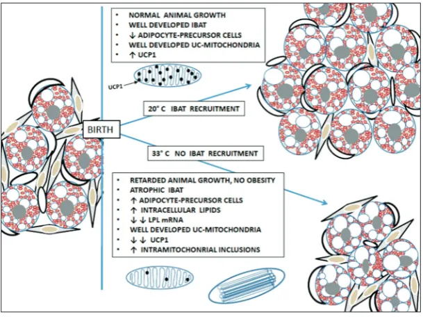
In summary (Fig. 4), rats delivered and raised at 33°C have a minimal metabolic rate. The animals re-mained euthermic. To prevent hyperthermia, IBAT was not recruited and nonshivering thermogenesis did not ensue. Without recruitment, the BAT remained disproportionally small and atrophic, having fewer mature brown adipocytes but more adipocyte pre-cursor cells. Mature thermoneutral brown adipocytes were smaller but contained more lipids compared to control brown adipocytes. Considering the low level of LPL mRNA expression, the increased lipid volume in the adipocytes could be the result of absent recruit-ment, or the reflection of de novo lipid synthesis in brown adipocytes. The UC-mitochondria were well-developed. However, without the recruitment, UCP1 mRNA expression was significantly reduced and the amount of UCP1 protein in UC-mitochondria was drastically decreased. Despite significant underexpres-sion of UCP1, the thermoneutral animals not only did not become obese due to low metabolic rate, but their growth, increase in size, weight and volume was significantly suppressed. Intramitochondrial tubular inclusion in UC-mitochondria was more frequent, perhaps because of the small amount of UCP1 pro-tein in the mitochondrial cristae, or as a result of an imbalance between the mitochondrial proteins and/ or substrates, all due to a lack of recruitment.
Acknowledgments: The authors gratefully acknowledge the tech-nical support and invaluable discussions from the leaders and members of the laboratories of Anton Svajger, Barbara Cannon and Bjorn A. Afzelius.
Funding: This work was supported by grants from the Self-Man-aging Community of Interest in Science of the Republic of Croatia (SIZ-V), through funds made available to the U.S.-Yugoslav Joint Board on Scientific and Technological Cooperation (No. 02-094-N), and by grants from the Swedish Natural Science Research Council. Some analyses of the samples were performed while Dragutin Lončar was a PhD student and recipient of a European Science Foundation grant, working at the Wenner-Gren Institute.
Author contributions: All authors contributed equally to this work.
Conflict of interest disclosure: The authors declare that they have no conflict of interest.
REFERENCES
1. Kajimura S, Spiegelman BM, Seale P. Brown and beige fat: physiological roles beyond heat generation. Cell Metabolism. 2015;22(4):546-59.
2. Valente A, Jamurtas AZ, Koutedakis Y, Flouris AD. Molecu-lar pathways linking non-shivering thermogenesis and obe-sity: focusing on brown adipose tissue development. Biol Rev Camb Philos Soc. 2015;90(1):77-88.
3. Nechad M. Structure and development of brown adipose tissue. In: Trayhurn P, Nicholls DG, editors. Brown adipose tissue. London: Edward Arnold; 1986. p. 1-30.
4. Loncar D. Development of thermogenic adipose tissue. Int J Develop Biol. 1991;35:321-33.
5. Cannon B, Nedergaard J. Brown adipose tissue: Function and physiological significance. Physiol Rev. 2004;84(1):277-359.
6. Klingenspor M, Bast A, Bolze F, Li Y, Maurer S, Schweizer S, Willershauser M, Fromme T. Brown adipose tissue. In: Symonds ME, editor. Adipose tissue biology. 2nd ed. New York: Springer; 2017. p. 91-147.
7. Cypress AA, Lehman S, Williams G, Tal I, Rdoman D, Goldfine AB, Kuo F, Palmer EL, Tseng YH, Doria A, Kolodny GM, Kahn CR. Identification and importance of brown adipose tissue in adult humans. N Engl J Med. 2009;360(15):1509-17.
8. Smith RE, Hock RJ. Brown Fat: Thermogenic effector of arousal in hibernators. Science. 1963;140(3563):199-200. 9. Gordon CJ. Thermal biology of the laboratory rat. Physiol
Behavior. 1990;47(5):963-91.
10. Gordon CJ. Thermal physiology of laboratory mice: defining thermoneutrality. J Thermal Biol. 2012;37:654-85.
11. Glossary of terms for thermal physiology 3rd ed. J Thermal
Biol. 2003;28:75-106.
12. Romanovsky AA, Ivanov AI, and Shimansky YP. Ambi-ent temperature for experimAmbi-ents in rats: a new method for determining the zone of thermal neutrality. J Appl Physiol. 2002;92:2667-79.
13. Nichelmann M, Tzschentke B. Thermoneutrality: traditions, problems, alternatives. In: Nagasaka T, Milton AS, editors. Body Temperature and Metabolism. Tokyo: IPEC; 1995. p. 77-82.
14. Eliason HL, Fewell JE. Thermoregulatory control during pregnancy and lactation in rats. J Appl Physiol. 1997;83:837-44.
15. Farrell WJ, Alberts JR. Rat behavioral thermoregulation integrates with nonshivering thermogenesis during post-natal development. Behavioral Neurosci. 2007;121:1333-41. 16. Kingma B, Frijns A, van Marken Lichtenbelt W. The ther-moneutral zone: implications for metabolic studies. Front Biosci. 2012;4:1975-85.
17. Satinof E. Thermoregulation. In: Whishaw IQ, Kolb B, edi-tors. Behavior of the Laboratory Rat. Oxford: Oxford Univ Press; 2014; p. 226-35.
18. Villarreal JA, Schlegel WM, Prange HD. Thermal environ-ment affects morphological and behavioral developenviron-ment of Rattus norvegicus. J Thermal Biol. 2007;91:26-35.
20. Loncar D. Immunoelectron microscopical studies on syn-thesis and localization of uncoupling protein in brown adi-pocytes. J Struct Biol. 1990;105:133-45.
21. Loncar D. Brown adipose tissue as a derivative of mesoderm grafted below the kidney capsule. A model for differentiation of isolated rat mesoderm. Int J Dev Biol. 1992;36:265-74. 22. Loncar D, Afzelius AA, Cannon B. Epididymal white adipose
tissue after cold stress in rats. I. Nonmitochondrial changes. J Ultrastruct Molecular Struct Res. 1988;101:109-22. 23. Cannon B, Hedin A, Nedergaard J. Exclusive occurrence of
thermogenin antigen in brown adipose tissue. FEBS Lett. 1982;150:129-32.
24. Slot JW, Geuze HJ. A new method of preparing gold probes for multiple-labeling cytochemistry. Eur J Cell Biol. 1985;38(1):87-93.
25. Weibel ER. Stereological Methods. Practical Methods for Biological Morphometry. Vol 1. London, New York: Aca-demic Press; 1979.
26. Loncar D, Bedrica L, Mayer J, Cannon B, Nedergaard J, Afze-lius AA, Svajger A. The effect of intermittent cold treatment on the adipose tissue of the cat: Apparent transformation from white to brown adipose tissue. J Ultrastruct Molec Struct Res. 1986;97(1-3):119-29.
27. Loncar D, Afzelius AA, Cannon B. Epididymal white adi-pose tissue after cold stress in rats II. Mitochondrial changes. J Ultrastruct Molec Struct Res. 1988;101(2-3):199-209. 28. Loncar D. Convertible adipose tissue in mice. Cell Tiss Res.
1991;266(1):149-61.
29. Jacobsson A, Stadler U, Glotzer MA, Kozak LP. Mitochon-drial uncoupling protein from mouse brown fat: molecular cloning, genetic mapping, and mRNA expression. J Biol Chem. 1985;260:16250-4.
30. Kirchgessner TG, Svenson KL, Lusis AJ, Schotz MC. The sequence of cDNA encoding lipoprotein lipase a member of a lipase gene family. J Biol Chem. 1987;262:8463-6. 31. Gwosdow AR, Besch EL. Effect of thermal history on the
rat’s response to varying environmental temperature. J Appl Physiol. 1985;59:413-19.
32. Yamauchi C, Fujita S, Obara T. Effects of room tem-perature on reproduction, body and organ weights, food and water intake, and hematology in rats. Lab Anim Sci. 1981;31(3):251-8.
33. Barnard T, Skala J. The development of brown adipose tis-sue. In: Lindberg O, editor. Brown Adipose Tistis-sue. New York, London, Amsterdam: Elsevier; 1970. p. 33-72. 34. Loncar D, Afzelius B. Ontogenetical changes in adipose
tis-sue of the cat: Convertible adipose tistis-sue. J Ultrastruct Molec Struct Res. 1989;102:9-23.
35. Herron D, Rehnmark S, Néchad M, Loncar D, Cannon B, Nedergaard J. Norepinephrine-induced synthesis of the uncoupling protein thermogenin (UCP) and its mitochon-drial targeting in brown adipocytes differentiated in culture. FEBS Lett. 1990;268:296-300.
36. Rossmeisl M, Barbatelli G, Flachs P, Brauner P, Zingaretti MC, Marelli M, Janovska P, Horakova M, Syrovy I, Cinti S, Kopecky J. Expression of the uncoupling protein 1 from the aP2 gene promoter stimulates mitochondrial bio-genesis in unilocular adipocytes in vivo. Eur J Biochem. 2002;269(1):19-28.
37. Kang HW, Ribich S, Kim BW, Hagen SJ, Bianco AC, Cohen DE. Mice lacking Pctp /StarD2 exhibit increased adaptive thermogenesis and enlarged mitochondria in brown adipose tissue. J Lipid Res. 2009;50:2212-21.
38. Wikstrom JD, Mahdaviani K, Liesa M, Sereda SB, Si Y, Las G, Twig G, Petrovic N, Zingaretti C, Graham A, Cinti S, Corkey BE, Cannon B, Nedergaard J, Shirihai OS. Hormone-induced mitochondrial fission is utilized by brown adipo-cytes as an amplification pathway for energy expenditure. EMBO J. 2014;33:418-36.
39. Altshuler-Keylin S, Kajimura S. Mitochondrial homeostasis in adipose tissue remodeling. Science Signaling. 2017;10:1-10. 40. Hemon P, Ricquier D, Mory G. The lipoprotein lipase
activ-ity of brown adipose tissue during early post-natal develop-ment of the normal and hypothyroid rat. Horm Metab Res. 1975;7:481-4.
41. Mitchell JR, Jacobsson A, Kirchgessner TG, Schotz MC, Cannon B, Nedergaard J. Regulation of expression of the lipoprotein lipase gene in brown adipose tissue. Am J Physiol - Endocrin Metabol. 1992;263:E500-E506.
42. Kersten S. Physiological regulation of lipoprotein lipase. Bio-chem Biophys Acta. 2014;1841(7):919-33.
43. Ricquier D: UCP1, the mitochondrial uncoupling protein of brown adipocyte: A personal contribution and a historical perspective. Biochimie. 2017;134:3-8.
44. Villarroya F, Peyrou M, Giralt M. Transcriptional regulation of the uncoupling protein-1 gene. Biochimie. 2017;134:86-92. 45. Loncar D. Uncoupling protein in the life cycle of rats: an
immunoelectron microscopical study of brown adipose tis-sue. Inst Phys Conference Ser. 1989;98:703-6.
46. Cousin B, Cinti S, Morroni M, Raimbault S, Ricquier D, Pénicaud L, Casteilla L. Occurrence of brown adipocytes in rat white adipose tissue: molecular and morphological characterization. J Cell Sci. 1992;103:931-42.
47. Porras A, Pefias M, Fernandez M, Benito M. Development of the uncoupling protein in the rat brown-adipose tissue dur-ing the perinatal period Its relationship with the mitochon-drial GDP-binding and GDP-sensitive ion permeabilities and respiration. Eur J Biochem. 1990;187:671-5.
48. Xiao XQ, Williams SM, Grayson BE, Glavas MM, Cowley MA, Smith MS, Grove KL. Excess weight gain during the early postnatal period is associated with permanent repro-gramming of brown adipose tissue adaptive thermogenesis. Endocrinology. 2007;148(9):4150-9.