CSEIT18317 | Received : 01 Jan 2018 | Accepted : 09 Jan 2018 | January-February-2018 [(3) 1 : 21-30]
International Journal of Scientific Research in Computer Science, Engineering and Information Technology © 2018 IJSRCSEIT | Volume 3 | Issue 1 | ISSN : 2456-3307
21
A Review on Various Image Segmentation Techniques for Brain
Tumor Detection
Munmun Saha, Chandrasekhar Panda
Department of Computer Science and Application, Sambalpur University, Jyoti Vihar, Sambalpur, Odisha, India
ABSTRACT
Segmentation is consider as one of the main step in image processing and it plays and important role in image processing. It is the process of subdividing an image into its constituent parts. In this paper we have reviewed various methods of segmentation and its application in medical image processing i.e. MRI image Ultrasound Image etc, we have focused on Brain Tumor MRI image. Recent medical imaging research faces the challenge of detecting brain tumor through MRI(Magnetic Resonance Image). There is a high diversity in the appearance of tumor tissue among different patients and in many cases similarity with the usual tissue. We have used MRI because it provide accurate visualize of anatomical structure of tissue. In this paper various method that have been used for segmentation of MRI for detecting brain tumor is reviewed.
Keywords: MRI, Segmentation, Clustering, K-means algorithm, Fuzzy C-mean algorithm, edge detection, Thresholding, Region Growing, Region Splitting, Watershed Segmentation Algorithm, Entropy, SVM.
I.
INTRODUCTION
Main organ in human nervous system is human brain and it is located in human head and covered by skull. Brain Tumor is defined as the abnormal growth of the tissues, which is a cluster of nerve cells, or neurons, in the brain.
Magnetic Resonance Imaging (MRI) is the test that makes use of pulses of radio wave energy and magnet to build the pictures containing the structure inside the body and the organs. A particular area of the body is placed inside the machine that contains strong magnet field at the time of MRI test.
Brain tumor detection and segmentation is one of the most challenging and time consuming task in medical image processing MRI provides plentiful information about the human soft tissue, which helps in the diagnosis of brain tumor. Accurate segmentation of MRI image is important for the diagnosis of brain tumor by computer aided clinical tool.
Process of segmentation
Firstly the original brain MRI image is found. Then the original Brain MRI image is Preprocessed. Processing is necessary as it enhanced the image and prepare for segmentation. Preprocessing includes resizing the image, noise removal from the image, image enhancement, image normalization and background segmentation. Preprocessing and enhancement techniques are used to improve the detection of the suspicious region from Magnetic Resonance Image (MRI). Secondly, the removal of high frequency components using filtering technique that includes median filter, Adaptive filter and Spatial filter. Other artifacts like text removed by some morphological operations. RGB to grey conversion and reshaping also takes place here.
The segmentation is the most important stage for analyzing image properly since it affects the accuracy of the subsequent steps.
Segmentation Technique
Approaches of segmentation
Isoled Point Detection: A point is the most basic type of discontinuity in a digital image. The most common approach for finding discontinuities is to run a (n x n) mask over each point in the image. The point is detected at a location (x, y) in an image where the mask is centered. If the corresponding value of R such that ||R||>T
Where R is the response of the mask at any point in the image and T is non-negative threshold value.
Line Detection Approach: Line detection is the next level of complexity in the direction of image discontinuity. For any point in the image, a response can be calculated that will show which direction the point of a line is most associated with. For line detection, we use two masks, i and j. [11]
Edge Detection: Since isolated points and lines of unitary pixel thickness are infrequent in most practical application, edge detection is the most common approach in gray level discontinuity segmentation. An edge is a boundary between two regions having distinct intensity level.[12] It is very useful in detecting of discontinuity in an image. When the image changes from dark to white or vice-versa. There are few operator Sobel edge operator, Laplacian Edge Operator, Prewitt Edge Operator.
Similarity Approach: It is also known as region based segmentation technique. The main goal of this method is to group pixels in the image, which are similar in some sense.
Thresholding: Threshold technique is one of the most used techniques in image segmentation. This technique can be expressed as: T=T[x, y, p(x, y), f(x, y)]
Where: T is the threshold value x, y are the coordinates of the threshold value point. p(x,y) f(x,y) are points the gray level image pixels. Threshold image g(x,y) can be define
Region Growing: The region-based segmentation is partitioning of an image into similar/homogenous areas of connected pixels through the application of homogeneity/similarity criteria among candidate sets of pixels. Each of the pixels in region is similar with respect to some characteristics or computed property such as color, intensity and/or texture. Failure to adjust the homogeneity/similarity criteria accordingly will produce undesirable results.
The segmented region might be smaller or larger than the actual
Over or under-segmentation of the image (arising of pseudo objects or missing objects) Fragmentation
Region growing is a simple region-based image segmentation method. It is also classified as a pixel-based image segmentation method since it involves the selection of initial seed points. This approach of segmentation
examines neighboring pixels of initial “seed points” and determines whether the pixel neighbors should be added to the region.[13]
Region Splitting Approach: The basic idea of region splitting is to break the image into a set of disjoint regions which are coherent within themselves:
Initially take the image as a whole to be the area of interest.
Look at the area of interest and decide if all pixels contained in the region satisfy some similarity constraint. [14]
If TRUE then the area of interest corresponds to a region in the image.
If FALSE split the area of interest (usually into four equal sub-areas) and consider each of the sub-areas as the area of interest in turn.
This process continues until no further splitting occurs. In the worst case this happens when the areas are just one pixel in size.
This is a divide and conquer or top down method.
If only a splitting schedule is used then the final segmentation would probably contain many neighboring regions that have identical or similar properties.
Thus, a merging process is used after each split, which compares adjacent regions and merges them if necessary. Algorithms of this nature are called split and merge algorithms.[14]
Watershed Transform
The watershed Transform considers the gradient magnitude of an image as a topographic surface. Pixels having the highest gradient magnitude intensities (GMIs) correspond to watershed lines, which represent the region boundaries. Water placed on any pixel
enclosed by a common watershed line flows downhill to a common local intensity minimum (LIM). Pixels draining to a common minimum form a catch basin, which represents a segment.[15]
Clustering
Clustering is a process whereby a data set is replaced by clusters, which are collections of data points that “belong together”. It is natural to think of image segmentation as clustering; we would like to represent an image in terms of clusters of pixels that “belong together”. The specific criterion to be used depends on the application. Pixels may belong together because they have the same color and/or they have the same texture and/or they are nearby, etc.[16]
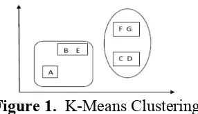
K-Mean Clustering Algorithm
K-means algorithm is the most well-known and widely used unsupervised clustering technique in partitioned clustering algorithms. Purpose of this algorithm is to minimize the distances of all the elements to their cluster centers. Inspiring or improving k-means develops most of the algorithms in this field. The algorithm upgrades the clusters iteratively and runs in a loop until it reaches to optimal solution. Performance of K-means algorithm depends on initial values of cluster centers. Therefore the algorithm should be tested for different outcomes with different initial cluster centers by multi-running.
Figure 1.
K-Means Clustering
Fuzzy Clustering
algorithm. Traditional clustering approaches generate partition, each pattern belongs to one and merely single cluster. That’s why, the clusters in a hard clustering technique are dislodged. Fuzzy clustering enlarges this notion to connect each pattern with every cluster by means of a membership function. The outcome of such algorithms is a clustering, although not a partition.
Figure 2. Fuzzy Cluster
Segmentation Using Genetic Algorithm (GA) Thangavel and Karnan (2005) said a genetic algorithm (GA) [10] is an optimization technique for obtaining the best possible solution in a vast solution space. Genetic algorithms operate on populations of strings, with the string coded to represent the parameter set. The intensity values of the tumor pixels are considered as initial population for the genetic algorithm.[10]The intensity values of the suspicious regions are then converted as 8 bit binary strings and these values are then converted as population strings and intensity values are considered as fitness value for genetic algorithm. Now the genetic operator’s reproduction, crossover and mutation are applied to get new population of strings.
Level Set Approach
Lavel-set method (LSM) are a conceptual framework for using level sets as a tool for numerical analysis of surface and spaces. The advantage of the level-set model is that one can perform numerical computations involving curves and surface on a fixed Cartesian grid without having to parameterized these object( this is called the Eulerian approach). [18] Also, the level-set method makes it very easy to follow shapes that change topology, for example, when a shape splits in two, develops holes, or the reverse of these operations. All these make the level-set method a great tool for modelling time-varying objects, like inflation of an airbag, or a drop of oil floating in water. The Level set method was initially proposed to track moving interface by Osher and Sethainin 1988 and gas spread across various imaging domains in late90s.It can be used to efficiently address the problem of curve/surface/etc.[18] propagation in an implicit
manner. The central idea is to represent the evolving contour using a signed function whose zero corresponds to the actual contour. Then, according to the motion equation of the contour, one can easily derive a similar flow for the implicit surface that when applied to the zero level will reflect the propagation of the contour.
Literature Review
A plenty of work has been proposed by researchers for the MRI brain image segmentation and tumor detection techniques. A brief review of some of the research work is presented here.
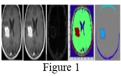
Swapnil R.Telrandhe ,Amit Pimpalkar and Ankita Kendhe [1] found a Technique of detecting brain tumor using similarity based segmentation algorithm and SVM for pattern recognition.In this paper, they implemented an automated system for brain tumor detection, the main functionality of this system is divided in some parts and Segmentation, Object Labeling, HOG (Histogram Oriented Gradient), feature extraction and linear SVM is implemented. [20]For Segmentation they have used K-means algorithm, for Object Labeling HOG is use, HOG also use to extract texture feature, shape context feature and color feature. Then they have implemented the SVM based on this feature they can train the SVM and further test is on other infected MRI images.
Figure 1
The experimental result of the above proposed experiment is shown in the figure.In above figure the result of proposed system is explain there, the first is original MRI image which is having some noise in it. The noise is removed with median filter the result of median filter is shown in the second image. Then the morphological filtering is used on the preprocessed image and the masks are used to remove the fatty tissues and skull bone.
also does the extraction of texture, color and shape context feature. [1]
Padmakant Dhage, Prof. M.R.Phegade, Dr.S.K.Shah [2] has proposed an paper which illustrates the ability of watershed segmentation technique to separate the abnormal tissue from the normal surrounding tissue to get a real identification of involved and noninvolved area that help the surgeon to distinguish the involved area precisely. At the end of the process tumor is extracted from the MRI image and its exact position and shape are determined and various parameters like perimeter, eccentricity, entropy and centroid have been calculated.The proposed system has four modules namely preprocessing, segmentation, connected component leveling(CCL) and multi parameter calculation. Preprocessing includes filtering of image. Segmentation is carried out by watershed algorithm.[2] Using CCL location is found out and area is calculated of the tumor in MRI image.
Figure
The above figure gives the experimental result of the proposed algorithm step by step.The input image (a) was processed. Noise is added in (b). It is denoise using median filter, resulting image is obtained shown in (c). Original MRI image having binary, distance transform, and watershed superimposed image shown in (d), (e) and (f) respectively. Compute CCL to dilated image whose object area is less than 500, resulting image shown in (g). Assign the RGB label for connected components shown in (h). Final detected tumor from MRI image is shown in (i).
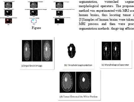
Anam Mustaqeem, Ali Javed, Tehseen Fatima [3] has proposed an efficient algorithm in there paper for tumor detection based on segmentation and morphological operators. Firstly quality of scanned image is enhanced and then morphological operators are applied to detect the tumor in the scanned image. They conducted a research to detect brain tumor using medical imaging techniques. The main technique used was segmentation, which is done using a method based on threshold segmentation, watershed segmentation and morphological operators. The proposed segmentation method was experimented with MRI scanned images of human brains, thus locating tumor in the images. [3]Samples of human brains were taken, scanned using MRI process and then were processed through segmentation methods .thugiving efficient end results.
The above images gives different observation in different entropy method. And they propose that Havrda Chavat gives the most accurate result.
N. Manasa, G. Mounica, B.Divya Tejaswi [5] proposed a method where the tumor is segmented based on canny
edge detection algorithm. It is the only procedure capable of finding the best contours while eliminating all edges associated with the gray matter in original image as compared to other edge detection algorithm [5] Finally thresholding is applied to the image and based on this tumor area and shape is calculated. The proposed method involves four modules. Preprocessing, Segmentation, Feature Extraction and Approximate Reasoning. Firstly preprocessing is done by filtering techniques such as Gaussian and Median filters. Image acquisition involves preprocessing such as scaling, Image Enhancement filtering and noise reduction. Secondly segmentation is done by canny algorithm. Thirdly Feature Extraction, in pattern recognition and in image processing, feature extraction is to obtain the most relevant information in a lower dimensionality space. When image sizes are large and a reduce feature representation is required to quickly complete tasks. Fourthly Approximate Reasoning, in this step thresholding is applied to the image. Finally tumor shape and area is obtained.
The Above figure shows all the experimental result.
J.selvakumar, A.Lakshmi T.Arivoli [6] proposed a paper, which deals with the implementation of Simple Algorithm for detection of range and shape of tumor in brain MRI images. However this method of detection resists the accurate determination of stage & size of tumor. To avoid that, this project uses computer-aided method for segmentation (detection) of brain tumor based on the combination of two algorithms. This method allows the segmentation of tumor tissue with accuracy and reproducibility comparable to manual segmentation. In addition, it also reduces the time for
the tumor shape and position in MRI image using edge detection method.[6]
The Above Images Shows all the step of the proposed algorithm respectively.
A.R.Kavitha, Dr.C.Chellamuthu, Ms.Kavin Rupa [7] has proposed Region growing is an important application of image segmentation in medical research for detection of tumor. In this paper, they proposed an effective modified region growing technique for detection of brain tumor. Modified region growing includes an orientation constraint in addition to the normal intensity constrain. The performance of the proposed technique is systematically evaluated using the MRI brain image received from the public sources. [7] For validating the effectiveness of the modified region growing, the quantity rate parameter has been considered. For the evaluation of the proposed technique of tumor detection, the sensitivity, specificity and accuracy values were used. Comparative analyses were made for the normal and the modified region
growing both the Feed Forward Neural Network (FFNN) and Radial Basis Function (RBF) neural network. The results shows that the modified region growing achieved better results when compared to the normal technique.[7] The proposed technique consists of four phases namely pre-processing, extended region growing, feature extraction and classification of tumor. In pre processing, Gaussian filtering and RGB to Grey image conversion is done. In the extended region growing they have produced an additional constraint of 'orientation' apart on the normal 'intensity' constraint used in the normal region growing techniques. In the feature extraction phase, certain features like area, orientation, mean, correlation and co-variance of the region is extracted. In the final classification they use the Neural network classifier to detect tumor or not.
Results. (a) MRI image with tumor, (b) gridded image, (c) seed point selected image, (d) segmented image
K.B Vishnavee, K.Amshakala [8] in there has proposed Self Organizing Map (SOM) clustering for MRI brain image segmentation. Before segmentation the Histogram Equalization is utilized for feature extraction, which will improve the segmentation accuracy. After the segmentation process, the feature extraction using Gray Level Co-occurrence Matrix is utilized which avoids the formation of misclustered regions. The Principle Component Analysis (PCA) method is used for the feature selection to improve the classifier accuracy. An effective classifier Proximal Support
Vector Machines (PSVM) is used to automatically detect the tumor from MRI brain image. This method is faster and computationally more efficient than the existing method SVM. While the SOM clustering with Histogram Equalization is a fast procedure for the segmentation of the whole volume and provides a way to model tissue classes, the PSVM-GLCM-PCA approach is a more robust scheme under noisy or bad intensity normalization conditions, which produces better results using high-resolution images.
Sangram Keshari Nayak and Dr Chandra Sekhar Panda [9] proposed an algorithm which deal withpartition of an image to get mutually exclusive and exhausted regions. Further the regions need to possess homogeneous pixels with respect to predefined
criterion. Segmentation algorithms are area oriented instead of pixel oriented. The result of segmentation is the splitting up of the image into connected areas. Thus segmentation is concerned with dividing an image in to meaningful regions.
The above image gives the experimental result.
Comparison Table
Sr No
Year Author Technique Used Result Advantage Reference
1. 2016 Swapnil R.Telrandhe, Amit Pimpalkar and Ankita Kendhe
K-means algorithm, HOG,SVM
The extracted features used to train SVM and the database and is use for pattern matching and test the system.
It gives a faire result for the input and high accuracy rate.
[1]
2. 2015 Padmakant Dhage, Prof M.R Phegade, Dr. S.K Shah
Median Filter, Watershed Transformation, CCL
Different parameter like Area, Perimeter, Eccentricity, Entropy, Centroid By using Watershed
Transformation
Its simplicity and efficiency. It can remove high frequency
component without
disturbing edges. [2]
3 2012 Anam
Mustaqeen, Ali Javed, Tehseen Fatima
Threshold segmentation Watershed Transformation, Morphological Operation
The portion with high intensity value is calculated and tumor is segmented and detected.
The portion with high intensity values are calculated and tumor is detected and segmented
[3]
4 2016 Devendra Somwanshi,
Threshold technique,
After comparing and analyzing through
Color is
II.
CONCLUSION
The paper reviews and summarise some existing method of segmentation for brain tumor detection from MRI image. Though this paper is not so much rich in describing many algorithm there r many other methods and algorithm present. Although there is no perfect method for image segmentation because the result of image depends on many factors i.e pixel, color, texture, intensity etc. Therefore it is not possible to consider a single method for all type of image. Hence it is good to use hybrid solution.
III.
REFERENCES
[1]. Swapnil R. Telerandhe et al,"Implementaton of
brain tumor detection using segmentation algorithm and SVM", International Journal on computer science and engineering,vol.8 no. 7 Jul 2016
[2]. Padmakant Dhage, Prof. M.R.Phegade,
Dr.S.K.Shah,"Watershed Segmentation Brain
Tumor Detection", International Conference on Pervasive Computing", year 2015
[3]. Anam Mustaqeem, Ali Javed, Tehseen Fatima," An
Efficient Brain Tumor Detection Algorithm Using Watershed & Thresholding Based Segmentation", I.J. Image, Graphics and Signal Processing, 2012, 10, 34-39 Published Online September 2012 in MECS.
[4]. Devendra Somwanshi, Ashutosh Kumar, Pratima
Sharma, Deepika Joshi," An efficient Brain Tumor Detection from MRI Images using Entropy Measures", IEEE International Conference on Recent Advances and Innovations in Engineering (ICRAIE-2016), December 23-25, 2016, Jaipur, India.
[5]. N. Manasa, G. Mounica, B.Divya Tejaswi,"Brain
Tumor Detection Based on Canny Edge Detection Algorithm and it’s area calculation",International Journal of Computer & Mathematical Sciences, IJCMS ISSN 2347 – 8527 Volume 5, Issue 3 March 2016
[6]. J.selvakumar, A.Lakshmi T.Arivoli," Brain Tumor
Segmentation and Its Area Calculation in Brain MR Images using K-Mean Clustering and Fuzzy C-Mean Algorithm", IEEE International Conference On Advances In Engineering, Science And Management (ICAESM -2012) March 30, 31, 2012
[7]. A.R.Kavitha, Dr.C.Chellamuthu, Ms.Kavin Rupa,"
An Efficient Approach for Brain Tumour Detection
Based on Modified Region Growing and Neural Network in MRI Images", 2012 International Conference on Computing, Electronics and Electrical Technologies [ICCEET].
[8]. K.B Vishnavee, K.Amshakala," An Automated
MRI Brain Image Segmentation
[9]. and Tumor Detection using SOM-Clustering and
Proximal Support Vector Machine Classifier",
[10]. Sangram Keshari Nayak and Dr Chandra Sekhar
Panda,"Segmentation of Brain MR image Containing Tumor using Extended Maxima Transform, Regional Transform and Image Model Techniques", IJRSCSE. Volume 2, Issue 9, September 2015, PP 4-13 ISSN 2349-4840 (Print) & ISSN 2349-4859 (Online)
[11]. K.Selvanayaki, Dr.P.Kalugasalam "Intelligent
Brain Tumor Tissue Segmentation From Magnetic Resonance Image (MRI) Using Meta Heuristic Algorithms", Journal Of Global Research In Computer Science Volume 4, No. 2, February 2013
[12]. Ahmed M. Ayash," Digital Image processing Lab",
Islamic University – Gaza Engineering Faculty Department of Computer Engineering April 27, 2013
[13]. Rajiv Kumar, M.Arthanari, M.Sivakumar," Image
Segmentation using Discontinuity-Based
Approach", International Journal Multimedia and Image Processing (IJMIP), Volume 2, Issues 1/2, March/June 2012
[14]. Shilpa Kamdi , R.K.Krishna," Image Segmentation
and Region Growing Algorithm", International Journal of Computer Technology and Electronics Engineering (IJCTEE) Volume 2, Issue 1
[15]. https://users.cs.cf.ac.uk/dave/Vision_lecture/node3
4.html
[16]. https://people.cs.uct.ac.za/~mgallott/honsproj/water
shed.html
[17]. Barghout, Lauren; Sheynin, "Real-world scene
perception and perceptual organization: Lessons from Computer Vision". Journal of Vision. 13 (9):709–709. doi:10.1167/13.9.709
[18]. Dhriti Sharma," Image processing and image
segmentation", ICRTEDC, Vol. 1, Spl. Issue 2 (May, 2014)
[19]. Sethian, James,"Level Set Methods and Fast