Karolina Sterzyńska
1, Aldona Kasprzak
1, Piotr Dzięgiel
1,2, Maciej Zabel
1,2Immuno- and Hybridocytochemical Analysis
of the Expression of Interleukin 2 and Its Receptor
in Lung Cancers*
Immuno- i hybrydocytochemiczna analiza ekspresji interleukiny 2
i jej receptora w nowotworach płuc
1 Department of Histology and Embryology, Poznań University of Medical Sciences, Poznań, Poland 2 Department of Histology and Embryology, Wroclaw Medical University, Wrocław, Poland
Abstract
Background. Interleukin 2 (IL-2) plays a significant role in the activation, differentiation and proliferation of hemopoietic cells, acting through a specific receptor complex (IL-2R). It is mainly produced by activated T helper cells (CD4+), but also by T cytotoxic lymphocytes (CD8+). Studies on IL-2/IL-2R in lung tumors focus mainly on detecting markers on immunocompetent cells isolated from patients’ blood. Only a few studies describe tissue expression of IL-2 and its receptor in lung cancers.
Objectives. This study aimed at examining the expression of IL-2 and its receptor (IL-2Rα and IL-2Rβ) in lung tumor cells and in T lymphocytes that infiltrate the tumors (TILs).
Material and Methods. The material for studies included archival paraffin blocks with the following types of lung tumors: typical and atypical carcinoids (TC, AC), small-cell lung carcinoma (SCLC), non-small-cell lung carci-noma, squamous carcinoma (NSCLC-SQ) and adenocarcinoma (ADENO). Expression of the markers was dem-onstrated using immunocytochemical and in situhybridization techniques, and the obtained data were subjected to statistical analysis.
Results. Expression of IL-2, IL-2Rα and IL-2Rβ was noted in all the studied types of pulmonary tumors. A positive immunocytochemical reaction was observed both in the tumor cells and in the tumor-infiltrating lymphocytes. The presence of IL-2 and IL-2Rα was confirmed by the detection of mRNA for both markers. IL-2Rβ expression was detected mainly in cell cytoplasm, and IL-2Rα on cell membranes of both neoplastic cells and TILs. The most pronounced total expression of IL-2 and IL-2Rβ was demonstrated in lung carcinoids, and the most pronounced expression of IL-2Rα was noted in squamous cell carcinomas. The most pronounced “neoplastic” expression of IL-2 was observed in TC, and of IL-2Rα in atypical carcinoids. The most pronounced “neoplastic” expression of the IL-2Rβ subunit was detected in atypical pulmonary carcinoids, while the least pronounced expression of this subunit was detected in small-cell lung cancer.
Conclusions. The prevalent expression of IL-2 and IL-2Rβ in tumor cells and of IL-2Rα in tumor-infiltrating lym-phocytes, regardless of the lung tumor involved, may suggest their auto- and/or paracrine activity in the microenvi-ronment of lung tumors. A direct relationship between the expression of the β subunit of IL-2R in tumor cells and tumor-infiltrating lymphocytes is observed in tumors of high proliferative activity (atypical carcinoids, small-cell and squamous pulmonary carcinomas) (Adv Clin Exp Med 2011, 20, 2, 137–148).
Key words: interleukin 2, receptor of interleukin 2, lung cancers.
Streszczenie
Wprowadzenie. Interleukina 2 (IL-2) odgrywa istotną rolę w aktywacji, różnicowaniu i proliferacji komórek hematopoetycznych, działając za pośrednictwem specyficznego kompleksu receptorowego (IL-2R). Jest wydzie-lana głównie przez aktywowane limfocyty T pomocnicze (CD4+), ale również przez limfocyty T cytotoksyczne (CD8+). Badania dotyczące IL-2/IL-2R w nowotworach płuc są głównie skupione na wykrywaniu tych markerów
Adv Clin Exp Med 2011, 20, 2, 137–148 ISSN 1230-025X
ORIgINAL PAPERS
© Copyright by Wroclaw Medical University
Interleukin 2 (IL-2) is one of the most impor-tant cytokines which control the proliferation and differentiation of cells in the immune system [1]. The cytokine is produced mainly by activated T helper lymphocytes (CD4+). Its expression can
also be detected in T CD8+ lymphocytes, dendritic
cells and thymocytes in the thymus [2].
The biological activity of IL-2 resulting in signal transduction and specific effects in the cell takes place due to specific interaction of IL-2 with a specific receptor. The receptor of IL-2 (IL-2R) consists of three subunits: the α chain (IL-2Rα, CD25, Tac, p55), β chain (IL-2Rβ, CD122, p70) and γ chain (IL-2Rγ, CD132). Depending on the links between individual subunits, three forms of IL-2R receptor with varying affinity of interaction with IL-2 can be distinguished [2–6].
Studies of the expression of IL-2 and its recep-tor have been conducted mainly on T and B cell leukemias, lymphomas and solid tumors such as cancers of the larynx, lungs, mammary gland, large intestine, liver, ovary, prostate and melanomas [4, 7–12]. The authors of these studies have fo-cused their attention on the role of the IL-2/IL-2R complex in controlling cell growth and on the evaluation of the cytokine as a potential marker of tumors. It is intriguing that exogenous IL-2 in-hibits the growth of certain human neoplastic cells via IL-2R, while the proliferation of other cells remains unchanged in the presence of a similar expression of IL-2Rβ on cell membranes [4, 13]. Some authors have presented evidence that endog-enous IL-2 is in fact pre-required for the prolifera-tion of certain tumor cells (such as squamous cell cancer of head and neck), while growth inhibition
could have resulted from the use of anti-sense IL-2 [14]. Evidence is also available indicating that endogenous IL-2 may be involved as a growth fac-tor for human neoplastic cells [4, 15].
Primary lung cancer is the most frequent malignant tumor in men and the second most frequent malignant tumor in women (following breast cancer), and is the most frequent cause of cancer deaths in both genders [16].
Studies on the role of IL-2 and its receptor in pulmonary tumors have dealt mainly with non-small cell lung cancers (NSCLC) [4, 9, 12, 17, 18]. The production of cytokines in tumors has mainly been examined in patients’ immunocompetent blood cells (lymphocytes, monocytes) using vari-ous types of ELISA (Enzyme-Linked Immunosor-bent Assay) test. Significantly reduced IL-2 pro-duction by those cells has been noted [17]. Lower production of IL-2 was documented in patients with metastasized forms of cancer than in patients without metastases [9, 11]. Moreover, patients with lowered ratios of CD4/CD8 lymphocytes manifested lower levels of IL-2 as compared to pa-tients with normal CD4/CD8 ratios.
Also, in cases of small cell lung cancers (SCLC), the synthesis of certain cytokines (including inter-leukin-2) by immunocompetent blood cells was found to be significantly lower than in controls following stimulation with a mitogen. In addition, the secretion of IL-2 depended on the tumor load; lowered production of this cytokine was demon-strated in more advanced clinical stages of lung cancers. Lowered production of IL-2 in blood cells at the moment of SCLC diagnosis was found to be an important indicator of a poor prognosis for
w komórkach immunokompetentnych izolowanych z krwi pacjentów. Nieliczne prace opisują tkankową ekspresję IL-2 i jej receptora w nowotworach płuc.
Cel pracy. Zbadano ekspresję IL-2 i jej receptora (IL-2Rα i IL-2Rβ) w komórkach zmienionych neoplastycznie (komórki guza płuc) oraz w limfocytach T naciekających guzy płuca (TILs).
Materiał i metody. Materiał do badań stanowiły archiwalne bloczki parafinowe z następującymi typami guzów płuc: rakowiaki typowe i atypowe (TC, AC), rak drobnokomórkowy (SCLC), rak płaskonabłonkowy (NSCLC-SQ) i rak gruczołowy (ADENO). Ekspresję markerów wykrywano z wykorzystaniem technik immunocytochemicznych i hybrydyzacji in situ. Wszystkie uzyskane dane poddano analizie statystycznej.
Wyniki. Ekspresję IL-2, IL-2Rα oraz IL-2Rβ wykrywano we wszystkich typach badanych nowotworów płuc. Pozytywną reakcję immunocytochemiczną obserwowano zarówno w komórkach nowotworowych, jak również w limfocytach infiltrujących guzy. Obecność IL-2 i IL-2Rα potwierdzono wykrywaniem mRNA dla obu markerów. Ekspresję IL-2Rβ umiejscowiano głównie w cytoplazmie komórek, a IL-2Rα na błonach komórkowych i to zarówno komórek neoplastycznych, jak i TILs. Największą całkowitą ekspresję IL-2 i IL-2Rβ wykazywały rakowiaki płuc, a IL-2Rα raki płaskonabłonkowe. Największą „guzową” ekspresję IL-2 obserwowano w TC, a IL-2Rα w atypowym rakowiaku płuc. Ekspresja „guzowa“ podjednostki β IL-2R również była największa w atypowym rakowiaku płuc, a najmniejsza dotyczyła drobnokomórkowego raka płuc.
Wnioski. Przeważająca ekspresja IL-2 i IL-2Rβ w komórkach guzowych, a IL-2Rα w limfocytach naciekających guz niezależnie od typu nowotworu płuc, może sugerować ich auto- i/lub parakrynowe działanie w mikrośrodowisku guza płuc. Wprost proporcjonalna zależność między ekspresją podjednostki β IL-2R w komórkach guza i w TILs dotyczy nowotworów płuc o dużej aktywności proliferacyjnej (atypowe rakowiaki, drobnokomórkowe i płaskona-błonkowe raki płuc) (Adv Clin Exp Med 2011, 20, 2, 137–148).
the patient’s survival, independent of other indica-tors such as staging, NSE (neuron specific enolase) concentration, LDH (lactate dehydrogenase) con-centration and the patient’s age and gender. Ac-cording to Fischer et al., the lowered production of IL-2 in small cell lung cancer may indicate immu-nosuppression in this type of cancer [19].
Studies on IL-2/IL-2R in lung tumors focus mainly on detecting markers on immunocompe-tent cells isolated from patients’ blood. Few inves-tigations describe tissue expression of IL-2 and its receptor in lung tumors.
Reports of detecting and localizing IL-2 in lung cancer tumor cells themselves are very rare. Studies on non-small cell lung cancer cell line cultures have found increased production of type 2 cytokines (mainly IL-5 and IL-10), but not of type 1 cytokines, including IL-2 [20, 21]. As in microenvironmen-tal lymphocytes, in NSCLC tumor cells them-selves, higher expression of IL-4, IL-10, TgF-α and TgF-β was demonstrated as compared to the ex-pression of IL-2, IL-12, IL-18 or IFN-γ [22].
Studies on the role of the IL-2 receptor (IL-2R) in lung tumors have shown that activated T lym-phocytes can produce the soluble α subunit of the receptor (sIL-2Rα), which is capable of effectively binding IL-2.
Recent studies related to the role of the IL-2 complex and its receptor have focused on the production of proteins in T regulatory lymphocytes (Treg), particularly those which are CD8-positive, in the contexts of immunomodulatory therapies and transplantological problems [23].
Lung cancer is the most frequently manifested tumor in men and the second most frequent in women (following breast cancer) [16]. The treat-ment of lung cancer still suffers from low efficacy. Numerous immunotherapy trials involving the signalling pathways of interleukin-2 and its re-ceptor are being undertaken; some of these trials include lung carcinomas [3, 10]. The data related to cytokine expression in tumor cells are relatively incoherent, and the role of tissue expression of IL-2 and its receptor (particularly of its β subunit) as markers of clinical significance remains poorly defined. Research into the specific cellular source of IL-2 and its receptor in various types of lung tu-mors seems potentially significant, due to numer-ous findings of IL-2Rα overexpression, which leads to increased cell proliferation, drug resistance and augmented transforming activity in lung tumors [3, 10]. Therefore, in this study we examined the immunocytochemical and hybridocytochemical expression of IL-2 and its receptor (IL-2Rα and IL-2Rβ) in neoplastically transformed cells (lung tumor cells) and in the tumor-infiltrating lym-phocytes (TILs).
Material and Methods
The experimental material for the study in-cluded archival paraffin blocks with the following types of lung cancers: typical and atypical car-cinoids (TC, n = 14; AC, n = 11), small cell lung cancer (SCLC, n = 23), non-small cell lung cancer – squamous cell lung cancer (NSCLC-SQ, n = 23) and adenocarcinoma (ADENO, n = 15). The avail-able clinical data on the patients included the his-topathological diagnosis, the degree of histologic malignancy or grading (g) (NSCLC group) and the age and gender of the patient. The histologi-cal type of lung tumor was determined in routine diagnostic tests conducted by two independent pa-thologists.
The control material consisted of archival par-affin blocks with fragments of lymphatic organs (lymph nodes, appendixes) with inflammatory le-sions and free of neoplasia (n = 10).
All the tissues were fixed of a buffered solution of 10% formalin, then embedded in paraffin, cut into 5 µm thick sections and deposited on Super-Frost/Plus glass microscope slides.
Paraffin sections were stained in the usual manner, using hematoxylin and eosin (H + E). The expression of markers (IL-2 – 1:100, R&D Systems; IL-2Rα – 1:100, Novocastra Labs; IL-2Rβ – 1:500, Abcam; CD3 – 1:500, Novocastra Labs; CD4 – 1:80, Dako; CD8 – 1:1, Dako) was detected using im-munocytochemistry (the ABC technique, in accor-dance with Hsu et al. [24]) and in situ hybridiza-tion. In the cases of IL-2Rα, IL-2Rβ, CD3, CD4 and CD8 antibodies, an additional high temperature antigen unmasking technique was required. All the reactions entailed overnight incubation of the applied primary antibodies. In order to compare the expression of proteins (IL-2, IL-2R subunits and molecules of CD3, CD4 and CD8) in tumor cells and in lung tumor-infiltrating lymphocytes (TILs), identical reactions were performed in tis-sue material of reactive lymphatic organs (n = 10, positive control). The tests in the positive control group were aimed only at verifying the quality of the applied antibodies and were not subjected to semi-quantitative analysis or statistical analysis.
Results
Comparison of IL-2, IL-2Rα and
IL-2Rβ Expression Among the
Examined Groups of Patients
Interleukin-2
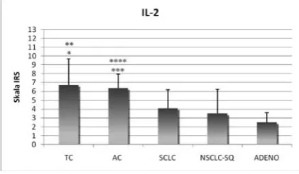
Statistical analysis of the results of immuno-cytochemical detection of the total IL-2 expression (in tumors and TILs) demonstrated the highest mean IL-2 expression in typical and atypical carci-noids (Fig. 1). Expression of IL-2 was significantly
higher in cases of TC and AC than in squamous cell lung cancer or pulmonary adenocarcinoma (Fig. 1, Table 1).
Similar differences in expression were found when IL-2 expression only in tumor cells (g) was considered: IL-2 expression was significantly higher in both types of lung carcinoid cells than in squamous cell lung cancer and adenocarcinoma (Fig. 2A, Fig. 3A, Table 1).
Interestingly, IL-2 expression in tumor-infil-trating lymphocytes (n) was most pronounced in atypical lung carcinoids (Fig. 3B), and it was sig-nificantly higher than the analogous expression of the cytokine in TILs in typical carcinoids or pul-monary adenocarcinomas. No significant differ-ences were noted in IL-2 expression in small-cell lung cancer, squamous cell lung cancer or adeno-carcinomas (Fig. 2B, Table 1).
IL-2 Receptor α Subunit (IL-2Rα)
The most pronounced expression of total IL-2Rα was noted in patients with squamous cell lung cancer, but it was significantly higher only as compared to that in pulmonary adenocarcinoma (p < 0.01) (Fig. 4, Table 1).
Considering tumor cells (g) only, the highest expression of IL-2Rα was demonstrated in patients with atypical lung carcinoid (Fig. 6A), but it was significantly higher only as compared to the anal-ogous expression in pulmonary adenocarcinoma (Fig. 5A). Relatively high mean IL-2Rα expression was also observed in cases of squamous cell lung cancer and SCLC; in both cases it was higher than Fig. 1. Comparison of mean values of total IL-2
expres-sion in particular groups of patients
Ryc. 1. Porównanie średnich wartości ekspresji IL-2 całkowitej w poszczególnych grupach chorych * TC vs NSCLC-SQ, p < 0.05.
** TC vs ADENO, p < 0.001. *** AC vs NSCLC-SQ, p < 0.05. **** AC vs ADENO, p < 0.001.
Fig. 2. Comparison of mean values of IL-2 expression in A) tumor cells (g) and B) tumor infiltrating lymphocytes (n) in particular groups of patients
Ryc. 2. Porównanie średnich wartości ekspresji IL-2 w komórkach nowotworowych guza (g) (A) i w limfocytach naciekających guzy (n) (B) między poszczególnymi grupami chorych
A) * TC vs NSCLC-SQ, p < 0.001. ** TC vs ADENO, p < 0.001. *** AC vs NSCLC-SQ, p < 0.05. **** AC vs ADENO, p < 0.05. B) * TC vs AC, p < 0.05. ** AC vs ADENO, p < 0.05.
strated that in both cases the highest expression was in atypical lung carcinoids. No significant differenc-es among individual groups were detected in terms of IL-2Rβ expression within the tumor (Fig. 9A) or in the tumor microenvironment (n) (Fig. 9B, Table 1).
Analysis of the Expression of
Markers Typical for T-Lymphocyte
Subpopulations (CD3, CD4 and CD8)
in the Microenvironment of Lung
Cancers
The expression of markers which differenti-ate pulmonary tumor-infiltrating lymphocytes pertained to subpopulations of T lymphocytes. Most frequently, the cells were present in greater numbers in the connective tissue sublayer of lung cancers. Occasional individual immunopositive T lymphocytes were also noted between tumor cells or scattered over the entire area of the histo-logical preparation.
The expression of CD3 (a marker molecule of T lymphocytes), CD4 (a subpopulation of T lym-phocytes: the so-called helper lymphocytes Th1 and Th2) and CD8 (a subpopulation of T lympho-cytes: cytotoxic T lymphocytes Tc) was analyzed on a 12-point IRS scale, scoring only cells with the entire circumference of cell membrane labelled.
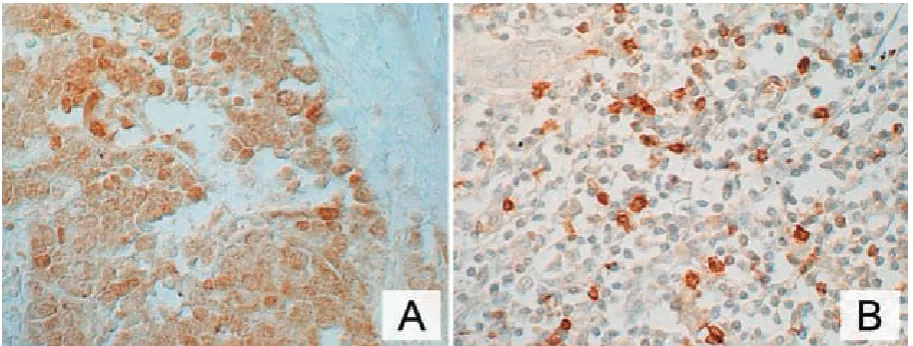
The presence of a positive reaction for T-lym-phocyte marker molecules was detected almost ex-clusively on cell membranes of tumor-infiltrating lymphocytes (Fig. 10A, B, C), which was comparable to the immunocytochemical reaction noted in reac-tive lymph nodes (the posireac-tive control) (Fig. 10D). As a rule, the immunocytochemical reaction was Fig. 3. Immunocytochemical localization of Il-2 in A) tumor cells in a typical carcinoid patient and B) individual cells of connective tissue stroma in an atypical carcinoid patient. ABC technique. Objective ×40; nuclei counterstained with hematoxylin (B)
Ryc. 3. Immunocytochemiczna lokalizacja Il-2 w komórkach nowotworowych u chorego z typowym rakowiakiem płuc (A) i w pojedynczych komórkach zrębu łącznotkankowego guza u chorego z atypowym rakowiakiem płuc (B). Technika ABC. Obiektyw 40×; jądra komórkowe podbarwione hematoksyliną (B)
Fig. 4. Comparison of mean values of total IL-2α expression in particular groups of patients
Ryc. 4. Porównanie średnich wartości ekspresji IL-2Rα całkowitej w poszczególnych grupach chorych
* NSCLC-SQ vs ADENO, p < 0.01.
the analogous expression in adenocarcinoma (Fig. 5A, Table 1).
In tumor-infiltrating lymphocytes (n), the most pronounced IL-2Rα expression was observed in NSCLC-SQ (Fig. 6B), which was significantly higher than the analogous expression in either AC or lung adenocarcinoma (Fig. 5B).
IL-2 Receptor β Subunit of (IL-2Rβ)
The most intense mean expression of the IL-2 receptor β subunit was noted in the group of patients with atypical lung carcinoid (Figure 7A); it was significantly higher than in small-cell lung cancer (SCLC) (Fig. 7B). The remaining groups of lung cancers did not differ significantly in total ex-pression of the marker (Fig. 8, Table 1).
demon-very intense, covering the entire membrane of T lymphocytes.
In patients with typical lung carcinoid, signifi-cant correlations were demonstrated between the expression of the CD4 molecule and the total IL-2 expression (r = 0.9380), and between CD8 expres-sion and the total IL-2Rβ expresexpres-sion (r = 0.8660). Similarly, in the SCLC group, a direct relationship was detected between CD3 expression and the to-tal tissue expression of IL-2Rβ (r = 0.6211).
Hybridocytochemical Studies
Studies using in situ hybridization in lung can-cers were conducted following the immunocyto-chemical examination of a given pulmonary tumor fragment. IL-2 and IL-2Rα mRNAs were detected using digoxygenin-labelled probes. Hybridocyto-chemical analysis of lung cancers was conducted on a total of 22 randomly selected representatives from the different patient groups; the data were not subjected to quantitative analysis. For all 22 of these patients, positive results were obtained in the hybridocytochemical reaction for both IL-2 mRNA Fig. 5. Comparison of mean values of IL-2α expression in A) tumor cells (g) and B) tumor infiltrating lymphocytes (n) in particular groups of patientsRyc. 5. Porównanie średnich wartości ekspresji IL-2α w komórkach guza (g) (A) i w limfocytach naciekających guzy (n) (B) między poszczególnymi grupami chorych
A) * AC vs ADENO, p < 0.01.
** SCLC vs ADENO, p < 0.05 NSCLC-SQ vs ADENO, p < 0.001. *** NSCLC-SQ vs ADENO, p < 0.001.
B) * NSCLC-SQ vs AC, p < 0.05. ** NSCLC-SQ vs ADENO, p < 0.01.
Fig. 6. Immunocytochemical localization of IL-2α in A) tumor cells in an atypical carcinoid patient and B) tumor-associated cells in a squamous cell carconoma patient. ABC technique. Objective ×40; nuclei counterstained with hematoxylin (B)
Ryc. 6. Immunocytochemiczna lokalizacja IL-2Rα w komórkach nowotworowych u chorego z atypowym rakowia-kiem płuc (A) i komórkach towarzyszących guzowi u chorego z płaskonabłonkowym rarakowia-kiem płuc (B). Technika ABC. Obiektyw 40×; jądra komórkowe podbarwione hematoksyliną (B)
and IL-2Rα mRNA. In the cases where IL-2 mRNA was examined, the positive hybridocytochemical reaction was in all cases (except one – a single pa-tient in the SCLC group) consistent with a positive result in the immunocytochemical reaction and with the presence of the IL-2 protein both in the tumor cells and in individual mononuclear cells in the tumor microenvironment. In cases of IL-2 mR-NA, nuclear localization prevailed, although the reaction product was also detected in the cell cy-toplasm (Fig. 11A). In cases where IL-2Rα mRNA was examined, a positive hybridocytochemical reaction was noted even when presence of the re-ceptor could not be shown using the immunocyto-chemical approach. In cases of IL-2Rα, the product of the hybridocytochemical reaction was in most cases noted mainly in cell nuclei, similar to the mRNA for IL-2 itself (Fig. 11B).
Fig. 7. Immunocytochemical localization of IL-2β in A) tumor cells in a typical carcinoid patient and B) tumor-infil-trating cells in a small cell lung cancer patient. ABC technique. Objective ×40; nuclei counterstained with hematoxylin
Ryc. 7. Immunocytochemiczna lokalizacja IL-2Rβ w komórkach nowotworowych u chorego z typowym rakowiakiem płuc (A) i komórkach infiltrujących guz u chorego z drobnokomórkowym rakiem płuc (B). Technika ABC. Obiektyw 40×; jądra komórkowe podbarwione hematoksyliną
Fig. 8. Comparison of mean values of total IL-2β expression in particular groups of patients
Ryc. 8. Porównanie średnich wartości ekspresji IL-2Rβ całkowitej w poszczególnych grupach chorych
* AC vs SCLC, p < 0.05
Fig. 9. Comparison of mean values of IL-2β expression in A) tumor cells (g) and B) tumor infiltrating lymphocytes (n) in particular groups of patients
Ryc. 9. Porównanie średnich wartości ekspresji IL-2β w komórkach nowotworowych guza (g) (A) i w TILs (n) (B) między poszczególnymi grupami pacjentów
Fig. 10. Immunocytochemical localization of A) CD3 in tumor-infiltrating cells in an adenocarcinoma patient; B) CD4 in tumor-infiltrating cells in a typical carcinoid patient; C) CD8 in tumor-infiltrating cells in a squamous cell carcinoma patient; and D), CD4 molecule in reactive lymph node cells (positive control). ABC technique. Objective ×40; nuclei counterstained with hematoxylin
Ryc. 10. Immunocytochemiczna lokalizacja: CD3 w komórkach infiltrujących guz u chorego z gruczołowym rakiem płuc (A), CD4 w komórkach naciekających guz u chorego z typowym rakowiakiem płuc (B), CD8 w komórkach naciekających guz u chorego z płaskonabłonkowym rakiem płuc (C) oraz lokalizacja cząsteczek CD4 w komórkach reaktywnego węzła chłonnego (kontrola pozytywna) (D). Technika ABC. Obiektyw 40×; jądra komórkowe podbar-wione hematoksyliną
Fig. 11. Hybridocytochemical localization of A) IL-2 in tumor cells and tumor-infiltrating cells in a non-small cell lung cancer patient; and B) IL-2Rα in tumor cells in an adenocarcinoma patient. Objective ×40; nuclei counterstained with hematoxylin
Discussion
The immunocytochemical and hybridocy-tochemical studies were conducted in the usual manner, on formalin-fixed and paraffin-embedded tissue material of typical and atypical lung carci-noids and two principal lung cancers: small-cell lung cancer (SCLC) and non-small cell lung cancer (NSCLC). In the latter histological subtype the tis-sue material included squamous cell lung cancer and adenocarcinoma. The credibility of the obtained re-sults was supported by statistical analysis.
The conducted analysis of cellular IL-2 expres-sion was focused first of all on establishing whether it was present in lung cancer cells (at the levels of protein and mRNA) and on comparing that to its analogous expression in lymphocytes infiltrating the tumors (TILs).
Studies in vitro and in vivo havedemonstrated that, like lymphocytes infiltrating pulmonary tu-mors, neoplastic cells themselves are also capable of producing cytokines, growth factors, chemot-actic molecules and proteases, which influence tu-mor growth, its immunogenicity and the immune response of the host [4, 15].
In lung cancers, IL-2 and its receptor have mainly been studied in patients’ immunocompe-tent blood cells (lymphocytes, monocytes), and the studies have been focused primarily on non-small cell lung cancers [4, 9, 12, 17, 18].
The current study has demonstrated that the cellular expression of IL-2 varies depending on the
histologic subtype of lung cancer. In the majority of the tumors studied, cytoplasmic expression of the IL-2 protein was detected in tumor cells and in TILs. It was only in some individual typical lung carcinoids that the product of immunocyto-chemical reaction was also detected in cell nuclei; this might have corresponded to growth control in the tumor cells [4]. IL-2 mRNA expression was confirmed in all the studied patients’ tissue mate-rial. The transcript for IL-2 and for the α chain of its receptor was localized mainly in the cell nuclei of both tumor cells and TILs. This confirmed the results of Li et al., who used in situhybridization to detect the presence of IL-2 mRNA in lung can-cers (NSCLC), albeit in lower quantities than the mRNAs for IL-4, IL-10 and TgF [22].
Comparing the „quantitative“ intensity of IL-2 expression in the various types of tumors ex-amined in this study, significantly more IL-2 was detected in typical lung carcinoids (TC), followed by atypical lung carcinoids (AC), than in squamous cell lung cancers and adenocarcinomas. TCs mani-fested an intense immunocytochemical reaction located mainly in the tumor cells themselves (and in individual cell nuclei). ACs mostly presented a moderately intense immunocytochemical reac-tion in the neoplastic cells and TILs; once again the predominant expression was in the neoplastic cells. However, the statistical analysis showed that TILs associated with ACs manifest the most pro-nounced IL-2 expression, as compared to other types of lung tumors. Atypical lung carcinoids Table 1. Comparative analysis of total IL-2, IL-2Rα and IL-2Rß expression, their expression in tumors and in the infiltrating cells in the analyzed groups of patients (Dunn’s test)
Tabela 1. Analiza porównawcza ekspresji IL-2, IL-2Rα, IL-2Rß całkowitych oraz w guzie i w nacieku między badanymi grupami chorych (test Dunna)
IL-2g IL-2n IL-2c IL-2Rαg IL-2Rαn IL-2Rαc IL-2Rßg IL-2Rßn IL-2Rßc
TC vs AC ns p < 0.05 ns ns ns ns ns ns ns
TC vs SCLC ns ns ns ns ns ns ns ns ns
TC vs SQ p < 0.001 ns p < 0.05 ns ns ns ns ns ns
TC vs ADENO p < 0.001 ns p < 0.001 ns ns ns ns ns ns
AC vs SCLC ns ns ns ns ns ns ns ns p < 0.05
AC vs SQ p < 0.05 ns p < 0.05 ns p < 0.05 ns ns ns ns
AC vs ADENO p < 0.05 p < 0.05 p < 0.001 p < 0.01 ns ns ns ns ns
SCLC vs SQ ns ns ns ns ns ns ns ns ns
SCLC vs ADENO ns ns ns p < 0.05 ns ns ns ns ns
SQ vs ADENO ns ns ns p < 0.001 p < 0.01 p < 0.01 ns ns ns
are among the neuroendocrine lung cancers with worse survival prognoses than typical lung carci-noids [26].
Among the lung cancers studied, the least pro-nounced expression of IL-2 in TILs was detected in adenocarcinoma – significantly lower than in TCs and ACs. This might result from suppressed production of the cytokine by TILs in this type of cancer and may indirectly indicate a poor prog-nosis. Reduced production of IL-2, indicating im-munosuppression, has been demonstrated in the case of SCLC [19]. In the current study, SCLC and NSCLC-SQ did not differ significantly from each other either in total IL-2 expression or in its expression in tumor/TIL compartments. Other authors have described a selective suppression of cytokine secretion (IL-2, IFN-α and γ) in pulmo-nary carcinomas, dependent on the size of the tu-mor and differing among patients with SCLC or NSCLC [17]. In the present study no significant differences between SCLC and NSC LC were de-tected in tissue expression of the cytokine.
It is worth emphasizing that in the adenocar-cinomas examined in this study, a positive corre-lation was documented between IL-2 expression in TILs and the expression of the α subunit of its receptor (IL-2Rα, CD25) in the same cells (indica-tive of autocrine action?) and in tumor cells of the same patients. This may indirectly indicate a po-tential for paracrine action of IL-2 on adenocarci-noma tumor cells.
IL-2 expression in TILs was associated with the T lymphocytes CD3+. On the other hand, the total expression of IL-2 itself in the patients stud-ied manifested a positive correlation with the ex-pression of the marker molecule of CD4+ helper T-lymphocytes, but only in typical lung carcinoids. Such relationships were not detected in any other type of the cancers.
Subunit α of the IL-2 receptor participates in binding IL-2, but it is not involved in the processes of internalization and signal transduction. Never-theless, the presence of the subunit in the IL-2R complex is a pre-condition for high-affinity bind-ing of IL-2 and the cytokine’s full biological activ-ity [27].
Cellular expression of the IL-2R α subunit (CD25) was more pronounced in squamous cell lung cancers than in the other tumors analyzed in this study, but a significant difference was docu-mented only in relation to adenocarcinomas, which manifested the lowest expression.
Some recent studies have been aimed at establish-ing a correlation between the expression of IL-2Rα (CD25) in tumor cells and their growth in vivo [3]. The results of those studies prove that overexpression of IL-2Rα is responsible for intensifying the
prolifera-tion of neoplastic cells. It is assumed that neoplastic IL-2Rα-positive cells proliferate more rapidly and demonstrate chromosomal instability, which may promote increased tumor aggressiveness, more pronounced resistance to drugs and a less favorable prognosis for the patient [3, 10, 28]. Elevated expres-sion of IL-2Rα mRNA has also been demonstrated in non-small cell lung cancers (squamous cell lung cancer and adenocarcinoma) as compared to such expression in a normal tissue [3].
The current study documented that the pres-ence of the IL-2R α chain (IL-2Rα, CD25) in all subtypes of lung cancers varies in terms of detecta-bility and intensity of expression. Production of the IL-2R subunit α and IL-2R (mRNA and protein) by all cells in all the representatives of lung cancers studied was confirmed, with the most pronounced expression in cells of squamous cell lung cancer and the least intense production in pulmonary ad-enocarcinoma cells (with a significant difference between them).
Using in situ hybridization, production of IL-2Rα mRNA was confirmed in all the lung can-cers studied, although the product was not evalu-ated in a quantitative manner. Nuclear localization of mRNA for the α chain of IL-2R was most often noted.
The immunohistochemical approach also showed that the presence of CD25 was prevalent in TILs accompanying squamous cell lung cancer, and that its expression was significantly higher than in TILs accompanying adenocarcinoma and/or atyp-ical lung carcinoids. A strong positive correlation was documented between the expression of IL-2Rα in tumors and TILs in TCs and adenocarcinoma (r = 0.7032). Naturally, CD25 expression was also documented in neoplastic cells in NSCLC-SQ, but the highest quantities were produced by atypical lung carcinoid tumor cells. CD25 molecules in TILs showed an exclusive membrane localization. In immunopositive neoplastic cells in pulmonary cancers, the product of immunocytochemical re-action was usually detected in the cytoplasm of the cells. In the tumor cells themselves the most pronounced tissue expression of the IL-2R subunit was observed in the group of atypical lung carcino-ids, followed by NSCLC-SQ and SCLC, and these expressions were significantly higher than the ex-pression in lung adenocarcinoma.
unequivocally. Further research, including func-tional studies, is required for an accurate assess-ment of the essence of tumor cell/TIL interactions in relation to the role of IL-2/IL-2R complex com-ponents. The prevalent immunocytochemical and hybridocytochemical expression of IL-2 and of IL-2Rβ (CD122) in tumor cells and of IL-2Rα in TILs regardless of the type of lung cancer involved may suggest their auto- and/or paracrine activity in the microenvironment of pulmonary tumors.
Due to its intra- and extracellular domains, IL-2Rβ plays the most important role in the trans-duction of a cellular signal following interaction with IL-2 [6].
The most pronounced expression of IL-2Rβ was demonstrated by atypical lung carcinoids (comparable to that in typical lung carcinoids); it was the least pronounced in small-cell lung can-cers (SCLC). The difference was significant.
The role of IL-2Rβ (CD122) expression in human neoplastic cells has not been widely docu-mented in the available literature, and the few ex-isting papers on the subject pertained not to lung cancers but to other solid tumors [4]. IL-2Rβ seems to play a significant role in growth control in neo-plastic cells, although the mechanisms involved have not been clearly identified. Some authors have noted distinct exogenous IL-2 activity, medi-ated by IL-2Rβ and γ subunits (growth inhibition), and the distinct effect of endogenous IL-2 effect on processes of cellular growth (growth control or stimulation of growth?) [4]. The authors of those studies have also suggested that IL-2Rβ can also be used by other cytokines than IL-2 and/or by growth factors.
In this study, a cytoplasmic localization of IL-2Rβ was most frequently documented in neoplastic cells, and a membraneous/cytoplasmic localization in tu-mor-infiltrating lymphocytes; the authors have not been able to confirm other studies’ documentation of a nuclear localization of the marker. According
to Lin et al. (1995), constitutively produced IL-2 is linked to its receptor on the cell surface. Subse-quently, it is internalized and translocated to the cell nucleus along with the β and γ chains. However, using flow cytometry Lin et al. also showed a cyto-plasmic localization of IL-2Rβ [4].
In the current study, immunocytochemical in-vestigations made it possible to demonstrate the variable expression of this type of IL-2 receptor in tumor cells as well as in tumor-infiltrating cells. No significant differences were found between tu-mors and TILS in terms of the localization of the expression in the lung cancers studied.
In none of the studied cases were there signifi-cant positive correlations between the expression of IL-2 itself and the expression of IL-2Rβ in lung cancers. On the other hand, almost ideal positive correlations were found between the “tumorous” and “infiltrative” expression of the IL-2Rβ subunit in AC, SCLC and squamous cell lung cancer. In typical lung carcinoids a very high positive correla-tion was detected between the expression of IL-2Rβ (CD122) and the expression of the cytotoxic T-lym- phocyte (CD8) marker molecule. In the case of SCLC, expression of this form of IL-2 receptor correlated with the expression of T-lymphocyte marker molecule (CD3). This may indirectly in-dicate the presence of IL-2Rβ epitopes on T lym-phocytes, including the subpopulation of suppres-sor/cytotoxic cells (CD8+), in accordance with data in the literature [2].
Cellular expression of IL-2 and both subu-nits of its receptor in neoplastic cells and TILs in the principal groups of lung cancers (carcinoids, SCLC, NSCLC), may indicate the markers’ in-volvement in growth control of both tumor cells and lymphocytes of the tumor microenvironment. To evaluate the prognostic significance of the ob-tained data further studies are required, and com-plete clinical data on patients with various types of lung cancers.
References
[1] Morgan DA, Ruscetti F, Gallo R: Selective in vitro growth of T lymphocytes from normal human bone marrow. Science 1976, 193, 1007–1008.
[2] Nelson BH: IL-2 regulatory T cells, and tolerance. J Immunol 2004, 172, 3983–3988.
[3] Kuhn DJ, Ping Dou Q: The role of interleukin-2 receptor alpha in cancer. Front Biosci 2005, 10, 1462–1474.
[4] Lin WC, Yasumura S, Suminami Y, Sung MW, Nagashima S, Stanson J, Whiteside TL: Constitutive produc-tion of IL-2 by human carcinoma cells, expression of IL-2 receptor, and tumor cell growth. J Immunol 1995, 155, 4805–4816.
[5] Minami Y, Oishi I, Liu Z, Nakagawa S, Miyazaki T, Taniguchi T: Signal transduction mediated by the reconsti-tuted IL-2 receptor. J Immunol 1994, 152, 5680–5690.
[6] Taniguchi T, Minami Y: The IL-2/IL-2 receptor system: A current overview. Cell 1993, 73, 5–8.
[7] Baier PK, Wolff-Vobeck G, Eggstein S, Baumgartner U, Hopt UT: Cytokine expression in colon carcinoma. Anticancer Res 2005, 25, 2135–2139.
[9] De Vita F, Turitto G, di Grazia M, Frattolillo A, Catalano G: Analysis of interleukin-2/interleukin-2 receptor system in advanced non-small cell lung cancer. Tumori 1998, 84, 33–38.
[10] Kuhn DJ, Smith DM, Pross S, Whiteside TL, Dou QP: Overexpression of interleukin-2 receptor in a human squamous cell carcinoma of the head and neck cell line is associated with increased proliferation, drug resistance, and transforming ability. J Cell Biochem 2003, 89, 824–836.
[11] Lissoni P, Barni S, Rovelli F, Rescaldani R, Rizzo V, Biondi A, Tancini G: Correlation of serum interleukin-2 levels, soluble interleukin-2 receptors and T lymphocyte subsets in cancer patients. Tumori 1990a, 76, 14–17.
[12] Tisi E, Lissoni P, Angeli M, Arrigoni C, Corno E, Cassina E, Ballabio D, Benenti C, Barni S, Tancini G:
Postoperative increase in soluble interleukin-2 receptor serum levels as predictor for early recurrence in non-small lung carcinoma. Cancer 1992, 69, 2458–2462.
[13] Olejniczak K, Kasprzak A: Biological properties of interleukin 2 and its role in pathogenesis of selected diseases – a review. Med Sci Monit 2008, 14, 179–189.
[14] Reichert TE, Watkins S, Stanson J, Johnson JT, Whiteside TL: Endogenous IL-2 in cancer cells: a marker of cel-lular proliferation. J Histochem Cytochem 1998, 46, 603–611.
[15] Żeromski J: Significance of tumor-cell receptors in human cancer. Arch Immunol Therap Exp 2002, 50, 105–110.
[16] Jemal A, Siegel R, Ward E, Hao Y, Xu J, Thun MJ: Cancer Statistics, 2009. Ca Cancer J Clin 2009, 59, 225–249.
[17] Fischer JR, Schindel M, Stein N, Lahm H, Gallati H, Krammer PH, Drings P: Selective suppression of cytokine secretion in patients with small-cell lung cancer. Ann Oncol 1995, 6, 921–926.
[18] Lissoni P, Barni S, Rovelli F, Viviani S, Maestroni GJ, Conti A, Tancini G: The biological significance of soluble interleukin-2 receptors in solid tumors. Eur J Cancer 1990b, 26, 33–36.
[19] Fischer JR, Schindel M, Bulzebruck H, Lahm H, Krammer PH, Drings P: Decrease of interleukin-2 secretion is a new independent prognostic factor associated with poor survival in patients with small-cell lung cancer. Ann Oncol 1997, 8, 457–461.
[20] Huang M, Wang J, Lee P, Sharma S, Mao JT, Meissner H, Uyemura K, Modlin R, Wollman J, Dubinett SM:
Human non-small cell lung cancer cells express a type 2 cytokine pattern. Cancer Res 1995, 55, 3847–3853.
[21] Ortegel JW, Staren ED, Faber LP, Warren WH, Braun DP: Modulation of tumor-infiltrating lymphocyte cytoli-tic activity against human non-small cell lung cancer. Lung Cancer 2002, 36, 17–25.
[22] Li R, Rüttinger D, Li R, Si LS, Wang YL: Analysis of the immunological microenvironment at the tumor site in patients with non-small cell lung cancer. Langenbecks Arch Surg 2003, 388, 406–412.
[23] Yanelli JR, Tucker JA, Hidalgo G, Perkins S, Kryscio R, Hirschowitz EA: Characteristics of PBMC obtained from leukapheresis products and tumor biopsies of patients with non-small cell lung cancer. Oncol Rep 2009, 22, 1459–1471.
[24] Hsu S, Raine L, Fanger H: Use a avidin-biotin peroxidase complex (ABC) in immunoperoxidase techniques. J Histochem Cytochem 1981, 29, 577–580.
[25] Remmele W, Stegner HE: Vorschlag zur einheitlichen Definition eines immunreaktiven Score (IRS) fur den Immunohistochemichen Ostrogenrezeptor-Nachweis (ER-ICA) im Mammikarzinomgewebe. Patologie 1987, 8, 138–140.
[26] Yesner R: Heterogeneity of so-called neuroendocrine lung tumors. Exp Mol Pathol 2001, 70, 179–182.
[27] Gutgsell NS, Malek TR: Formation of high affinity IL-2 receptors is dependent on a nonligand binding region of the α subunit. J Immunol 1994, 153, 3899–3907.
[28] Garcia-Tunnon J, Ricote M, Ruiz A, Fraile B, Paniagua R, Royuela M: Interleukin-2 and its receptor complex in situ and infiltrative human breast cancer: an immunohistochemical comparative study. Breast Cancer Res 2004, 6, 1–7.
[29] Marc MM, Korosec P, Kern I, Sok M, Ihan A, Kosnik M: Lung tissue and tumor-infiltrating T lymphocytes in patients with non-small cell lung carcinoma and chronic obstructive pulmonary disease (COPD): moderate/severe versus mild stage of COPD. Scand I Immunol 2007, 66, 694–702.
[30] Trentin L, Zambello R, Bulian P, Cerutti A, Milani A, Pirone E, Nitti D, Agostini C, Semenzato G: Functional role of IL-2 receptors on tumor-infiltrating lymphocytes. Br J Cancer 1994, 69, 1046–1051.
Address for correspondence:
Karolina Sterzyńska
Department of Histology and Embryology Poznań University of Medical Sciences Święcickiego 6
60-781 Poznań, Poland Tel.: +48 61 854 64 52
E-mail: [email protected] Conflict of interest: None declared Received: 21.12.2010