Received 1 April 2019 Accepted 9 April 2019
Edited by M. Weil, Vienna University of Technology, Austria
Keywords:crystal structure; hydrogen bond; C—H (ring) interactions; pyrazolo-pyrimidine; Hirshfeld surface analysis.
CCDC reference:1909062
Supporting information:this article has supporting information at journals.iucr.org/e
Crystal structure and Hirshfeld surface analysis of
3-(4-methoxyphenyl)-1-methyl-4-phenyl-1
H
-pyrazolo[3,4-
d
]pyrimidine
Mohamed El Hafi,a* Sevgi Kansiz,b* Sanae Lahmidi,aMohammed Boulhaoua,a Youssef Ramli,cNecmi Dege,b El Mokhtar Essassiaand Joel T. Magued
aLaboratoire de Chimie Organique He´te´rocyclique, Centre de Recherche Des Sciences des Me´dicaments, Poˆle de
Compe´tence Pharmacochimie, Av Ibn Battouta, BP 1014, Faculte´ des Sciences, Universite´ Mohammed V, Rabat, Morocco,bOndokuz Mayıs University, Faculty of Arts and Sciences, Department of Physics, 55139, Kurupelit, Samsun,
Turkey,cLaboratory of Medicinal Chemistry, Faculty of Medicine and Pharmacy, Mohammed V, University Rabat,
Morocco, anddDepartment of Chemistry, Tulane University, New Orleans, LA 70118, USA. *Correspondence e-mail:
[email protected], [email protected]
In the title molecule, C19H16N4O, the planar pyrazolopyrimidine moiety is
inclined to the attached phenyl rings by 35.42 (4) and 54.51 (6). In the crystal,
adjacent molecules are linked into chains parallel to [110] and [110] by C— H O and C—H N hydrogen bonds. Additional C—H (ring) interactions lead to the formation of the final three-dimensional network structure. The Hirshfeld surface analysis of the title compound suggests that the most significant contributions to the crystal packing are from H H (48.2%), C H/ H C (23.9%) and N H/H N (17.4%) contacts.
1. Chemical context
Pyrazolo[3,4-d]pyrimidine derivatives represent an important class of compounds because of their potent biological activ-ities and thus find applications as antiproliferative (Tallaniet al., 2010), antibacterial (Rostamizadeh et al., 2013) or anti-tumor agents (Tintoriet al., 2015). The present contribution is a continuation of the investigation of pyrazolo[3,4-d ]pyrimi-dine derivatives recently published by us (El Hafiet al., 2017, 2018a,b). We report herein the synthesis, molecular and crystal structures of the title compound, 3-(4-methoxyphenyl)-1-methyl-4-phenyl-1H-pyrazolo[3,4-d]pyrimidine (Fig. 1), along with the results of a Hirshfeld surface analysis.
2. Structural commentary
The heterocyclic ring system is planar (r.m.s. deviation of the fitted atoms = 0.0194 A˚ ) with a maximum displacement of 0.0329 (10) A˚ from the mean plane for atom C1. The attached
benzene rings (C6–C11 and C13–C18) are inclined to the above plane by 35.42 (4) and 54.51 (6), respectively.
3. Supramolecular features
In the crystal, a combination of C9—H9 N2 hydrogen bonds between aromatic hydrogen atoms and one of the pyrimidine N atoms as well as C12—H12B O1 hydrogen bonds between a methyl H atom and the methoxy O atoms of adjacent mol-ecules lead to the formation of chains extending alternately parallel to [110] and [110] (Table 1 and Fig. 2). Centrosym-metric dimers with an R2
2(8) graph-set motif are formed by
pairwise C17—H17 O1 hydrogen bonds. The chains are linked into layers parallel to (001) by C19—H19C Cg1 interactions, and pairs of layers are joined into thicker slabs by C19—H19B Cg4 interactions (Table 1 and Figs. 2–4).
4. Database survey
A search of the Cambridge Structural Database (CSD, version 5.40, update November 2018; Groom et al., 2016) for the 1-methyl-1H-pyrazolo[3,4-d]pyrimidine skeleton yielded seven hits. In all of these structures, the pyrazolo[3,4-d
]pyrimi-research communications
Acta Cryst.(2019). E75, 638–641 El Hafiet al. C
[image:2.610.316.564.67.367.2]19H16N4O
639
Table 1Hydrogen-bond geometry (A˚ ,).
Cg1 andCg4 are the centroids of the C3/C4/C5/N4/N3 and C13–C18 rings, respectively.
D—H A D—H H A D A D—H A
C9—H9 N2i 0.988 (18) 2.579 (17) 3.3995 (19) 140.3 (14) C12—H12B O1ii 0.98 (2) 2.49 (2) 3.2694 (19) 136.4 (15) C17—H17 O1iii 0.983 (17) 2.618 (9) 3.4973 (17) 149.3 (14) C19—H19B Cg4iv 1.02 (2) 2.74 (2) 3.5928 (19) 141.9 (14) C19—H19C Cg1v 0.995 (19) 2.947 (19) 3.9072 (19) 162.0 (15)
[image:2.610.46.297.118.186.2]Symmetry codes: (i)x1;yþ1;z; (ii)xþ1;y1;z; (iii) x;yþ1;zþ1; (iv) xþ1;yþ1;zþ1; (v)x;yþ1;z.
Figure 2
[image:2.610.316.568.442.563.2]Details of the intermolecular interactions in a view along [100]. C— H O and C—H N hydrogen bonds are shown, respectively, as black and light-purple dashed lines while the C—H (ring) interactions are shown as green dashed lines. [Symmetry codes: (i)x+ 1,y+ 1,z+ 1; (ii)x+ 1,y1,z; (iii)x1,y+ 1,z.]
Figure 3
[image:2.610.54.284.514.721.2]Packing of the crystal viewed along [100] with intermolecular interactions depicted as in Fig. 2.
Figure 1
The title molecule with the labeling scheme and displacement ellipsoids drawn at the 50% probability level.
Figure 4
[image:2.610.314.566.611.722.2]dine rings are planar, as in the title compound. In FEWVIP (El Hafiet al., 2018a), centrosymmetric dimers with an R2
2(8)
graph set motif are formed by pairwise N—H O hydrogen bonds; the dimers are connected into chains parallel to [001], similar to those in the title compound. Neighbouring mol-ecules in FAXFEP (Sheldrick & Bell, 1987a) and in FOGXAA, FOGXEE, FOGXII, JAGROY (Sheldrick & Bell, 1987b) are linked by N—H O hydrogen bonds, whereas in XAXRUM (El Falet al., 2017), C—H N hydrogen bonds are responsible for the formation of double chains running parallel to [100].
5. Hirshfeld surface analysis
CrystalExplorer17 (Turner et al., 2017) was used to perform the Hirshfeld surface analysis (Spackman & Jayatilaka, 2009) and obtain the associated two-dimensional fingerprint plots (McKinnonet al., 2007). Fig. 5 showsdnorm,di,de, shape-index,
curvedness and electrostatic potential mapped over the Hirshfeld surface for the title compound while Fig. 6 illustrates the Hirshfeld surface of the molecule in the crystal, with the evident hydrogen-bonding interactions indicated as intense red spots.
Fig. 7ashows the two-dimensional fingerprint of the sum of the contacts contributing to the Hirshfeld surface represented in normal mode. The fingerprint plots provide information about the percentage contributions of various interatomic
contacts in the structure. The blue color refers to the frequency of occurrence of the (di, de) pair with the full
fingerprint outlined in gray. Individual fingerprint plots (Fig. 7b) reveal that the H H contacts clearly give the most significant contribution to the Hirshfeld surface (48.2%). In addition, C H/H C, N H/H N, O H/H O and C N/N C contacts contribute 23.9%, 17.4%, 5.3% and 2.6%, respectively, to the Hirshfeld surface. In particular, the N H/H N and O H/H O contacts indicate the presence of intermolecular C—H N and C—H O inter-actions, respectively. Much weaker C C (2.2%) and C O/ O C (0.5%) contacts also occur.
A view of the molecular electrostatic potential, in the range
[image:3.610.313.566.67.236.2]0.0500 to 0.0500 a.u. using the 6-31G(d,p) basis set (DFT method), for the title compound is shown in Fig. 8. The donors
Figure 5
The Hirshfeld surfaces of the title compound mapped over (a)dnorm, (b)
[image:3.610.44.295.69.215.2]di, (c)de, (d) shape-index, (e) curvedness and (f) electrostatic potential.
Figure 6
dnormmapped on Hirshfeld surfaces for visualizing the intermolecular interactions of the title compound.
Figure 7
[image:3.610.348.528.493.718.2]Two-dimensional fingerprint plots for the title structure, with adnormview and relative contribution of the atom pairs to the Hirshfeld surface.
Figure 8
[image:3.610.45.293.614.723.2]and acceptors for C—H O and C—H N hydrogen bonds are shown as blue and red areas around the atoms related with positive bond donors) and negative (hydrogen-bond acceptors) electrostatic potentials, respectively.
6. Synthesis and crystallization
Under an atmosphere of argon, a mixture of 1-methyl-4-phenyl-1H-pyrazolo[3,4-d]pyrimidine (0.1 g, 0.47 mmol), 4-iodoanisole (0.22 g, 0.95 mmol), Cs2CO3(0.46g, 1.42 mmol),
K3PO4 (0.25 g, 1.18 mmol), 1,10-phenanthroline (0.034 g,
0.19 mmol), and Pd(OAc)2 (0.021 g, 0.094 mmol) in DMA
(3 ml) was heated to 438 K for 48 h. After completion of the reaction, the mixture was allowed to cool to room temperature and the solvent was removed under reduced pressure. Water (15 ml) was added, and the resulting aqueous phase was extracted with CH2Cl2 (3 15 ml). The combined organic
layers were dried with MgSO4 and concentrated under
vacuum. The residue was purified by column chromatography on silica gel (EtOAc/petroleum ether). The title compound was recrystallized from ethanol at room temperature, giving colorless crystals (yield: 71%; m.p. 412–414 K).
7. Refinement
Crystal data, data collection and structure refinement details are summarized in Table 2. All H atoms were located in a difference-Fourier map and were freely refined.
Funding information
The support of NSF–MRI grant No. 1228232 for the purchase of the diffractometer and Tulane University for support of the Tulane Crystallography Laboratory are gratefully acknowl-edged.
References
Brandenburg, K. & Putz, H. (2012). DIAMOND, Crystal Impact GbR, Bonn, Germany.
Bruker (2016). APEX3 and SAINT. Bruker AXS, Inc., Madison, Wisconsin, USA.
El Fal, M., Mague, J. T., Taoufik, J., Essassi, E. M. & Ramli, Y. (2017).
IUCrData,2, x171042.
El Hafi, M., Boulhaoua, M., Lahmidi, S., Ramli, Y., Essassi, E. M. & Mague, J. T. (2018a).IUCrData,3, x180243.
El Hafi, M., Lahmidi, S., Boulhaoua, M., Ramli, Y., Essassi, E. M. & Mague, J. T. (2018b).IUCrData,3, x180483.
El Hafi, M., Naas, M., Loubidi, M., Jouha, J., Ramli, Y., Mague, J. T., Essassi, E. M. & Guillaumet, G. (2017).C. R. Chim.20, 927–933. Groom, C. R., Bruno, I. J., Lightfoot, M. P. & Ward, S. C. (2016).Acta
Cryst.B72, 171–179.
Krause, L., Herbst-Irmer, R., Sheldrick, G. M. & Stalke, D. (2015).J. Appl. Cryst.48, 3–10.
McKinnon, J. J., Jayatilaka, D. & Spackman, M. A. (2007).Chem. Commun.pp. 3814–3816.
Rostamizadeh, S., Nojavan, M., Aryan, R., Sadeghian, H. & Davoodnejad, M. (2013).Chin. Chem. Lett.24, 629–632.
Sheldrick, G. M. (2008).Acta Cryst.A64, 112–122. Sheldrick, G. M. (2015a).Acta Cryst.A71, 3–8. Sheldrick, G. M. (2015b).Acta Cryst.C71, 3–8.
Sheldrick, W. S. & Bell, P. (1987a).Z. Naturforsch. Teil B,42, 195– 202.
Sheldrick, W. S. & Bell, P. (1987b). Inorg. Chim. Acta, 137, 181– 188.
Spackman, M. A. & Jayatilaka, D. (2009).CrystEngComm,11, 19–32. Taliani, S., La Motta, C., Mugnaini, L., Simorini, F., Salerno, S., Marini, A. M., Da Settimo, F., Cosconati, S., Cosimelli, B., Greco, G., Limongelli, V., Marinelli, L., Novellino, E., Ciampi, O., Daniele, S., Trincavelli, M. L. & Martini, C. (2010).J. Med. Chem.53, 3954– 3963.
Tintori, C., Fallacara, A. L., Radi, M., Zamperini, C., Dreassi, E., Crespan, E., Maga, G., Schenone, S., Musumeci, F., Brullo, C., Richters, A., Gasparrini, F., Angelucci, A., Festuccia, C., Delle Monache, S., Rauh, D. & Botta, M. (2015).J. Med. Chem.58, 347– 361.
Turner, M. J., MacKinnon, J. J., Wolff, S. K., Grimwood, D. J., Spackman, P. R., Jayatilaka, D. & Spackman, M. A. (2017).
CrystalExplorer17. University of Western Australia. http://hirsh-feldsurface.net.
research communications
Acta Cryst.(2019). E75, 638–641 El Hafiet al. C
[image:4.610.43.296.88.365.2]19H16N4O
641
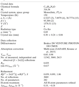
Table 2Experimental details.
Crystal data
Chemical formula C19H16N4O
Mr 316.36
Crystal system, space group Monoclinic,P21/n
Temperature (K) 150
a,b,c(A˚ ) 6.5227 (3), 7.8979 (4), 30.7774 (15)
() 95.389 (2)
V(A˚3) 1578.51 (13)
Z 4
Radiation type CuK
(mm1) 0.69
Crystal size (mm) 0.300.240.04 Data collection
Diffractometer Bruker D8 VENTURE PHOTON 100 CMOS
Absorption correction Multi-scan (SADABS; Krauseet al., 2015)
Tmin,Tmax 0.85, 0.98
No. of measured, independent and observed [I> 2(I)] reflections
11542, 3069, 2613
Rint 0.035
(sin/)max(A˚
1
) 0.617
Refinement
R[F2> 2(F2)],wR(F2),S 0.039, 0.091, 1.06 No. of reflections 3069
No. of parameters 282
H-atom treatment All H-atom parameters refined
max, min(e A˚
3
) 0.19,0.18
sup-1
Acta Cryst. (2019). E75, 638-641
supporting information
Acta Cryst. (2019). E75, 638-641 [https://doi.org/10.1107/S2056989019004894]
Crystal structure and Hirshfeld surface analysis of
3-(4-methoxyphenyl)-1-methyl-4-phenyl-1
H
-pyrazolo[3,4-
d
]pyrimidine
Mohamed El Hafi, Sevgi Kansiz, Sanae Lahmidi, Mohammed Boulhaoua, Youssef Ramli, Necmi
Dege, El Mokhtar Essassi and Joel T. Mague
Computing details
Data collection: APEX3 (Bruker, 2016); cell refinement: SAINT (Bruker, 2016); data reduction: SAINT (Bruker, 2016); program(s) used to solve structure: SHELXT (Sheldrick, 2015a); program(s) used to refine structure: SHELXL2018
(Sheldrick, 2015b); molecular graphics: DIAMOND (Brandenburg & Putz, 2012); software used to prepare material for publication: SHELXTL (Sheldrick, 2008).
3-(4-Methoxyphenyl)-1-methyl-4-phenyl-1H-pyrazolo[3,4-d]pyrimidine
Crystal data
C19H16N4O
Mr = 316.36
Monoclinic, P21/n
a = 6.5227 (3) Å
b = 7.8979 (4) Å
c = 30.7774 (15) Å
β = 95.389 (2)°
V = 1578.51 (13) Å3
Z = 4
F(000) = 664
Dx = 1.331 Mg m−3
Cu Kα radiation, λ = 1.54178 Å Cell parameters from 8514 reflections
θ = 2.9–72.2°
µ = 0.69 mm−1
T = 150 K Plate, colourless 0.30 × 0.24 × 0.04 mm
Data collection
Bruker D8 VENTURE PHOTON 100 CMOS diffractometer
Radiation source: INCOATEC IµS micro-focus source
Mirror monochromator
Detector resolution: 10.4167 pixels mm-1
ω scans
Absorption correction: multi-scan (SADABS; Krause et al., 2015)
Tmin = 0.85, Tmax = 0.98 11542 measured reflections 3069 independent reflections 2613 reflections with I > 2σ(I)
Rint = 0.035
θmax = 72.2°, θmin = 5.8°
h = −7→8
k = −9→8
l = −37→33
Refinement
Refinement on F2 Least-squares matrix: full
R[F2 > 2σ(F2)] = 0.039
wR(F2) = 0.091
S = 1.06 3069 reflections 282 parameters
0 restraints
Hydrogen site location: difference Fourier map All H-atom parameters refined
w = 1/[σ2(F
o2) + (0.0337P)2 + 0.5793P] where P = (Fo2 + 2Fc2)/3
supporting information
sup-2
Acta Cryst. (2019). E75, 638-641
Δρmin = −0.18 e Å−3 Extinction correction: SHELXL2018 (Sheldrick, 2015b), Fc*=kFc[1+0.001xFc2λ3/sin(2θ)]-1/4 Extinction coefficient: 0.0035 (3)
Special details
Geometry. All esds (except the esd in the dihedral angle between two l.s. planes) are estimated using the full covariance matrix. The cell esds are taken into account individually in the estimation of esds in distances, angles and torsion angles; correlations between esds in cell parameters are only used when they are defined by crystal symmetry. An approximate (isotropic) treatment of cell esds is used for estimating esds involving l.s. planes.
Refinement. Refinement of F2 against ALL reflections. The weighted R-factor wR and goodness of fit S are based on F2, conventional R-factors R are based on F, with F set to zero for negative F2. The threshold expression of F2 > 2sigma(F2) is used only for calculating R-factors(gt) etc. and is not relevant to the choice of reflections for refinement. R-factors based on F2 are statistically about twice as large as those based on F, and R- factors based on ALL data will be even larger.
Fractional atomic coordinates and isotropic or equivalent isotropic displacement parameters (Å2)
x y z Uiso*/Ueq
sup-3
Acta Cryst. (2019). E75, 638-641
C17 0.1517 (2) 0.39289 (19) 0.44698 (5) 0.0313 (3) H17 0.017 (3) 0.394 (2) 0.4587 (6) 0.041 (5)* C18 0.2157 (2) 0.25287 (19) 0.42500 (5) 0.0300 (3) H18 0.124 (2) 0.157 (2) 0.4201 (5) 0.033 (4)* C19 0.3310 (3) 0.8127 (2) 0.48265 (6) 0.0407 (4) H19A 0.252 (3) 0.891 (3) 0.5003 (6) 0.057 (6)* H19B 0.471 (3) 0.782 (2) 0.4976 (7) 0.054 (5)* H19C 0.356 (3) 0.862 (2) 0.4538 (6) 0.046 (5)*
Atomic displacement parameters (Å2)
U11 U22 U33 U12 U13 U23
O1 0.0375 (6) 0.0338 (6) 0.0346 (6) 0.0032 (4) 0.0009 (4) −0.0060 (4) N1 0.0310 (6) 0.0318 (7) 0.0298 (6) 0.0027 (5) 0.0009 (5) −0.0055 (5) N2 0.0314 (6) 0.0314 (6) 0.0314 (7) 0.0052 (5) 0.0035 (5) −0.0016 (5) N3 0.0262 (6) 0.0331 (7) 0.0304 (6) 0.0050 (5) −0.0010 (5) 0.0001 (5) N4 0.0277 (6) 0.0338 (7) 0.0297 (6) 0.0020 (5) 0.0001 (5) −0.0002 (5) C1 0.0248 (6) 0.0284 (7) 0.0240 (7) −0.0009 (5) 0.0034 (5) 0.0004 (5) C2 0.0348 (8) 0.0334 (8) 0.0330 (8) 0.0031 (6) 0.0027 (6) −0.0068 (6) C3 0.0261 (7) 0.0297 (7) 0.0258 (7) 0.0013 (5) 0.0039 (5) 0.0019 (6) C4 0.0251 (6) 0.0295 (7) 0.0254 (7) −0.0005 (5) 0.0012 (5) 0.0013 (5) C5 0.0247 (6) 0.0265 (7) 0.0238 (7) 0.0006 (5) 0.0031 (5) 0.0008 (5) C6 0.0272 (7) 0.0298 (7) 0.0201 (6) 0.0023 (5) 0.0022 (5) 0.0008 (5) C7 0.0340 (8) 0.0304 (7) 0.0265 (7) 0.0003 (6) −0.0006 (6) 0.0004 (6) C8 0.0489 (9) 0.0295 (8) 0.0312 (8) 0.0066 (7) 0.0029 (7) 0.0009 (6) C9 0.0402 (8) 0.0421 (9) 0.0320 (8) 0.0161 (7) 0.0060 (6) 0.0047 (7) C10 0.0274 (7) 0.0465 (9) 0.0302 (8) 0.0059 (6) 0.0024 (6) 0.0043 (7) C11 0.0277 (7) 0.0333 (8) 0.0251 (7) 0.0008 (6) 0.0025 (5) 0.0006 (6) C12 0.0273 (8) 0.0407 (9) 0.0421 (10) 0.0082 (6) −0.0004 (7) 0.0055 (7) C13 0.0274 (7) 0.0304 (7) 0.0216 (7) 0.0007 (5) −0.0017 (5) −0.0004 (5) C14 0.0268 (7) 0.0338 (8) 0.0285 (7) −0.0015 (6) 0.0009 (6) −0.0003 (6) C15 0.0322 (7) 0.0304 (8) 0.0301 (8) −0.0036 (6) 0.0001 (6) −0.0010 (6) C16 0.0330 (7) 0.0312 (7) 0.0219 (7) 0.0049 (6) −0.0030 (5) −0.0006 (5) C17 0.0252 (7) 0.0395 (8) 0.0286 (7) 0.0013 (6) 0.0001 (6) −0.0010 (6) C18 0.0270 (7) 0.0330 (8) 0.0294 (7) −0.0034 (6) −0.0010 (6) −0.0015 (6) C19 0.0536 (10) 0.0306 (8) 0.0374 (9) −0.0015 (7) 0.0012 (8) −0.0042 (7)
Geometric parameters (Å, º)
supporting information
sup-4
Acta Cryst. (2019). E75, 638-641
N4—C4 1.3310 (17) C13—C18 1.401 (2) C1—C5 1.4087 (19) C14—C15 1.393 (2) C1—C6 1.4778 (18) C14—H14 0.997 (17) C2—H2 0.993 (17) C15—C16 1.389 (2) C3—C5 1.4089 (18) C15—H15 1.025 (15) C4—C5 1.4351 (19) C16—C17 1.393 (2) C4—C13 1.4753 (19) C17—C18 1.381 (2) C6—C7 1.395 (2) C17—H17 0.983 (17) C6—C11 1.3982 (19) C18—H18 0.973 (17) C7—C8 1.387 (2) C19—H19A 1.00 (2) C7—H7 0.987 (17) C19—H19B 1.02 (2) C8—C9 1.384 (2) C19—H19C 0.995 (19) C8—H8 0.990 (18)
sup-5
Acta Cryst. (2019). E75, 638-641
C9—C10—C11 119.94 (14) H19B—C19—H19C 106.3 (15)
C3—N3—N4—C4 −0.08 (15) C5—C1—C6—C7 36.6 (2) C12—N3—N4—C4 −179.99 (13) N1—C1—C6—C11 34.01 (18) C2—N1—C1—C5 −2.4 (2) C5—C1—C6—C11 −147.05 (14) C2—N1—C1—C6 176.60 (13) C11—C6—C7—C8 1.8 (2) C3—N2—C2—N1 2.3 (2) C1—C6—C7—C8 178.07 (13) C1—N1—C2—N2 −1.2 (2) C6—C7—C8—C9 −0.1 (2) C2—N2—C3—N3 −179.20 (14) C7—C8—C9—C10 −1.6 (2) C2—N2—C3—C5 0.1 (2) C8—C9—C10—C11 1.6 (2) N4—N3—C3—N2 −179.83 (13) C9—C10—C11—C6 0.1 (2) C12—N3—C3—N2 0.1 (2) C7—C6—C11—C10 −1.8 (2) N4—N3—C3—C5 0.79 (16) C1—C6—C11—C10 −178.18 (13) C12—N3—C3—C5 −179.31 (14) N4—C4—C13—C14 52.12 (18) N3—N4—C4—C5 −0.66 (15) C5—C4—C13—C14 −130.58 (16) N3—N4—C4—C13 177.20 (11) N4—C4—C13—C18 −124.27 (15) N1—C1—C5—C3 4.26 (19) C5—C4—C13—C18 53.0 (2) C6—C1—C5—C3 −174.66 (12) C18—C13—C14—C15 0.7 (2) N1—C1—C5—C4 −179.91 (16) C4—C13—C14—C15 −175.81 (13) C6—C1—C5—C4 1.2 (3) C13—C14—C15—C16 0.7 (2) N2—C3—C5—C1 −3.2 (2) C19—O1—C16—C15 2.7 (2) N3—C3—C5—C1 176.16 (12) C19—O1—C16—C17 −177.26 (13) N2—C3—C5—C4 179.52 (13) C14—C15—C16—O1 178.62 (13) N3—C3—C5—C4 −1.11 (15) C14—C15—C16—C17 −1.4 (2) N4—C4—C5—C1 −175.01 (16) O1—C16—C17—C18 −179.36 (13) C13—C4—C5—C1 7.5 (3) C15—C16—C17—C18 0.7 (2) N4—C4—C5—C3 1.10 (15) C16—C17—C18—C13 0.8 (2) C13—C4—C5—C3 −176.36 (14) C14—C13—C18—C17 −1.4 (2) N1—C1—C6—C7 −142.30 (14) C4—C13—C18—C17 175.00 (13)
Hydrogen-bond geometry (Å, º)
Cg1 and Cg4 are the centroids of the C3/C4/C5/N4/N3 and C13–C18 rings, respectively.
D—H···A D—H H···A D···A D—H···A
C9—H9···N2i 0.988 (18) 2.579 (17) 3.3995 (19) 140.3 (14) C12—H12B···O1ii 0.98 (2) 2.49 (2) 3.2694 (19) 136.4 (15) C17—H17···O1iii 0.983 (17) 2.618 (9) 3.4973 (17) 149.3 (14) C19—H19B···Cg4iv 1.02 (2) 2.74 (2) 3.5928 (19) 141.9 (14) C19—H19C···Cg1v 0.995 (19) 2.947 (19) 3.9072 (19) 162.0 (15)